Department of Pediatrics, University of São Paulo School of Medicine, São Paulo SP, Brazil. (FMUSP): 1Livre-Docente em Pediatria pela FMUSP, Médica Assistente encarregada de Setor da Unidade de Cuidados Intensivos Neonatal do Instituto da Criança do Hospital das Clínicas da FMUSP; 2Doutora em Ciências pela FMUSP, Médica Chefe do Laboratório de Investigação Médica-36 da FMUSP; 3Professor Titular do Departamento de Pediatria da FMUSP; 4Professor Titular do Departamento de Pediatria da FMUSP. This work was sup-ported by a grant from Fundação de Auxílio à Pesquisa do Estado de São Paulo (FAPESP).
Received 3 June 2004. Accepted 11 September 2004.
Dra. Vera Lúcia Jornada Krebs - Rua Cristiano Viana 450/122 - 05411-000 São Paulo SP - Brasil. E-mail: [email protected]
TUMOR NECROSIS FACTOR-
α
a
, INTERLEUKIN-1
β
b
AND INTERLEUKIN-6 IN THE CEREBROSPINAL
FLUID OF NEWBORN WITH MENINGITIS
Vera Lúcia Jornada Krebs1, Thelma Suely Okay2, Yassuhiko Okay3, Flávio Adolfo Costa Vaz4
ABSTRACT - Objective: To analyze the usefulness of determining the cerebrospinal fluid(CSF) levels of tumor necrosis factor-α(TNF-α),interleukin-1β(IL-1β) and interleukin-6(IL-6)for the early diagnosis and evaluation of the prognosis of neonatal meningitis. Method:We studied 54 newborn that underwent lumbar punc-ture.Thirty patients had meningitis and 24 were the control group.CSF and sera were obtained at the moment of suspicion of meningitis and stored at -700C.Cytokines were performed by enzyme-linked
immunosor-bent assay method. Results:CSF cytokines were detected in all the newborn with meningitis.TNF-αwas detected in the CSF in 63.3% of the neonates, IL-1βin 73.3% and IL-6 in 96.6%.The CSF levels were signifi-cantly higher than serum in neonates with meningitis.There was no correlation between the CSF levels of cytokines and neurologic complications. Conclusion: The detection of TNF-α, IL-1βand IL-6 in the CSF is of great value in order to achieve a early diagnosis of neonatal meningitis.Among the three cytokines ana-lyzed, IL-6 was the best indicator of meningeal inflammation.
KEY WORDS: meningitis, cerebrospinal fluid, newborn, tumor necrosis factor-α, interleukin-1β, interleukin-6.
Fator de necrose tumoral-αa, interleucina-1βbe interleucina-6 no líquido cefalorraqueano de recém-nascidos com meningite
RESUMO - Objetivo:Analisar a utilidade da dosagem dos níveis de fator de necrose tumoral-α(TNF-α), inter-leucin-1β(IL-1β) e interleucina-6(IL-6) no líquido cefalorraqueano (LCR) para o diagnóstico precoce e avalia-ção do prognóstico da meningite neonatal. Método:Foram estudados 54 recém-nascidos submetidos à punção lombar.Trinta pacientes apresentavam meningite e 24 constituíram o grupo controle. As amostras de LCR e sangue foram obtidas no momento da suspeita clínica de meningite e estocadas a - 700C.A
dosa-gem de citocinas foi feita pelo método ELISA (enzyme-linked immunosorbent assay). Resultados: Foram detectadas citocinas no LCR em todos os neonatos com meningite. O TNF-αfoi detectado em 63,3% dos casos, a IL-1βem 73,3% e a IL-6 em 96,6%. Os níveis liquóricos foram significativamente mais elevados do que os séricos nos neonatos com meningite. Não houve correlação entre os níveis de citocinas no LCR e complicações neurológicas. Conclusão: A detecção de TNF-α, IL-1βe IL-6 no LCR é de grande valor para o diagnóstico precoce de meningite neonatal. Entre as três citocinas analisadas, a IL-6 foi o melhor indica-dor de inflamação meníngea.
PALAVRAS-CHAVE: meningite, líquido cefalorraqueano, recém-nascido, fator de necrose tumoral-α, interleu-cin-1β, interleucina-6.
The incidence of neonatal meningitis varies from 0.22 to 2.66/1000 live births with mortality between 17% to 29% and sequelae in 15% to 68% of the survivors1-3.The unfavorable course of the disease and frequency of sequelae are directly related to the difficulty in its diagnosis, due to the nonspeci-fic signs and symptoms, especially at the beginning of the infectious process. Since the cell count and
8 Arq Neuropsiquiatr 2005;63(1)
CSF can represent a valuable resource for the rap-id recognition of the disease and for evaluation of the degree of neurological involvement5,6. Regar-ding immunologically immature newborn with me-ningitis, little is known about either the pattern of elaboration of the inflammatory mediators or its rela-tionship with the course of the infectious process. The objective of the present study was to ana-lyze the usefulness of determining the CSF levels of tumor necrosis factor-α(TNF-α), interleukin-1β (IL-1β) and interleukin-6 (IL-6) for the early diag-nosis and evaluation of the progdiag-nosis of neonatal meningitis.
METHOD
Fifty-four newborn that underwent lumbar puncture due to clinical suspicion of meningitis were studied.These had been admitted to the Neonatal Intensive Care Unit of the Children’s Institute, Hospital das Clínicas, University of São Paulo, during January 1, 1997 to March 31, 2001. Thirty newborn presented meningitis and 24, without meningitis, constituted the control group.The diagnosis of meningitis was based on either the presence of bacte-ria in the CSF or CSF with an increase in the number of cells (>20 cells/mm3), predominance of neutrophiles,
in-crease in the concentration of protein (>100 mg/dL) and reduction in the concentration of glucose (<50% of the concomitant glycemia)7.Neonates with toxoplasmosis,
syphilis, cytomegalia, rubella, meningomyelocele, hydro-cephalus, perinatal asphyxia, and those with a prior his-tory of surgical procedure in the central nervous system (CNS) were excluded from the study.Ampicillin and third-generation cephalosporin was begun immediately after the lumbar puncture was performed, being modified,
when necessary, in accordance with the result of the cul-tures and maintained during a minimum period of 21days.Skull ultrasound scan and encephalic computed tomography were performed for all of the newborn with meningitis.
Samples of CSF and blood for TNF-α, IL-1βand IL-6 detection were obtained at the time of clinical suspicion of meningitis. A quantity of 1.5 ml of blood and CSF was collected, immediately centrifuged under refrigeration and stored in a freezer at -70°C.All samples were test-ed by enzyme-linktest-ed immunosorbent assay (ELISA) in du-plicate. ELISA tests were performed following the manu-facturer instructions (R&D Systems, Minneapolis, USA, Quantikine human IL-1β, human IL-6 and human TNF-α). The lowest limit of sensitivity for the detection of the cytokines in the serum and CSF was 4.4 pg/ml (TNF-α), 1.0 pg/ml (IL-1β) and 0.7 pg/ml (IL-6). Free and informed consent of the parents was obtained for all of the in-fants. The study was approved by the Commission of Ethics for Analysis of Research Projects (Hospital das Clí-nicas,University of São Paulo School of Medicine).
Student’s t test was utilized for the comparison of the two groups regarding age and birth weight and chi-squared test to compare the distribution between sex-es. Mann-Whitney test was used to compare the groups regarding the levels of TNF-α, IL-1βand IL-6 in the CSF and blood, and for comparisons of the levels of cytokines in CSF and for the presence of complications.The values below detection limits were considered to be equal to zero.The serum and CSF values in the same group (me-ningitis or control) were compared by the Wilcoxon test. Spearman’s coefficient of correlation was used to ana-lyze the correlation between the CSF cell count, protein and glucose concentrations and levels of the cytokines;
pvalues < 0.05 were considered statistically significant.
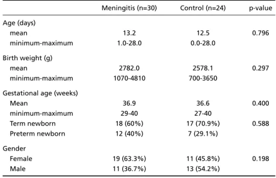
Table 1. Comparision between the values of age, birth weight, gestational age and gen-der in the group with meningitis and the control group.
Meningitis (n=30) Control (n=24) p-value Age (days)
mean 13.2 12.5 0.796
minimum-maximum 1.0-28.0 0.0-28.0
Birth weight (g)
mean 2782.0 2578.1 0.297
minimum-maximum 1070-4810 700-3650
Gestational age (weeks)
Mean 36.9 36.6 0.400
minimum-maximum 29-40 27-40
Term newborn 18 (60%) 17 (70.9%) 0.588
Preterm newborn 12 (40%) 7 (29.1%)
Gender
Female 19 (63.3%) 11 (45.8%) 0.198
Arq Neuropsiquiatr 2005;63(1)
9
cells protein glucose CSF Serum CSF serum CSF serum
(/mm3) (mg/dL) (mg/dL)
1 640 105 56 47.7 <4.4 49.1 6.5 18.1 <1.0 Staphylococcus coagulase negative (blood) No
2 4550 82 9 74.2 35.7 190.8 36.1 561.2 74 Streptococcus pyogenes (CSF-blood) Thrombosis of saggital sinus, ventriculitis, convulsions 3 2000 93 35 21.2 <4.4 38.9 <1.0 35.7 <1.0 Klebsiella pneumoniae (urine)*** No
4 43 121 31 <4.4 <4.4 <1.0 <1.0 2.3 <1.0 Staphylococcus aureus (sinovial fluid) Convulsions
5 47 54 36 16.4 50.1 38.9 43.3 14.9 11.7 — No
6 185 839 29 18.8 <4.4 95.3 13.8 21.3 <0.7 Staphylococcus coagulase negative (blood) Ventriculitis
7 87 2272 81 <4.4 <4.4 6.8 10.4 504.8 6.6 — Convulsions
8 1680 237 23 28.4 <4.4 103.0 52.7 11.7 6.4 — Ventriculitis, hydrocephalus
9 385 460 9 274.9 <4.4 307.0 52.3 900.2 8.2 Acinetobacter baumannii (CSF-blood) Ventriculitis, hydrocephalus
10 176 148 34 38.7 <4.4 40.8 10.6 483.2 325.0 — Thrombosis of saggital sinus,
convulsions
11 3669 177 23 71.6 <4.4 34.7 32.8 964.6 7.5 — No
12 143 85 45 <4.4 <4.4 1.2 35.7 190.0 7.2 — No
13 110 53 83 <4.4 22.1 <1.0 <1.0 9.4 27.9 Staphylococcus aureus (blood) No
14 570 282 31 <4.4 <4.4 5.8 <1.0 479.9 12.5 Escherichia coli (urine)*** Convulsions, intrventricular hemorrhage
15 32 180 67 42.9 21.2 195.0 341.0 16.5 45.3 — No
16 1370 78 53 273.4 38.6 31.0 6.6 659.2 68.4 Staphylococcus aureus (blood) Ventriculitis
17 3 49 54 <4.4 28.5 <1.0 <1.0 0.8 <0.7 Streptococcus viridans (CSF) No
18 2720 303 34 52.0 <4.4 61.6 1.1 197.7 21.0 Staphylococcus aureus (blood) No
19 137 313 5 14.6 <4.4 29.8 0.4 208.7 26.5 Staphylococcus aureus (sinovial fluid) Ventriculitis, hydrocephalus 20 5 178 82 1920.7 134.5 506.0 31.5 1022.0 446.0 Alcaligenes xilosoxidans (CSF) Pseudomonas Coma, death
aeruginosa (blood)
21 240 282 9 1229.4 82.7 242.0 <1.0 1000.0 33.2 Gram-negative diplococcus* *(CSF) Coma, death
22 3 121 38 <4.4 <4.4 <1.0 <1.0 22.3 9.3 Klebsiella pneumoniae (CSF) No
23 35 76 — <4.4 <4.4 <1.0 <1.0 146.5 <0.7 Enterobacter cloacae (blood) No
24 60 134 59 58.8 <4.4 3.3 9.8 43.0 19.0 — Thrombosis of saggital sinus
25 126 50 — 42.1 <4.4 3.3 <1.0 3.8 24.0 Staphylococcus coagulase negative (blood) No
26 22 151 24 65.7 46.7 11.5 5.5 123.6 46.5 Klebsiella pneumoniae (blood) Periventricular leukomalacia
27 20 116 34 55.9 <4.4 <1.0 <1.0 <0.7 <0.7 Streptococcus viridans (CSF) No
28 67 346 52 <4.4 <4.4 <1.0 <1.0 21.3 17.8 Staphylococcus aureus (CSF) Subdural collection,hearing loss,convulsions
29 101 11 47 <4.4 <4.4 14.9 <1.0 162.1 12.2 Escherichia coli (urine)*** Periventricular leukomalacia
30 80 90 47 <4.4 <4.4 <1.0 <1.0 82.3 <0.7 — No
*Mean values: leukocytes=643.6cells/mm3; protein=249.6mg/dL; glucose=40.3mg/dl. **Bacteria identified in Gram-stained smear. ***Urine obtained by supra-pubic puncture. **** Sixteen (53.4%) newborn
RESULTS
The two groups were considered to be similar regarding age, gender, birth weight and gestation-al age (Table 1).In most of the neonates with me-ningitis (63.3%) the signs and symptoms were non-specific: fever, lethargy, irritability, poor feeding, vomiting, abdominal distention, jaundice, tachyp-nea, poor skin perfusion, aptachyp-nea, and tachycardia. Neurological signs and symptoms occurred in 36.7% of the neonates, these presented convulsions, bul-ging anterior fontanel, coma, tremors, hypertonia and cranial nerve signs. The mean values for CSF find-ings, Gram-stained smears, cultures, neurological complications and deaths are presented in Table 2. TNF-αwas detected in the CSF in 63.3% of the neonates, IL-1βin 73.3% and IL-6 in 96.6%.
In the newborn of the control group mean val-ues for CSF findings were: leukocytes= 5.7 cells/mm3, protein=80.9 mg/dL and glucose=50.8 mg/dl. Gram-stained smears and culture of the CSF were nega-tive in all of the newborn.The defininega-tive diagnoses
10 Arq Neuropsiquiatr 2005;63(1)
were sepsis (50.0%), dehydration (16.7%), infec-tion of the urinary tract (8.3%), fever without source (8.3%), convulsive syndrome (8.3%), hyperekplex-ia (4.2%) and polycythemhyperekplex-ia (4.2%).There was one death (4.2%).
The difference between CSF cytokines levels in the group with meningitis and the control group are shown in Figures 1 to 3.The comparison between the CSF and serum levels of cytokines in the group with meningitis and in the control group are shown in Table 3.The comparison between the levels of cytokines in the CSF and the presence of neurologi-cal complications, and values of cells, protein, and glucose in the newborn with meningitis are shown in Table 4.
DISCUSSION
We demonstrated the presence of cytokines in the CSF of all the newborn with meningitis, a fact which was not observed in the control group. Fur-thermore, the levels of the three cytokines analyzed were significantly higher in the newborn with me-ningitis.The detection of cytokines in the CSF was possible regardless of gestational age, indicating that even preterm newborn responded to stimula-tion of the infecstimula-tion with increased producstimula-tion of cytokines in the CNS. The wide variation of the CSF levels, also reported by other authors8, probably re-flects the timing of the collection of the sample and magnitude of the inflammatory response to the in-fection.
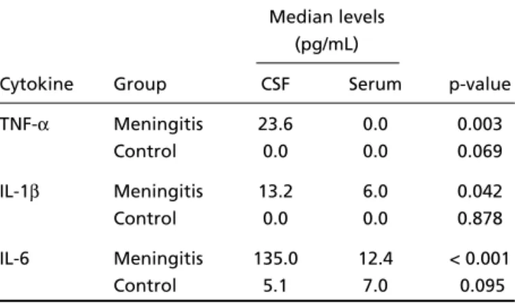
One of the questions to be discussed is whether the TNF-α, IL-1βand IL-6 detected in the CSF of neo-nates with meningitis were produced by cells of the CNS or if they originated from the systemic compart-Table 3. Comparison between the CSF and serum levels of
TNF-α, IL-1βand IL-6 in the group with meningitis and the control group.
Median levels (pg/mL)
Cytokine Group CSF Serum p-value TNF-α Meningitis 23.6 0.0 0.003
Control 0.0 0.0 0.069
IL-1β Meningitis 13.2 6.0 0.042
Control 0.0 0.0 0.878
IL-6 Meningitis 135.0 12.4 < 0.001
Control 5.1 7.0 0.095
Table 4. Comparison between the levels of cytokines in the CSF and the presence of neurological com-plications, and values of cells, protein, and glucose in 30 neonates with meningitis.
CSF CSF CSF
TNF-α(pg/mL)* IL-1β(pg/mL)* IL-6(pg/mL)* Neurological complication
Present (n=16) 33.5 30.4 344.3
Absent (n=14) 18.8 2.3 20.2
P-value** 0.510 0.467 0.061
Spearman's coefficient (P-value)***
Cells 0.312 (0.093) 0.495 (0.005) 0.414 (0.023)
Protein 0.215 (0.253) 0.403 (0.027) 0.361(0.050)
Glucose –0.281 (0.148) –0.302 (0.118) –0.255 (0.189)
ment. Although the cytokines present in the blood circulation can reach the CSF through an impaired or even an intact blood-CSF barrier9, it is known that the subarachnoid space and the circulation are separate compartments in respect to the production of these.High concentrations of TNF-α, IL-1βand IL-6 were detected in the CSF of rabbits after intracis-ternal inoculation of lipopolysaccharide (LPS) of
Haemophilus influenzaeor Neisseria meningitidis, yet no activity was detected in the serum10. In our patients the analysis of serum levels, obtained con-comitantly with CSF levels, allowed us to establish the origin of the cytokines in the CSF. In neonates with meningitis the CSF levels of the three cytokines analyzed were significantly higher than the serum, a fact not observed in the control group. This behavi-or indicates the existence of local production of cy-tokines in the presence of meningitis, in response to the stimulus presented by the microorganism or their products. In older children, Ohga et al.11reported simi-lar findings.
The production of TNF-αby the CNS seems to play a part of great importance in the pathogenesis of the initial lesion of the blood-CSF barrier. Although the mechanism of the action of TNF-αis still not ful-ly clarified, experimental studies suggest that this cytokine is directly cytotoxic to the neurons. The cyto-toxicity could also occur indirectly, through the release of secondary mediators, such as O2free
rad-icals, that attack the double bonds of the polyun-satureted fatty acids of cellular membranes in the process of lipid peroxidation of the membrane. The brain of the newborn, in a phase of accelerated growth, contains high concentrations of polyun-saturated fatty acids, thus is particularly suscepti-Fig 1. Levels of TNF-αin the CSF of 30 neonates with
meningi-tis and 24 neonates of the control group. Neonates with me-ningitis=144.9 pg/ml±404.9(mean ± SD); 0.0-1920.7 pg/ml (mini-mum-maximum); 24.80 pg/ml (median). Control group= 5.3 pg/ml ±7.7(mean ± SD); 0.0-23.6 pg/ml (minimum-maximum);0.00 pg/ml (median); Mann-Whitney test: p = 0.002.
Fig 2. Levels of IL-1βin the CSF of 30 neonates with meningi-tis and 24 neonates of the control group. Neonates with menin-gitis =67.0 pg/ml ± 114.8(mean ± SD); 0.0-505.7 pg/ml (minimum-maximum); 22.4 pg/ml (median). Control group = 8.2 pg/ml ±13.8 (mean±SD); 0.0-38.9 pg/ml (minimum-maximum); 0.00 pg/ml (median); Mann-Whitney test: p = 0.002.
ble to the lesion induced by O2 free radicals. In addition, these factors coincide also with the fact that the lowest levels of oxygen free radical scav-engers and antioxidant enzymes are occurring in the neonatal period12. Experimental studies have demonstrated the sequential emergence of the bioactivity of TNF-α, IL-1β and IL-6 in CSF after injection of PLS or viable meningococci in the sub-arachnoid space13. Probably the increase of the pro-duction of TNF-αat the beginning of the inflam-matory reaction12,14 induces the secretion of IL-1β, both cytokines being responsible for the first steps of the inflammation cascade that leads to the des-truction of tissue.
We observed that TNF-αwas present in the CSF less frequently than IL-1βand IL-6. Similar findings
have been reported by Mustafa et al.,15who
de-monstrated the presence of IL-1β in the CSF in
90% of 21 neonates with ventriculitis, but in the absence of TNF-α. This behavior can be attributed in part to the timing of the collection of CSF.It is kno-wn that the duration of the activity of TNF-αis short, such that it is impossible to detect this cytokine when the infectious process is in a more advanced phase.
The presence of TNF-α in the CSF was registered
45 minutes after the intracisternal inoculation of lipo-oligosaccharide of Haemophilus influenzae, in rabbits, with a peak at two hours and persistence in the CSF for approximately five hours10. The increa-se of TNF-αin the CSF can be observed even before the increase of cell number, protein concentration and decrease of glucose levels16.
IL-1βwas detected in the CSF of 73.3% of new-born with meningitis. This finding is similar to that observed by other authors and suggests the role of this cytokine as mediator of meningeal inflam-mation8,17. López-Cortés et al.17 considered this cytokine to be the best biochemical indicator of the disease in patients with neurosurgical patholo-gies, whose CSF IL-1βconcentrations above 90 pg/ml were highly indicative of the disease. We observed that 23.4% of the neonates with meningitis and none of the control group presented such CSF lev-els. In the literature, only McCracken et al.8 analy-zed the levels of TNF-αand IL-1βin the CSF from a group of patients composed exclusively of neo-nates. In 42 newborn with meningitis due to enter-obacteria, those authors observed detectable lev-els of IL-1βin the CSF in 95% of the cases at the time of the diagnosis in concentrations varying from 20 pg/ml to 2616 pg/ml.
12 Arq Neuropsiquiatr 2005;63(1)
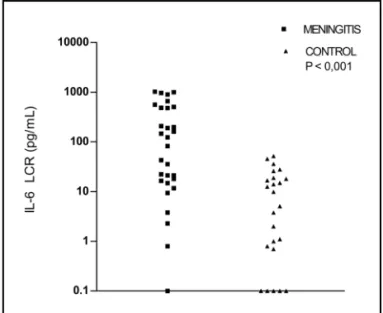
The presence of IL-6 in 96.6% of the neonates with meningitis, and their CSF levels showing a high-ly significant difference in relation to the control group, indicate that CSF IL-6 detection is highly sug-gestive of meningeal inflammation. Similar results were obtained in studies of infants, older children and adults6,18,19. According to Dulkerian et al.20 , young infants respond to the bacterial invasion of the CNS with the release of cytokines at comparable lev-els to those observed in older children and adults. The results we have obtained demonstrate that neo-nates with meningitis are capable of producing
TNF-α, IL-1βand IL-6 in a magnitude comparable to that observed in older age groups.
Bearing in mind that the increase in the CSF lev-els of cytokines can occur even before the onset of inflammatory alterations, its detection in the CSF can be of great value in the diagnosis of bacterial meningitis among patients with a normal or incon-clusive CSF exam. Matsuzono et al.19consider that the CSF level of IL-6 is the only parameter capable of detecting the inflammatory process when the routine findings in the CSF are normal. Azuma et al.21highlighted that the measurement of IL-6 in the CSF might be particularly useful for the early diagnosis of meningitis in situations of immunolo-gical depression. Among our patients, four new-born (cases 17, 20, 22 and 27) with bacterial growth in the culture of CSF, presented a normal CSF exam or few alterations. In case 17, the CSF cells, protein and glucose were normal, but with growth of Strep-tococcus viridansin the culture and a detectable level of IL-6. In case 20, who died just a few hours after hospitalization, the CSF presented only pro-tein 178 mg/dl, however an impressive increase was observed in TNF-α, IL-1βand IL-6. CSF culture, who-se result was obtained after death of the patient, showed Alcaligenes xilosoxidans. Case 22 present-ed glucose 38 mg/dl, protein 121 mg/dl, Klebsiel-la pneumoniaein the culture of the CSF and detec-table levels of IL-6. Case 27 presented protein 116 mg/dl, glucose 34 mg/dl, culture isolated Strepto-coccus viridansand detectable CSF levels of
TNF-αand IL-1β.These results indicate that the detec-tion of cytokines in the CSF is useful to confirm the diagnosis of meningitis, prior to obtaining a result from the cultures.
the study of Harvey et al.1showing that mortali-ty due to neonatal bacterial meningitis, in England, has decreased from 50%, in the 1970s, to less than 10%, in 1997.It is possible that the use of third-gene-ration cephalosporin, in the treatment of the dis-ease, could be related to the greater survival. In spite of the mortality in our study being relatively low, the frequency of serious complications was high, demonstrating the need to develop methods of ear-ly detection and evaluation of the clinical course of the disease. Low et al.18 observed a correlation between the simultaneous presence of TNF-α, IL-1βand IL-6 in the CSF, with low values of glucose in five children with meningitis, and between the detection of TNF-αor IL-1βin the CSF and the du-ration of fever, convulsions, spasticity and death. McCracken et al.8verified a relationship between levels of IL-1βin CSF of over 200 pg/ml and the num-ber of days in which endotoxin, antigen K1and bac-teria persisted in 42 neonates with meningitis due to enterobacteria. We observed a positive correla-tion between the number of cells in the CSF and the levels of IL-1βand IL-6, as well as between the protein values and the levels of these cytokines, sug-gesting that these also are important in the eval-uation of the magnitude of the inflammatory pro-cess in the central nervous system.
We conclude that the detection of TNF-α, IL-1β and IL-6 in the CSF is of great value in order to achie-ve a rapid and early diagnosis of neonatal meningi-tis, both for term and preterm newborn.Among the three cytokines analyzed, IL-6 can be considered the best indicator of meningeal inflammation.
Acknowledgments -We thank Mr.Roberto Mariz for his technical assistance.
REFERENCES
1. Harvey D, Holt DE, Bedford H. Bacterial meningitis in the newborn: a prospective study of mortality and morbidity. Semin Perinatol 1999; 23:218-225.
2. Nel E. Neonatal meningitis: mortality, cerebrospinal fluid, and microbio-logical findings. J Trop Pediatr 2000;46:237-239.
3. Krebs VLJ, Diniz EMA, Vaz FAC, Marques-Dias MJ, Takiguti C, Ramos JLA. Meningite bacteriana neonatal: estudo prospectivo da evolução a longo prazo de 55 crianças. Arq Neuropsiquiatr 1996;54:75-81. 4. Sarman G, Moise AA, Edwards MS. Meningeal inflammation in
neona-tal gram-negative bacteremia. Pediatr Infect Dis J 1995;14:701-704. 5. Mustafa MM, Ramilo O, Sáez-Llorens X, Olsen KD, Magness RR,
McCracken Jr GH.Cerebrospinal fluid prostaglandins, interleukin-1βand tumor necrosis factor in bacterial meningitis. AJDC 1990;144:883-887. 6. Rusconi F, Parizzi F, Garlaschi L, et al. Interleukin-6 activity in infants
and children with bacterial meningitis. The Collaborative Study on Me-ningitis. Pediatr Infect Dis J 1991;10:117-121.
7. Overall JC Jr. Neonatal bacterial meningitis: analysis of predisposing factors and outcome compared with matched control subjects. J Pediatr 1970;76:499-511.
8. McCracken GH Jr, Mustafa MM, Ramilo O, Olsen KD, Risser RC. Cere-brospinal fluid interleukin-1 beta and tumor necrosis factor concentra-tions and outcome from neonatal gram-negative enteric bacillary menin-gitis. Pediatr Infect Dis J 1989;8:155-159.
9. 0Savman K, Blennow M, Gustafson K, Tarkowski E, Hagberg H. Cyto-kine response in cerebrospinal fluid after birth asphyxia. Pediatr Res 1998;43:476-451.
10. Ramilo O, Sáez-Llorens X, Mertsola J, et al. Tumor necrosis factor-α /cachectin and interleukin-1βinitiate meningeal inflammation. J Exp Med 1990;172:497-507.
11. Ohga S, Aoki T, Okada K, et al.Cerebrospinal fluid concentrations of interleukin-1β, tumor necrosis factor-αand interferon gamma in bacte-rial meningitis. Arch Dis Child 1994;70:123-125.
12. Park WS, Chang YS, Lee M. The efficacy of pentoxifylline as an anti-inflammatory agent in experimental Escherichia colimeningitis in the newborn piglet. Biol Neonate 2000;77:236-242.
13. Waage A, Brandtzaeg P, Halstensen A, Kierulf P, Espevik T. The com-plex pattern of cytokines in serum from patients with meningococcal septic shock. J Exp Med 1989;169:333-338.
14. Glimäker M, Kragsbjerg P, Forsgren M, Olcén P. Tumor necrosis fac-tor-α(TNF-α) in cerebrospinal fluid from patients with meningitis of different etiologies: high levels of TNF-αindicate bacterial meningitis. J Infect Dis 1993;16:882-889.
15. Mustafa MM, Mertsola J, Ramilo O, Sáez-Llorens X, Risser RC, McCracken GH Jr. Increased endotoxin and interleukin-1β concentra-tions in cerebrospinal fluid of infants with coliform meningitis and ventri-culitis associated with intraventricular gentamicin therapy. J Infec Dis 1989;160:891-895.
16. Mustafa MM, Ramilo O, Mertsola, J et al. Modulation of inflammation and cachectin activity in relation to treatment of experimental Haemophilus influenzaetype B meningitis. J Infect Dis 1989;160:818-825. 17. López-Cortés LF, Marquez-Arbizu R, Jimenez-Jimenez LM, et al. Cerebrospinal fluid tumor necrosis factor-αinterleukin-1β, interleukin-6, and interleukin-8 as diagnostic markers of cerebrospinal fluid infec-tion in neurosurgical patients. Crit Care Med 2000;28:215-219. 18. Low PS, Lee BW, Yap HK, et al. Inflammatory response in bacterial
me-ningitis: cytokine levels in the cerebrospinal fluid. Ann Trop Paediatr 1995;15:55-59.
19. Matsuzono Y, Narita M, Akutsu Y, Togashi T. Interleukin-6 in cerebros-pinal fluid of patients with central nervous system infections. Acta Paediatr 1995;84:879-883.