RESUMO
Objetivos: Avaliar as diferenças de desempenho cognitivo entre pacientes com glau coma primário de ângulo aberto (POAG), glaucoma de pressão normal (NTG) e controle de indivíduos saudáveis (C).
Métodos: Um total de 60 pessoas (20 POAG, 20 NTG e 20 indivíduos saudáveis) foram incluídos neste estudo. Um exame oftalmológico detalhado foi realizado em todos os participantes. Um sistema de tomografia de coerência óptica de domínio espectral (SDOCT ) foi utilizado para medir as espessuras da camada de células ganglionares plexiforme interna (GCIPL) e da camada de fibras nervosas da retina (RNFL). Para avaliar o desempenho cognitivo de todos os participantes, foi realizado pelo mesmo neurologista um exame neurológico detalhado, incluindo miniexame do estado mental (MMSE).
Resultados: Não houve diferenças significativas entre os grupos em termos de idade (p=0,348) e sexo (p=0,935). Espessuras médias da RNFL foram significativamente di ferentes, sendo 85,2 ± 14,7, 76,8 ± 10,3 e 91,4 ± 7,7 µm nos grupos POAG, NTG e controles, respectivamente (p<0,001). As espessuras médias da GCIPL observadas foram 77.5 ± 9.7 μm no grupo POAG, 73,4 ± 7,8 µm no grupo NTG e 78,8 ± 3,8 µm nos controlos. As diferenças entre os grupos não foram estatisticamente significantes (p=0,085). Graduações do MMSE foram 26,1 ± 1,4, 25,7 ± 2,3 e 28,8 ± 0,9 nos grupos POAG, NTG e controles, respectivamente. Houve diferenças significativas entre os três grupos (p<0,001). Houve diferença significativa entre NTG e saudáveis (p<0,001). Houve diferença significativa entre POAG e saudáveis (p=0,001). Não houve diferença significativa entre o POAG e NTG (p=0,595).
Conclusões: Parecem haver fatores de risco semelhantes no glaucoma e nos distúrbios neurodegenerativos que causam deterioração no desempenho cognitivo. Comparando a baixa graduação do MMSE de pacientes com POAG e NTG com controles saudáveis referenda nossa hipótese. Consequentemente recomendase que um neurologista também examine os pacientes de glaucoma.
Descritores: Glaucoma de ângulo aberto; Glaucoma de baixa tensão; Transtornos cog nitivos; Demencia
ABSTRACT
Purpose: To assess cognitive performance differences among primary open-angle glaucoma (POAG) patients, normal-tension glaucoma (NTG) patients, and healthy control (C) subjects.
Methods: A total of 60 participants (20 POAG, 20 NTG, and 20 C subjects) were included in this study. A detailed ophthalmologic examination was performed on all participants. A spectral domain-optical coherence tomography (SD-OCT ) system was used to measure the ganglion cell-inner plexiform layer (GC-IPL) and retinal nerve fiber layer (RNFL) thicknesses. To assess the cognitive performance of all participants, detailed neurological examinations, including the mini-mental state examination (MMSE), were performed by the same neurologist.
Results: There were no significant differences among the groups in terms of age (p=0.348) or gender (p=0.935). The mean RNFL thicknesses were significantly diffe rent among the groups (85.2 ± 14.7, 76.8 ± 10.3, and 91.4 ± 7.7 µm in the POAG, NTG, and C subjects, respectively; p<0.001). The mean GC-IPL thicknesses were 77.5 ± 9.7 µm in the POAG group, 73.4 ± 7.8 µm in the NTG group, and 78.8 ± 3.8 µm in the C group. Differences among the groups were not statistically significant (p=0.085). MMSE scores were 26.1 ± 1.4, 25.7 ± 2.3, and 28.8 ± 0.9 in the POAG, NTG, and C groups, respectively. There were significant differences among the three groups (p<0.001). Specifically, there were significant differences between the NTG and C groups (p<0.001), and between the POAG and C groups (p=0.001). There was no significant difference between the POAG and NTG groups (p=0.595). Conclusions: There appear to be similar risk factors in glaucoma and neurodege-nerative disorders that cause deterioration in cognitive performance. Comparing the low MMSE scores of the POAG and NTG patients with the scores of healthy C participants supports our hypothesis. Consequently, it is recommended that a neurologist should also examine glaucoma patients.
Keywords: Glaucoma, Open-angle; Low-tension glaucoma; Cognition disorders; Dementia
INTRODUCTION
Glaucoma is characterized by a progressive loss of retinal ganglion cells (RGCs) and atrophy of the optic nerve, leading to a characteris-tic pattern of visual ield loss(1). It is the leading cause of irreversible
visual loss worldwide(2), and without treatment, glaucoma can cause
blindness(3). Primary open-angle glaucoma (POAG) is the most common
type of glaucoma(4). While the pathogenesis of POAG is not yet known,
there are several known risk factors, one of which is elevated
intrao-cular pressure (IOP)(5). To decrease the IOP with this treatment slows
down the progression of the disease(1). However, visual loss may still
continue despite reducing the IOP in some patients(1).
Another type of glaucoma is normal-tension glaucoma (NTG), in which there is optic nerve degeneration without IOP elevation. Patients with statistically normal IOP who develop the characteristic changes of progressive retinal nerve iber layer (RNFL) loss, RGC loss, and visual ield defects are grouped as having NTG, an important
Cognitive performance of primary open-angle glaucoma and
normal-tension glaucoma patients
Desempenho cognitivo em pacientes com glaucoma primário de ângulo aberto
e glaucoma de pressão normal
MehMet Bulut1, Aylin yAMAn2, MuhAMMet KAziM erol1, Fatma Kurtuluş2, DevriM toslAK1, Deniz turgut CoBAn1, Ebru Kaya başar3
Submitted for publication: August 24, 2015 Accepted for publication: January 7, 2016
1 Ophthalmology Department, Antalya Training and Research Hospital, Antalya, Turkey. 2 Neurology Department, Antalya Training and Research Hospital, Antalya, Turkey.
3 Department of Animal Science Biometry and Genetics Unit, Faculty of Agriculture, Akdeniz Uni-versity, Akdeniz, Turkey.
Funding: No specific financial support was available for this study.
Disclosure of potential conflicts of interest: None of the authors have any potential conflicts of interest to disclose.
Corresponding author: Muhammet Kazim ErolD. Meydan kavağı mah. Avni tolunay cad. Yerge daran sit. C blok d: 22 - Muratpaşa, Antalya - Turkey - E-mail: [email protected] Approved by the following research ethics committee: Antalya Training and Research Hospital
subset of open-angle glaucoma (OAG)(6). POAG and NTG patients
form the subgroups of OAG patients.
Dementia, which afects 24 million people worldwide, with dou-ble the number of afected people every 20 years, is an important health problem for the aging population(7). It is a term used to
des-cribe a group of conditions that can afect a person’s ability to think, remember, understand, make judgments, communicate, and interact socially(8). Alzheimer-type dementia (ATD) is known as the most
wi-despread form of dementia and is an important health problem in every country(9). It is a progressive neurodegenerative disorder
cha-racterized by cognitive deterioration and deterioration in memory, changes in personality, behavioral disturbances, and impaired ability to perform the activities of daily life(10).
Glaucoma and dementia (especially ATD) share several features. Both become more severe with advanced age, and they occur more frequently in women than in men(11,12). Common genetic risk factors
have been reported in ATD and glaucoma, and similar pathological changes in the optic nerves of glaucoma patients and the brains of patients with ATD have been demonstrated(13). At the molecular level,
caspase activation was shown in a rat study of chronic ocular hyper-tension to induce abnormal amyloid precursor protein formation, which is the key event in the pathogenesis of ATD(14).
Both ATD and glaucoma demonstrate early structural changes in the visual cortex and lateral geniculate nucleus. Both diseases afect magnocellular visual processing(15). Spectral domain-optical
coheren-ce tomography (SD-OCT) can demonstrate these early changes, so it is reasonable to use OCT when assessing for cognitive decline in glaucoma patients to analyze the possible correlations between OCT measures and cognitive parameters.
Although several clinical studies have demonstrated an increased prevalence of glaucoma in dementia patients(16), large population-based
studies have not revealed an association between glaucoma and de-mentia(17). Since there have been very few studies of the relationship
between glaucoma and dementia, more studies are necessary to irmly establish this relationship. Since the MMSE is one of the most frequen-tly used screening tools for the assessment of cognitive function, the aim of our study was to determine whether there are diferences among the POAG, NTG, and control (C) groups in terms of MMSE scores, and also to assess the relationship between the MMSE scores and SD-OCT parameters.
METHODS
The study was performed in compliance with the Helsinki Decla-ration and with the approval of the Ethics Committee of the Antalya Education and Research Hospital. A total of 60 people, including 20 POAG patients (aged between 44 and 73 years), 20 NTG patients (aged between 45 and 73 years), and 20 healthy C participants (aged between 47 and 69 years), participated in this study, and the age and gender proportions of the groups were similar. We recruited our patients in the hospital outpatient setting. C subjects meeting the inclusion criteria were recruited in the same setting from the neurology and ophthal-mology outpatient clinics. Glaucoma patients were taking at least one topical medication (beta-blockers, carbonic anhydrase inhibitors, prostaglandin analogs, sympathomimetic drugs, and parasympatho-mimetic drugs). All of our participants had graduated from elementary school. Since most of the subjects in the patient and C groups had attained this level of education, illiterate subjects and high school and university graduates were excluded to provide homogeneity. The majority of the subjects were on a similar level of socioeconomic sta-tus. Patients who had neurological diseases that could have afected cognitive performance, such as ATD, vascular dementia, and mild cog-nitive impairment (MCI), were excluded from this study. None of the patients had subjective complaints concerning cognitive impairment. Further, at the time of the study, none of the participants had been using systemic medications that could have afected cognition such
as benzodiazepines, opiates, tricyclic antidepressants, anticonvulsants, and dopamine agonists, etc. Subjects who did not meet these criteria were excluded. Subjects with moderate-severe depression and chronic systemic diseases such as diabetes mellitus, arthritis, hypertension, heart disease, stroke, and cancer were excluded. Additionally, patients who smoked cigarettes, had a best-corrected visual acuity of less than 1.0 according to Snellen chart, or had eye diseases other than glauco-ma that could have afected RNFL and ganglion cell-inner plexiform layer (GC-IPL) thicknesses, such as macular degeneration and optic neuropathy, were excluded from this study.
O
PHTHALMOLOGICALEXAMINATIONAll participants in this study underwent an ophthalmological exa-mination including visual acuity assessment with a Snellen chart, IOP with Goldman’s applanation tonometer after application of a local anesthetic (hydrochloric proxymetacaine 0.5%), measurement of cen-tral corneal thickness (CCT) with an optic pachymeter (Lenstar LS 900; Haag-Streit, Koeniz, Switzerland), slit-lamp-assisted biomicroscopy of the anterior and posterior segments of the eye, gonioscopy, and photography of the fundus (Visucam NM-FA; Carl Zeiss Meditec Inc., Oberkochen, Germany). SD-OCT (Cirrus HD OCT model 5000; Carl Zeiss Meditec Inc., Dublin, CA, USA) was used to measure the GC-IPL (macu-lar cube 512 × 128) and RNFL (optic disc cube 200 × 200) thicknesses. The examination was concluded with a check of the visual ields using a static perimeter apparatus type Octopus 900 (Haag-Streit). These examinations were used for the glaucoma diagnosis and classiication. The diagnostic criteria for POAG were high IOP (>21 mmHg, corrected by corneal thickness), normal iridocorneal open angle, glaucomatous changes in the visual ield with optic nerve cupping, and the absence of other optic neuropathies. Except for evidence of high IOP (≤21 mmHg), we used the same diagnostic criteria of POAG to diagnose NTG. All POAG and NTG diagnoses were applied according to the guidelines of the European Glaucoma Society(18).
N
EUROLOGICALEXAMINATIONDetailed neurological examination, including the MMSE test which evaluates cognitive function, was applied to all participants. All of the patients had intact neurological examination indings, including those of the motor, sensory, and cerebellar systems and the cranial nerves, except for the optic nerve. The MMSE is the most commonly used test for evaluating cognitive function and scanning for dementia. Orientation, attention, memory, language, and shape copying are evaluated in this test. The maximum number of points is 30. The study for the validity and reliability of the MMSE in Turkey was performed by Güngen(19) and his associates in 2002, and the cut-of value was
determined to be 23/24. This test is afected by education, and is considered to be reliable for identifying the degree of mild dementia. There is also a modiied version for uneducated people. The MMSE is used for both diagnosis and treatment follow-up. Long-term memory, short-term memory, attention span, calculation, the naming of items, performance of a task with three steps, reading, writing, and assem-bling abilities are all measured in this test.
S
TATISTICALANALYSISnot provided assumptions for the parametric tests (RNFL thickness, GC-IPL thickness, MD, and MMSE scores). Correlations among the va-riables were assessed using Spearman’s correlation coeicients. P<0.05 was regarded as statistically signiicant.
RESULTS
The mean ages of group one (POAG), group two (NTG), and group three (C) were 59.8 ± 10.1, 61.9 ± 9.8, and 60.1 ± 8.5 years, respectively. There were no signiicant diferences with regard to age among the groups (p=0.348). All subjects involved in the study were Caucasian. There were 11 males and nine females in the POAG group, 11 males and nine females in the NTG group, and 10 males and 10 females in the C group (p=0.935) (Table 1). The mean IOP value was 16.2 ± 2.9 mmHg in the POAG group, 14.7 ± 1.9 mmHg in the NTG group, and 14.8 ± 2.9 mmHg in the C group (p=0.270).
The CCT was 545.6 ± 29.7 µm in the POAG group, 519 ± 25.7 µm in the NTG group, and 551.9 ± 26.2 µm in the C group. The diferences observed between the NTG group and the two other groups were statistically signiicant (p<0.001). When we compared the groups pairwise, we found statistically signiicant diferences between the NTG and POAG groups (p=0.001), and the NTG and healthy C groups (p<0.001). The MD in the worse eye was 4.8 ± 3.1 dB in the POAG group, 7.9 ± 3.8 dB in the NTG group, and 1.2 ± 1.1 dB in the C group; there were signiicant diferences among the three groups (p<0.001). The MD in the better eye was 3.5 ± 2.1 dB in the POAG group, 4.6 ± 3.1 dB in the NTG group, and 0.7 ± 0.7 dB in the C group; there were signiicant diferences among the three groups (p=0.001). The IOP, CCT, MD in the worse-eye, and MD in the better-eye measurement values, are shown in table 1.
Among the POAG patients, three were taking beta-blockers, se-ven were taking prostaglandin analogs, four were taking beta-blockers plus carbonic anhydrase inhibitors, two were taking beta-blockers plus prostaglandin analogs, two were taking beta-blockers plus sympa-thomimetic drugs, one was taking beta-blockers plus carbonic anhydrase inhibitors as well as parasympathomimetic drugs, and one was taking beta-blockers plus carbonic anhydrase inhibitors plus sympathomimetic drugs. Among the NTG patients, three were taking beta-blockers, eight were taking prostaglandin analogs, two were taking sympathomimetic drugs, two were taking beta-blockers plus carbonic anhydrase inhibitors, four were taking beta-blockers and prostaglandin analogs, and one was taking beta-blockers plus sympathomimetic drugs.
The mean RNFL thickness measurements were 85.2 ± 14.7 µm for the POAG group, 76.8 ± 10.3µm for the NTG group, and 91.4 ± 7.7 µm for the C group; statistically signiicant diferences were found among the three groups (p<0.001). When the groups were compared pairwise, statistically signiicant diferences were found between the NTG and POAG groups (p=0.034), and the NTG and C groups (p<0.001). The
diference between the POAG and C groups was not statistically sig-niicant (p=0.102). These measurements are shown in table 2.
The mean GC-IPL measurement was 77.5 ± 9.7 µm in the POAG group, 73.4 ± 7.8 µm in the NTG group, and 78.8 ± 3.8 µm in the con-trol group. No statistically signiicant diference could be found when all the groups were compared with each other (p=0.085). However, when the groups were compared pairwise, the diference between the NTG and C groups was statistically signiicant (p=0.023). When we compared the POAG group with the NTG and C groups separately, no signiicant diferences were found (p=0.143 and p=0.714, respectively). The above measurements are shown in table 2.
The MMSE scores were 26.1 ± 1.4 in the POAG group, 25.7 ± 2.3 in the NTG group, and 28.8 ± 0.9 in the C group. When all three scores were compared together, the diferences between the mea-surements were statistically signiicant (p<0.001). The measurements are shown in table 2. When the scores were compared pairwise, a statistically signiicant diference was found between both the NTG and C groups, and the POAG and C groups; however, the diference was not statistically signiicant between the NTG and POAG groups. The measurements are shown in table 3. A signiicant correlation was not found between the MMSE scores and either the RNFL (p=0.385) or GC-IPL (p=0.813) thicknesses.
DISCUSSION
In the present study, it was necessary to determine if there were any diferences in the MMSE scores (same age and gender) of the three groups (20 POAG, 20 NTG, and 20 healthy C participants). In this study, the hypothesis was that lower cognitive scores in glaucoma patients were anticipated. The pathogenesis of the dementia group diseases, such as ATD and MCI, which cause lower MMSE scores, is similar to that of glaucoma. Since both diseases have noticeable neuron loss, it evident that there is a relationship of some kind.
Studies have shown a higher incidence of glaucoma among ATD patients. In a previous study, the rate of glaucoma was found to be 25.9% in ATD patients and 5.2% in controls(17). Tamura et al. found
that 23.8% of patients with ATD had glaucoma, while only 9.9% of the age-matched controls had glaucoma(1). From these data, it was
proposed that the optic nerve of patients with ATD may be less resis-tant to elevated IOP(15). Based on this proposition, we suspected that
in the early stages of ATD, lower IOP may cause glaucomatous optic neuropathy; therefore, the NTG patients could be expected to attain
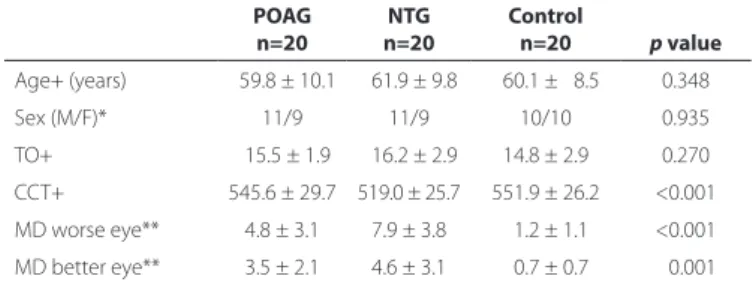
Table 1. Demographic and clinical characteristics of patients and control subjects
POAG n=20
NTG n=20
Control
n=20 p value
Age+ (years) 059.8 ± 10.1 061.9 ± 9.8 060.1 ± 08.5 0.348
Sex (M/F)* 11/9 11/9 10/10 0.935
TO+ 015.5 ± 1.9 016.2 ± 2.9 14.8 ± 2.9 0.270
CCT+ 545.6 ± 29.7 519.0 ± 25.7 551.9 ± 26.2 <0.001
MD worse eye** 4.8 ± 3.1 7.9 ± 3.8 01.2 ± 1.1 <0.001
MD better eye** 3.5 ± 2.1 4.6 ± 3.1 00.7 ± 0.7 <0.001
POAG= primary open-angle glaucoma; NTG= normotensive glaucoma; M= male; F= female; TO= tension ocular; CCT= central corneal thickness; MD= mean defect; += one-way ANOVA test with Bonferroni correction; *= Chi-square test; **= Kruskal-Wallis test.
Table 2. Comparison of the mean RNFL, GC-IPL, and MMSE complex among the POAG, NTG, and control groups
POAG n=20
NTG n=20
Control
n=20 p value
RNFL (mean)* 85.2 ± 14.7 76.8 ± 10.3 91.4 ± 7.7 <0.001
GC-IPL (mean)* 77.5 ± 09.7 73.4 ± 07.8 78.8 ± 3.8 <0.085
MMSE* 26.1 ± 01.4 25.7 ± 02.3 28.8 ± 0.9 <0.001
RNFL= retinal nerve fiber layer; GC-IPL= ganglion cell-inner plexiform layer; MMSE= mini-mental state examination; POAG= primary open-angle glaucoma; NTG= normotensive glaucoma; *= Kruskal-Wallis test.
Table 3. Pairwise comparison of the MMSE scores between the control subjects and the POAG and NTG patients
Control (n=20) MMSE: 28.8 ± 0.9
POAG (n=20) MMSE: 26.1 ± 1.4
p=0.001*
NTG (n=20) MMSE: 25.7 ± 2.3
p<0.001*
lower MMSE scores. However, we did not ind a signiicant diference in the MMSE scores between the POAG and NTG groups.
Few studies have explored cognitive impairment in glaucoma patients. Yochim et al. found cognitive impairment in 44% of 41 older glaucoma patients(20). Similarly, we found lower MMSE scores in the
glau coma patients than in the healthy C group. Hagerman et al. found that 32% of patients with low vision had cognitive impairment as determined by the MMSE(21). In contrast, Cumurcu et al. showed no
signiicant diferences in the MMSE score among pseudoexfoliative glaucoma patients, POAG patients, and healthy controls(22).
The dementia group of disorders (ATD, vascular dementia, mixed dementia) causes cognitive impairment(23). The MMSE is used to
eva-luate the cognitive condition of a person and scanning of dementia. Personal orientation, attention to detail, language ability, and the copying of shapes are evaluated in this test, and 30 is the maximum possible score. Further, general cognitive function can be assessed with the MoCA (Montreal Cognitive Assessment), which provides a more comprehensive assessment than MMSE. Executive function, short-term memory, attention, language skills, and visuospatial pro-cessing are the categories included in the MoCA test(24). Since the
educational level of our patients was low, we could not perform the MoCA test in the present study. In our study, we found statistically signiicant diferent MMSE scores among the three groups (26.1 ± 1.4 in the POAG group, 25.7 ± 2.3 in the NTG group, and 28.8 ± 0.9 in the C group; p<0.001). Similarly, Jeferis et al. found lower MMSE scores in glaucoma patients as opposed to healthy controls(25). Since MMSE
requires intact vision for eight of the 30 points, they also studied the MMSE scores of their subjects by extracting the part of the MMSE that requires intact vision. At this time, they found no signiicant diference in the MMSE scores between the glaucoma patients and healthy controls. In our study, we excluded patients with vision less than 1.0 according to Snellen chart, to minimize the efect of vision on MMSE scores.
Although we excluded patients with vision less than 1.0 accor-ding to the Snellen chart, vision may still have afected the MMSE scores in glaucoma patients, since glaucoma afects the magnocellular visual pathway. Visual function mediated by this part of the brain is diicult to assess by conventional ophthalmological examination. Motion process and contrast sensitivity are parts of these visual pro-cesses(26). In addition, ATD patients may have structural defects in the
magnocellular visual pathway even in the absence of plaques and neuroibrillary tangles in these brain areas(27).
In our study, we found that the mean thicknesses of RNFL and GC-IPL in both the glaucoma groups were thinner, as expected, compared with the normal C group. These results are consistent with those of previous studies(27,28). The CCT values were found to be signiicantly
thinner in the NTG group than in the other groups (p<0.05). These results are also consistent with those from the available literature(29).
We did not ind any signiicant correlations between the MMSE score and either the RNFL or GC-IPL thicknesses. The literature did not re-veal any studies with similar glaucoma and dementia parameters for glaucoma patients. In some studies performed on dementia-group diseases causing cognitive impairment, a signiicant correlation was found between the MMSE score and RNFL thickness(30,31). In addition,
in a study by Bayhan et al., a significant correlation was(32) found
between the MMSE score and the mean GC-IPL thickness. Conversely, in another study, no correlation was found between the MMSE score and RNFL thickness(33).
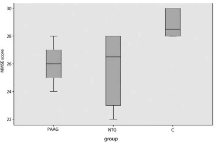
To minimize the efects of vision on the MMSE score, because we did not include patients with a visual acuity of less than 1.0, most of our patients had early-stage glaucoma. Since POAG patients are at an earlier stage of glaucoma than NTG patients, even though the GC-IPL and RNFL thicknesses were lower in these two groups than in the C group, this diference was not statistically signiicant. We found that NTG patients had lower MMSE scores than POAG patients. We believe that although there was no statistically signiicant diference
of the MMSE scores between the NTG and POAG patients, the lower values of MMSE in NTG patients might be attributed to the later stage of glaucoma in those patients. Figure 1 shows a box plot graph of the MMSE scores in each group.
Some research has shown that glaucoma has some of the same characteristics as ATD, which is the most common cause of dementia(1).
They are both chronic neurodegenerative diseases that are closely related to aging, and both progress very slowly. Recently, Yoneda et al.(34) suggested that beta-amyloid and tau (neuroibrillary tangles),
which have signiicance in ATD pathology, may also have important roles in glaucoma pathology. They also found a signiicant decrease in the levels of beta-amyloid and a signiicant increase in the level of tau in the vitreous of glaucoma patients when compared with that in the vitreous of a healthy control group. In a previous study, when ATD patients were compared with a control group, a signiicant decrease was found in the beta-amyloid levels of the cerebrospinal luid, as well as a signiicant increase in the level of tau(35). Based on these indings,
the neurodegenerative process causing neuron loss in glaucoma may have a mechanism similar to the process causing the same pathology in ATD. In addition, it was found that beta-amyloid accumulates in the RGCs of rats with glaucoma induced for experimental purposes(14). In
another experimental glaucoma study performed on rats, a hypothesis was proposed that chronic beta-amyloid neurotoxicity at the molecular level causes the death of retinal neurons and is similar to the death of neurons in the brains of ATD patients(36).
There were some limitations in this study. The number of subjects (60) was relatively low, and the glaucoma drugs used by the patients were not taken into account. Although all the patients in this study had good visual acuity, we could not conirm the efect of visual function on the MMSE scores. The absence of complete neuropsy-chologic data is another limitation. Unfortunately, we were unable to perform more sensitive tests to evaluate the cognitive status of the patients, such as the MoCA, due to the low educational level of the participants. This could be considered a limitation of our study. Un-doubtedly, longitudinal studies should be performed to obtain more data on this topic. A strength of this study is that the neurologist who performed the neurologic examinations on the patients was com-pletely unaware of the ophthalmological diagnoses of the patients.
In conclusion, since the glaucoma group of diseases and the de-mentia group of diseases share similar neurodegenerative processes afecting cognitive impairment, our indings support our hypothesis. Since glaucoma patients have lower cognitive performance, glaucoma can take place in the group of neurodegenerative diseases. Therefore, it is incumbent upon the ophthalmologist to refer glaucoma patients to a neurologist.
MMSE= mini-mental state examination
REFERENCES
1. Tamura H, Kawakami H, Kanamoto T, Kato T, Yokoyama T, Sasaki K, et al. High frequency of open-angle glaucoma in Japanese patients with Alzheimer’s disease. J Neurol Sci. 2006;246(1-2):79-83.
2. Quigley HA, Broman AT. The number of people with glaucoma worldwide in 2010 and 2020. Br J Ophthalmol. 2006;90(3):262-7.
3. Kwon YH, Fingert JH, Kuehn MH, Alward WLM. Primary open-angle glaucoma. N Engl J Med. 2009;360(11):1113-24.
4. Ou Y, Grossman DS, Lee PP, Sloan FA. Glaucoma, Alzheimer disease and other demen-tia: a longitudinal analysis. Ophthalmic Epidemiol. 2012;19(5):285-92.
5. Cartwright MJ, Grajewski AL, Friedberg ML, Anderson DR, Richards DW. Immune-re-lated disease and normal-tension glaucoma. A case-control study. Arch Ophthalmol. 1992;110(4):500-2.
6. Yu M, Chen B, Gong B, Shuai P, Wu Z-Z, Lin W. Association of n3 and n6 polyunsa-turated fatty acids in red blood cell membrane and plasma with severity of normal tension glaucoma. Int J Ophthalmol. 2015;8(3):476-83.
7. Ferri CP, Prince M, Brayne C, Brodaty H, Fratiglioni L, Ganguli M, et al. Global preva-lence of dementia: A Delphi consensus study. Lancet. 2005; 366(9503):2112-7. 8. Mandas A, Mereu RM, Catte O, Saba A, Serchisu L, Costaggiu D, et al.Cognitive
impair-ment and age-related vision disorders: their possible relationship and the evaluation of the use of aspirin and statins in a 65 years-and-over Sardinian population. Front Aging Neurosci. 2014; 6:309.
9. Rocchi A, Pellegrini S, Siciliano G, Murri L. Causative and susceptibility genes for Alzheimer’s disease: a review. Brain Res Bull. 2003;61(1):1-24.
10. Lin I-C, Wang Y-H, Wang T-J, Wang I-J, Shen Y-D, Chi N-F, et al. Glaucoma, Alzheimer’s disease, and Parkinson’s disease: an 8-year population-based follow-up study. PLoS One. 2014;9(9):e108938.
11. Shiose Y, Kitazawa Y, Tsukahara S, Akamatsu T, Mizokami K, Futa R, et al. Epidemiology of glaucoma in Japan--a nationwide glaucoma survey. Jpn J Ophthalmol. 1991;35(2): 133-55.
12. Henderson VW. The epidemiology of estrogen replacement therapy and Alzheimer’s disease. Neurology. 1997;48(5 Suppl 7):S27-35.
13. Sadun AA, Bassi CJ. Optic nerve damage in Alzheimer’s disease. Ophthalmology. 1990;97(1):9-17.
14. McKinnon SJ, Lehman DM, Kerrigan-Baumrind LA, Merges CA, Pease ME, Kerrigan DF, et al. Caspase activation and amyloid precursor protein cleavage in rat ocular hypertension. Invest Ophthalmol Vis Sci. 2002;43(4):1077-87.
15. Valenti DA. Alzheimer’s disease and glaucoma: imaging the biomarkers of neurode-generative disease. Int J Alzheimers Dis. 2010;2010:793931.
16. Bayer AU, Ferrari F, Erb C. High occurrence rate of glaucoma among patients with Alzheimer’s disease. Eur Neurol. 2002;47(3):165-8.
17. Kessing L V, Lopez AG, Andersen PK, Kessing S V. No increased risk of developing Alzheimer disease in patients with glaucoma. J Glaucoma. 2007;16(1):47-51. 18. European Glaucoma Society. Terminology and Guidelines for Glaucoma. 3rd ed. Savano:
Dogma; 2008.
19. Güngen C, Ertan T, Eker E, Yaşar R, Engin F. [Reliability and validity of the standardized Mini Mental State Examination in the diagnosis of mild dementia in Turkish popula-tion]. Turk Psikiyatri Derg. 2002;13(4):273-81. Turkish.
20. Yochim BP, Mueller AE, Kane KD, Kahook MY. Prevalence of cognitive impairment, depression, and anxiety symptoms among older adults with glaucoma. J Glaucoma. 2012;21(4):250-4.
21. Hagerman KE, Taussig MJ, Coalter JD, Jay WM. Low-vision rehabilitation in patients with visual and cognitive impairment. Vis Impair Res. 2007;9(1):19-22.
22. Cumurcu T, Cumurcu BE, Celikel FC, Etikan I. Depression and anxiety in patients with pseudoexfoliative glaucoma. Gen Hosp Psychiatry.2006;28(6):509-15.
23. Wostyn P, Audenaert K, De Deyn PP. Alzheimer’s disease and glaucoma: is there a causal relationship? Br J Ophthalmol. 2009;93(12):1557-9.
24. Mok GS, Wu YY, Lu KM, Wu J, Chen LK, Wu TH. Evaluation of the screening power of Cognitive Abilities Screening Instrument for probable Alzheimer’s disease using voxel-based morphometry. Clin Imaging. 2012;36(1):46-53.
25. Jeferis JM, Taylor J-P, Collerton J, Jagger C, Kingston A, Davies K, et al. The association between diagnosed glaucoma and cataract and cognitive performance in very old people: cross-sectional indings from the newcastle 85+ study. Ophthalmic Epidemiol. 2013;20(2):82-8.
26. Hof PR, Morrison JH. Quantitative analysis of a vulnerable subset of pyramidal neurons in Alzheimer’s disease: II. Primary and secondary visual cortex. J Comp Neurol. 1990; 301(1):55-64.
27. Nouri-Mahdavi K, Nowroozizadeh S, Nassiri N, Cirineo N, Knipping S, Giaconi J, et al. Macular ganglion cell/inner plexiform layer measurements by spectral domain optical coherence tomography for detection of early glaucoma and comparison to retinal nerve iber layer measurements. Am J Ophthalmol. 2013;156(6):1297-307.e2. 28. Kimura Y, Hangai M, Matsumoto A, Akagi T, Ikeda HO, Ohkubo S, et al. Macular structu-re parameters as an automated indicator of paracentral scotoma in early glaucoma. Am J Ophthalmol. 2013;156(5):907-17.e1.
29. Park JH, Jun RM, Choi K-R. Signiicance of corneal biomechanical properties in patients with progressive normal-tension glaucoma. Br J Ophthalmol. 2015;99(6):746-51. 30. Shen Y, Shi Z, Jia R, Zhu Y, Cheng Y, Feng W, et al. The attenuation of retinal nerve
iber layer thickness and cognitive deterioration. Front Cell Neurosci. 2013;7:142. 31. Oktem EO, Derle E, Kibaroglu S, Oktem C, Akkoyun I, Can U. The relationship between
the degree of cognitive impairment and retinal nerve iber layer thickness. Neurol Sci. 2015;36(7):1141-6.
32. Bayhan HA, Aslan Bayhan S, Celikbilek A, Tanık N, Gürdal C. Evaluation of the chorio-retinal thickness changes in Alzheimer’s disease using spectral-domain optical coherence tomography. Clin Experiment Ophthalmol. 2015;43(2):145-51. 33. Gharbiya M, Trebbastoni A, Parisi F, Manganiello S, Cruciani F, D’Antonio F, et al.
Cho-roidal thinning as a new inding in Alzheimer’s disease: evidence from enhanced depth imaging spectral domain optical coherence tomography. J Alzheimers Dis. 2014;40(4):907-17.
34. Yoneda S, Hara H, Hirata A, Fukushima M, Inomata Y, Tanihara H. Vitreous luid levels of beta-amyloid((1-42)) and tau in patients with retinal diseases. Jpn J Ophthalmol. 2005;49(2):106-8.
35. Engelborghs S, De Vreese K, Van de Casteele T, Vanderstichele H, Van Everbroeck B, Cras P, et al. Diagnostic performance of a CSF-biomarker panel in autopsy-conirmed dementia. Neurobiol Aging. 2008;29(8):1143-59.