www.jbp.org.br
ISSN 1806-3713
Volume 44, Number 4 July | August 2018
HIGHLIGHT
V
olume 44, Number 4
July | A
ugus
t 20
18
Community-acquired
pneumonia
Patient-ventilator
asynchrony
www.jornaldepneumologia.com.br
FATOR
DE
IMPACTO
1.532
ISSN 1806-3713
Published once every two months J Bras Pneumol. v.44, number 4, p. 253-340 July/August 2018
Publicação Indexada em: Latindex, LILACS, Scielo Brazil, Scopus, Index Copernicus, ISI Web of Knowledge, MEDLINE e PubMed Central (PMC)
Disponível eletronicamente nas versões português e inglês: www.jornaldepneumologia.com.br e www.scielo.br/jbpneu
Associação Brasileira de Editores Científicos
I N T E R N A T I O N A L
EDITOR-IN-CHIEF
Rogerio Souza - Universidade de São Paulo, São Paulo - SP
DEPUTY EDITOR
Bruno Guedes Baldi - Universidade de São Paulo, São Paulo - SP
EXECUTIVE EDITORS
Caio Júlio Cesar dos Santos Fernandes - Universidade de São Paulo - São Paulo - SP
Carlos Roberto Ribeiro de Carvalho - Universidade de São Paulo, São Paulo - SP
Carlos Viana Poyares Jardim - Universidade de São Paulo, São Paulo - SP
ASSOCIATE EDITORS
Afrânio Lineu Kritski - Universidade Federal do Rio de Janeiro, Rio de Janeiro, RJ
Andre Luis Pereira de Albuquerque - Universidade de São Paulo - São Paulo - SP
Bruno Hochhegger - Universidade Federal do Rio Grande do Sul - Porto Alegre – RS
Edson Marchiori - Universidade Federal Fluminense, Niterói - RJ
Fernanda Carvalho de Queiroz Mello - Universidade Federal do Rio de Janeiro - Rio de Janeiro - RJ
Frederico Leon Arrabal Fernandes - Universidade de São Paulo - São Paulo - SP
Giovanni Battista Migliori - Director WHO Collaborating Centre for TB and Lung Diseases, Fondazione S. Maugeri, Care and Research Institute, Tradate - Italy
Giovanni Sotgiu - University of Sassari, Sassari - Italy
Irma de Godoy - Universidade Estadual Paulista, Botucatu - SP
Marcelo Alcântara Holanda - Universidade Federal do Ceará - Fortaleza - CE
Pedro Caruso - Universidade de São Paulo - São Paulo - SP
Pedro Rodrigues Genta - Universidade de São Paulo - São Paulo - SP
Renato Tetelbom Stein - Pontifícia Universidade Católica do Rio Grande do Sul, Porto Alegre - RS
Ricardo de Amorim Corrêa - Universidade Federal de Minas Gerais - Belo Horizonte - MG
Ricardo Mingarini Terra - Universidade de São Paulo - São Paulo - SP
Simone Dal Corso - Universidade Nove de Julho - São Paulo – SP
Tomás Pulido - Instituto Nacional de Cardiología Ignacio Chávez - México
Ubiratan de Paula Santos - Universidade de São Paulo, São Paulo - SP
Veronica Amado - Universidade de Brasília, Brasília - DF
EDITORIAL COUNCIL
Alberto Cukier - Universidade de São Paulo, São Paulo – SP
Álvaro A. Cruz - Universidade Federal da Bahia, Salvador, BA
Ana C. Krieger - Weill Cornell Medical College - New York – USA
Ana Luiza Godoy Fernandes - Universidade Federal de São Paulo, São Paulo - SP
Antonio Segorbe Luis - Universidade de Coimbra, Coimbra - Portugal
Ascedio Jose Rodrigues - Universidade de São Paulo - São Paulo - SP
Brent Winston - University of Calgary, Calgary - Canada
Carlos Alberto de Assis Viegas - Universidade de Brasília, Brasília - DF
Carlos Alberto de Castro Pereira - Universidade Federal de São Paulo, São Paulo - SP
Carlos M. Luna - Hospital de Clinicas, Universidad de Buenos Aires, Buenos Aires - Argentina
Carmen Silvia Valente Barbas - Universidade de São Paulo, São Paulo - SP
Celso Ricardo Fernandes de Carvalho - Universidade de São Paulo, São Paulo - SP
Dany Jasinowodolinski - Universidade de São Paulo, São Paulo - SP
Denis Martinez - Universidade Federal do Rio Grande do Sul, Porto Alegre - RS
Douglas Bradley - University of Toronto, Toronto, ON - Canadá
Emílio Pizzichini - Universidade Federal de Santa Catarina, Florianópolis - SC
Fábio Biscegli Jatene - Universidade de São Paulo, São Paulo - SP
Frank McCormack - University of Cincinnati School of Medicine, Cincinnati, OH - USA
Geraldo Lorenzi Filho - Universidade de São Paulo, São Paulo - SP
Gilberto de Castro Junior - Universidade de São Paulo, São Paulo - SP
Gustavo Javier Rodrigo - Hospital Central de las Fuerzas Armadas, Montevidéu – Uruguay
Ilma Aparecida Paschoal - Universidade de Campinas, Campinas - SP
C. Isabela Silva Müller - Vancouver General Hospital, Vancouver, BC - Canadá
J. Randall Curtis - University of Washington, Seattle, Wa - USA
John J. Godleski - Harvard Medical School, Boston, MA - USA
José Alberto Neder - Queen’s University - Ontario, Canada
José Antonio Baddini Martinez - Universidade de São Paulo, Ribeirão Preto - SP
José Dirceu Ribeiro - Universidade de Campinas, Campinas - SP
José Miguel Chatkin - Pontifícia Universidade Católica do Rio Grande do Sul, Porto Alegre - RS
José Roberto de Brito Jardim - Universidade Federal de São Paulo, São Paulo - SP
José Roberto Lapa e Silva - Universidade Federal do Rio de Janeiro, Rio de Janeiro - RJ
Kevin Leslie - Mayo Clinic College of Medicine, Rochester, MN - USA
Luiz Eduardo Nery - Universidade Federal de São Paulo, São Paulo - SP
Marc Miravitlles - University Hospital Vall d’Hebron - Barcelona, Catalonia, Spain
Marisa Dolhnikoff - Universidade de São Paulo, São Paulo - SP
Marli Maria Knorst - Universidade Federal do Rio Grande do Sul, Porto Alegre - RS
Mauro Musa Zamboni - Instituto Nacional do Câncer, Rio de Janeiro - RJ
Nestor Muller - Vancouver General Hospital, Vancouver, BC - Canadá
Noé Zamel - University of Toronto, Toronto, ON - Canadá
Oliver Augusto Nascimento - Universidade Federal de São Paulo - São Paulo - SP
Paul Noble - Duke University, Durham, NC - USA
Paulo Francisco Guerreiro Cardoso - Universidade de São Paulo, São Paulo - SP
Paulo Manuel Pêgo Fernandes - Universidade de São Paulo, São Paulo - SP
Peter J. Barnes - National Heart and Lung Institute, Imperial College, London - UK
Renato Sotto Mayor - Hospital Santa Maria, Lisboa - Portugal
Richard W. Light - Vanderbili University, Nashville, TN, USA
Rik Gosselink - University Hospitals Leuven - Bélgica
Robert Skomro - University of Saskatoon, Saskatoon - Canadá
Rubin Tuder - University of Colorado, Denver, CO - USA
Sérgio Saldanha Menna Barreto - Universidade Federal do Rio Grande do Sul, Porto Alegre - RS
Sonia Buist - Oregon Health & Science University, Portland, OR - USA
Talmadge King Jr. - University of California, San Francisco, CA - USA
Thais Helena Abrahão Thomaz Queluz - Universidade Estadual Paulista, Botucatu - SP
ISSN 1806-3713
Expediente
BRAZILIAN THORACIC SOCIETY
Office: SCS Quadra 01, Bloco K, Asa Sul, salas 203/204. Edifício Denasa, CEP 70398-900, Brasília, DF, Brazil. Tel. +55 61 3245-1030/+55 0800 616218. Website: www.sbpt.org.br. E-mail: sbpt@sbpt.org.br
The Brazilian Journal of Pulmonology (ISSN 1806-3713) is published once every two months by the Brazilian Thoracic Society (BTS). The statements and opinions contained in the editorials and articles in this Journal are solely those of the authors thereof and not of the Journal’s Editor-in-Chief, peer reviewers, the BTS, its officers, regents, members, or employees. Permission is granted to reproduce any figure, table, or other material published in the Journal provided that the source for any of these is credited.
BTS Board of Directors (2017-2018 biennium):
President: Fernando Luiz Cavalcanti Lundgren - PE Secretary-General: Benedito Francisco Cabral Júnior - DF CFO: Simone Chaves Fagondes - RS
Scientific Director: Ana Luisa Godoy Fernandes - SP
Director, Communications: Fernanda Miranda de Oliveira - GO Director, Education and Professional Practice:: Irma de Godoy - SP Director, Professional Advocacy: Marcelo Gervilla Gregório - SP President, BTS Congress 2018: Marcelo Fouad Rabahi - GO President Elect (2019/2020 biennium): José Miguel Chatkin - RS
Editor-in-Chief of the Brazilian Journal of Pulmonology: Rogério de Souza - SP
AUDIT COMMITTEE:
Active Members: Ronaldo Rangel Travassos Júnior - PB, Eduardo Felipe Barbosa Silva - DF, Filadélfia Passos Travassos Martins - CE
Alternates: Leandro Genehr Fitscher - RS, Ciléa Aparecida Victória Martins - ES, Eduardo Pamplona Bethlem - RJ
COORDINATORS, BTS DEPARTMENTS:
Thoracic Surgery – Darcy Ribeiro Pinto Filho -RS Sleep–disordered Breathing – Pedro Rodrigues Genta -SP Respiratory Endoscopy – Mauro Musa Zamboni -RJ Pulmonary Function – Silvia Carla Sousa Rodrigues -SP Imaging – Pablo Rydz Pinheiro Santana -SP
Lung Diseases – Vera Luiza Capelozzi -SP
Pediatric Pulmonology – Marina Buarque de Almeida -SP
COORDINATORS, BTS SCIENTIFIC COMMITTEES:
Asthma – Maria Alenita de Oliveira - SP Lung Cancer – Gustavo Faibischew Prado - SP Pulmonary Circulation – Marcelo Basso Gazzana -SP Advanced Lung Disease – Paulo Henrique Ramos Feitosa -DF Interstitial Diseases – José Antônio Baddini Martinez -SP
Environmental and Occupational Respiratory Diseases – Carlos Nunes Tietboehl-Filho -RS COPD – Frederico Leon Arrabal Fernandes -SP
Epidemiology – Juliana Carvalho Ferreira - SP Cystic Fibrosis – Rodrigo Abensur Athanazio – SP
Respiratory Infections and Mycoses – Mônica Corso Pereira - SP Pleura – Roberta Karla Barbosa de Sales -SP
Smoking – Maria da Penha Uchoa Sales - CE Intensive Care – Eduardo Leite Vieira Costa -SP Tuberculosis – Denise Rossato Silva -RS
ADMINISTRATIVE SECRETARIAT OF THE BRAZILIAN JOURNAL OF PULMONOLOGY
Address:SCS Quadra 01, Bloco K, Asa Sul, salas 203/204. Edifício Denasa, CEP 70398-900, Brasília, DF, Brazil. Tel. +55 61 3245-1030/+55 0800 616218.
Assistant Managing Editor: Luana Maria Bernardes Campos. E-mail:jbp@jbp.org.br | jbp@sbpt.org.br
Circulation:4.000 copies
Distribution: Free to members of the BTS and libraries Printed on acid-free paper
ISSN 1806-3713
Published once every two months J Bras Pneumol. v.44, number 4, p. 253-340 July/August 2018
Contents
EDITORIAL
253 - A teacher to remember and to emulate Ana Luisa Godoy Fernandes, Sonia Maria Faresin
254 - Community-acquired pneumonia: challenges of the situation in Brazil Mauro Gomes
257 - Six-minute walk test in patients with idiopathic pulmonary fibrosis José Antônio Baddini-Martinez
CONTINUING EDUCATION: IMAGING
259 - Nodular fissure
Edson Marchiori, Bruno Hochhegger, Gláucia Zanetti
CONTINUING EDUCATION: SCIENTIFIC METHODOLOGY
260 - Twelve tips to write an abstract for a conference: advice for young and experienced investigators
Juliana Carvalho Ferreira, Cecilia Maria Patino
ORIGINAL ARTICLE
261 - Clinical, epidemiological, and etiological profile of inpatients with community -acquired pneumonia in a public hospital in the interior of Brazil
Laura Fuchs Bahlis, Luciano Passamani Diogo, Ricardo de Souza Kuchenbecker, Sandra Costa Fuchs
267 - Six-minute walk distance and survival time in patients with idiopathic pulmonary fibrosis in Brazil
Eliane Viana Mancuzo, Maria Raquel Soares, Carlos Alberto de Castro Pereira
273 - Asthma control, lung function, nutritional status, and health-related quality of life: differences between adult males and females with asthma
Gabriele Carra Forte, Maria Luiza Hennemann, Paulo de Tarso Roth Dalcin
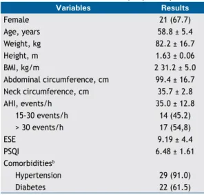
279 - Correlation of lung function and respiratory muscle strength with functional exercise capacity in obese individuals with obstructive sleep apnea syndrome Thays Maria da Conceição Silva Carvalho, Anísio Francisco Soares,
Danielle Cristina Silva Climaco, Isaac Vieira Secundo, Anna Myrna Jaguaribe de Lima
285 - Translation, cross-cultural adaptation, and reliability of the Understanding COPD questionnaire for use in Brazil
ISSN 1806-3713
Published once every two months J Bras Pneumol. v.44, number 4, p. 253-340 July/August 2018
Contents
292 - Hyperhidrosis: prevalence and impact on quality of life
Erica Nishida Hasimoto, Daniele Cristina Cataneo., Tarcísio Albertin dos Reis, Antonio José Maria Cataneo
299 - Spirometry in patients screened for coronary artery disease: is it useful? Frederico Leon Arrabal Fernandes, Regina Maria Carvalho-Pinto, Rafael Stelmach, João Marcos Salge, Carlos Eduardo Rochitte, Eliane Cardoso dos Santos Souza, Janaina Danielle Pessi, Alberto Cukier
307 - CT-guided percutaneous core needle biopsy of pulmonary nodules smaller than 2 cm: technical aspects and factors influencing accuracy
Juliano Ribeiro de Andrade, Rafael Dahmer Rocha, Priscila Mina Falsarella, Antonio Rahal Junior, Ricardo Sales dos Santos, Juliana Pereira Franceschini, Hiran Chrishantha Fernando, Rodrigo Gobbo Garcia
315 - Validation of a bioelectrical impedance analysis system for body composition assessment in patients with COPD
Fernanda Rodrigues Fonseca, Manuela Karloh, Cintia Laura Pereira de Araujo, Cardine Martins dos Reis, Anamaria Fleig Mayer
REVIEW ARTICLE
321 - Patient-ventilator asynchrony
Marcelo Alcantara Holanda, Renata dos Santos Vasconcelos, Juliana Carvalho Ferreira, Bruno Valle Pinheiro
IMAGING IN PULMONARY MEDICINE
334 - Presence of gas in an unusual place: spontaneous pneumomediastinum (Hamman’s syndrome)
Nicholas Oliveira Duarte, Camila Hino Verdelho, Rodolfo Mendes Queiroz
LETTER TO THE EDITOR
335 - Hyperimmunoglobulin E syndrome (Job syndrome): chest CT findings Pablo Rydz Pinheiro Santana, Augusto Kreling Medeiros, Cinthia Callegari Barbisan, Antônio Carlos Portugal Gomes, Edson Marchiori
337 - Left main coronary artery compression in a patient with portopulmonary hypertension
Iara Teixeira de Araújo, Pammela Jacomeli Lembi, Eduardo Belisario Falchetto, Ricardo de Amorim Corrêa
ISSN 1806-3713 © 2018 Sociedade Brasileira de Pneumologia e Tisiologia
http://dx.doi.org/10.1590/S1806-37562018000040003
A teacher to remember and to emulate
Ana Luisa Godoy Fernandes1,a, Sonia Maria Faresin1,b1. Departamento de Medicina, Escola Paulista de Medicina, Universidade Federal de São Paulo – Unifesp – São Paulo (SP) Brasil. a. http://orcid.org/0000-0003-3910-6016; b. http://orcid.org/0000-0001-9868-8015
Professor Manuel Lopes dos Santos was always an uncommon teacher. In addition to being a natural leader, he knew how to encourage and recognize the quality of each member of his team, as well as how to bring new members into the group. It was thus that he formed and directed the Discipline of Pulmonology of the Escola Paulista de Medicina (EPM, Paulista School of Medicine), in the city of São Paulo, Brazil, which is recognized as a center of excellence in pulmonology training, at
the national and international level. He was the first
preceptor of the residency program at the Hospital São Paulo (HSP), operated by the EPM, demonstrating the relevance of in-service training, which is the gold standard for professional training. He was a manager of the center of excellence, head of the discipline, director of the HSP, and director of the EPM, which he transformed into
the Federal University of São Paulo, becoming its first
rector. He founded the Paulista Society of Pulmonology and Phthisiology, after which he launched the Journal of Pulmonology (which would later become the JBP) and
was its first editor.
It was common to hear Professor Santos say, “São Paulo owes a debt to the country. We must train
pulmonologists as a group of critical thinkers who will disseminate knowledge throughout the country.” In addition to receiving students from other countries in Latin America, he taught students from every state in Brazil, making the EPM Discipline of Pulmonology the leading producer of pulmonology specialists in the country. He
also actively collaborated in the development of the first
graduate program in pulmonology in Brazil. His ability to impart knowledge was without par, due to the empathy he had for all who wanted to learn and work, as well as the unconditional love he had for the mission of teaching.
Although Professor Santos could have been treated at any other hospital, including one of those with virtually
infinite resources, he always chose the HSP, where, despite
a lack of material resources, he could be surrounded by the throng of critically thinking, humane professionals that he had helped educate. In addition, he always had the good sense to use the facility in which he believed and at which he had treated all of his own patients.
The loss of Professor Santos will felt for years to come.
During this moment of reflection and mourning, we ponder
how great Brazil would be if there were more individuals of his ilk in the country.
J Bras Pneumol. 2018;44(4):253-253
253
http://dx.doi.org/10.1590/S1806-37562018000040002
Community-acquired pneumonia:
challenges of the situation in Brazil
Mauro Gomes1,2,a1. Disciplina de Pneumologia, Faculdade de Ciências Médicas da Santa Casa de São Paulo, São Paulo (SP) Brasil. 2. Setor de Pneumologia, Hospital Samaritano, São Paulo (SP) Brasil.
a. http://orcid.org/0000-0002-5165-4501
Community-acquired pneumonia (CAP) is the leading cause of morbidity, hospitalization, and mortality worldwide and represents a diagnostic and treatment challenge.(1) In Brazil, CAP constitutes the
leading cause of hospitalization (excluding causes related to pregnancy, childbirth, and puerperium) via
the Brazilian Unified Health Care System. In 2017,
there were 598,668 CAP-related hospitalizations and
52,776 CAP-related deaths in our country.(2) Although
CAP is one of the most common infections, there have been few clinical or epidemiological studies of CAP in Brazil.
In the current issue of the JBP, Bahlis et al.(3)
publish a single-center cohort study of CAP conducted in the interior of Brazil and involving all patients hospitalized for CAP over a one-year period. Despite some limitations, the study has the great virtue of reinforcing some of our conceptions about CAP and alerting to the need for implementing measures to improve the management of CAP in our country. Chief among these conceptions are initial misdiagnosis of
pneumonia, difficulty in identifying the pathogen,
the advantages of using severity scores in the initial assessment of patients, and the importance of implementing measures to improve in-hospital management of cases.
We see in daily clinical practice that there is a lot of misdiagnosis when it comes to CAP. Clinical and laboratory signs that are characteristic of severe infection, such as tachycardia, fever, and altered
leukocyte counts, are nonspecific and are frequently
present in other acute conditions. Biomarkers, such as C-reactive protein and procalcitonin, are also
nonspecific and have more value in ruling out a diagnosis of infection than in establishing a definitive
diagnosis. It has long been known that errors in radiological interpretation made by non-pulmonary specialists working at emergency rooms occur relatively frequently.(4) As a result of these difficulties
in the initial clinical assessment, an accurate diagnosis of CAP can often be a challenge. It is of note in the study by Bahlis et al.(3) that practically one third of
the patients hospitalized for respiratory infection did not meet the diagnostic criteria for CAP and were excluded from the analysis. This may raise a debate as to whether the data for a large number of hospitalizations for CAP in Brazil are correct. Would
our statistics for CAP be inflated by inclusion of other
respiratory infections misdiagnosed as CAP? After diagnosis, major guidelines recommend that CAP severity should be assessed against criteria to determine the place of treatment and antibiotic therapy. The implementation of guidelines into care protocols has always been a major challenge, because, in daily practice, severity scores are not used by all emergency department attending physicians. However, despite assessment errors that are attributable to these scores, their implementation reduces mortality.
(5) Silveira et al.(6) reported that admission and
treatment criteria were guideline-concordant in
most of a sample of Brazilian patients (in 73.2% and 58.9%, respectively), and that guideline-concordant treatment was associated with lower 30-day mortality.
In addition, Bahlis et al.(3) found that major severity
scores showed good ability to predict in-hospital mortality, with no statistical differences between the Confusion, Urea, Respiratory rate, Blood pressure, and age > 65 years (CURB-65) score and the Pneumonia Severity Index (PSI). Therefore, especially with regard to using severity scores in the initial assessment of patients, we might apply the old saying “bad with them, worse without them.”
Difficulty in identifying the pathogen causing the
infection increases the challenge in CAP management.
Certain microorganisms are particularly difficult to cultivate, requiring specific culture media or a
particular environment. Although it is well established that Streptococcus pneumoniae is the pathogen most commonly associated with CAP, the fact is that studies of the etiology of CAP fail to identify the pathogen in most patients.(7) The currently published
study(3) is not different in this respect and identified
the pathogen in only 17% of the cases, with the
most commonly isolated agent being Streptococcus pneumoniae (in 36%).
Knowing the potential etiologic agents in CAP, which ones are increasing in incidence, their degree of resistance, and their lethality rate is critical to an effective therapy. Bahlis et al.(3) observed a high rate
of change in antibiotic regimen during hospitalization
(63%). In most cases, the initial treatment regimen
consisted of amoxicillin + clavulanate, and the most common change was the addition of azithromycin to the regimen, on the basis of clinical assessment by the attending physician. The mean length of hospital J Bras Pneumol. 2018;44(4):254-256
Gomes M
stay was 7.2 days (median, 5 days), and, over the patients’ course, 29% required ICU treatment and 15.5% died in hospital.
If early identification of the etiologic agent were
possible, a likely consequence would be a more rational use of antibiotics, with a reduction in complications and mortality from CAP. However, the current standard for diagnosis is blood culture,
which usually takes at least 48-72 h to obtain a
result, and cultures often remain negative even when bacterial or fungal infections are strongly suspected. This results in patients being initially treated empirically and often with broader spectrum antibiotics to increase the likelihood that a pathogenic organism will be adequately covered. This approach, despite being currently valid, has negative aspects, which include the potential for toxicity from use of multiple antibiotics, the high associated costs, and the development of resistance to antimicrobials.
Results after the recent development of rapid and highly sensitive molecular assays have produced surprising information about the causes of CAP.
For the first time, multiple agents that had not
been observed in older studies were detected. These methods increasingly identify respiratory
viruses, including rhinovirus and influenza virus,
in patients with CAP.(8) Pathogens were identified
in the bloodstream of infected patients within 6 h, with high sensitivity and with a three times higher
likelihood of identification than that of standard culture. These findings could potentially have
resulted in a change in antibiotic treatment in up
to 57% of the patients studied.(9)
Knowledge about CAP has undergone phases in which the presence of the so-called atypical pathogens was highly valued, and today we are going through a phase in which viruses are being
strongly considered. The true importance of finding
viruses in the respiratory secretions of patients with CAP has yet to be established, as it remains unknown whether viruses are effective pathogens
or copathogens in pneumonias. We also need to develop skills to handle molecular tests; they bring
the prospect of rapidly defining the cause of CAP
and changing the initial approach. This may allow targeted antibiotic therapy rather than empirical antibiotic therapy, which is what we currently use.
Another important challenge when it comes to treatment of CAP is the time from diagnosis to initiation of antibiotic therapy. Studies have established that a delay in initiation of antibiotic therapy of more than 4 h increases the potential risk of complications and death.(10) Current guidelines
on sepsis recommend that antibiotic therapy ideally
be initiated within the first hour, given that any
delay in therapy can result in decreased survival.
(11) The study published in the current issue of
the JBP(3) also raises this issue when it reports
the long elapsed time to initiation of antibiotic
therapy (mean, 10 h), with few patients receiving antibiotics within the first 4 h: only 19%. Because the vast majority of patients received the first
dose of antibiotics after quite a long time, the authors could not correlate this delay with the high in-hospital mortality rate and the great need for ICU admission in the study population. These data alert to the need for hospitals to implement
measures to expedite administration of the first
dose of antibiotics in cases of CAP in the emergency room, with the suggestion that this be done even for those as outpatients.
CAP constitutes a serious public health problem in Brazil. At a time when we are reviewing and updating the Brazilian guidelines for the management of CAP, studies that lay bare the situation in the country and alert to the need for measures to improve the diagnosis and therapeutic management of CAP are essential. Promoting the adoption of care protocols in health facilities, with measures tailored to the situation in our country, can reduce excessive hospitalizations and the rate of mortality from pneumonia in Brazil.
REFERENCES
1. Wiemken TL, Peyrani P, Ramirez JA. Global changes in the epidemiology of community-acquired pneumonia. Semin Respir Crit Care Med. 2012;33(3):213-9. https://doi.org/10.1055/s-0032-1315633 2. Departamento de Informática do SUS - DATASUS [homepage on the Internet]. Brasília: Ministério da Saúde. Brasília: Ministério da Saúde. [cited 2018 May 26]. Morbidade Hospitalar do SUS - por local de residência - Brasil. Available from: http://tabnet.datasus.gov.br/cgi/ tabcgi.exe?sim/cnv/obt10uf.def
3. Bahlis LF, Diogo LP, Kuchenbecker RS, Fuchs SC. Perfil clínico,
epidemiológico e etiológico de pacientes internados com pneumonia adquirida na comunidade em um hospital público do interior do Brasil. J Bras Pneumol. 2018;44(4):261-66.
4. Morrone N, Freire JA, Pereira CA, Saito M, Mendes ES, Dourado AD. Erros diagnósticos em pneumonia adquirida na comunidade: freqüência, causas e conseqüências. J Pneumol. 1987;13(1):1-6.
5. Menéndez R, Torres A, Zalacaín R, Aspa J, Martín-Villasclaras JJ, Borderías L, et al. Guidelines for the treatment of community-acquired pneumonia: predictors of adherence and outcome. Am J Respir Crit Care Med. 2005;172(6):757-62. https://doi.org/10.1164/ rccm.200411-1444OC
6. Silveira CD, Ferreira CS, Corrêa Rde A. Adherence to guidelines and its impact on outcomes in patients hospitalized with community-acquired pneumonia at a university hospital. J Bras Pneumol. 2012;38(2):148-57. https://doi.org/10.1590/S1806-37132012000200002
7. Welte T, Torres A, Nathwani D. Clinical and economic burden of community-acquired pneumonia among adults in Europe. Thorax. 2012;67(1):71-9. https://doi.org/10.1136/thx.2009.129502
8. Jain S, Self WH, Wunderink RG, Fakhran S, Balk R, Bramley AM, et al. Community-Acquired Pneumonia Requiring Hospitalization
Community-acquired pneumonia: challenges of the situation in Brazil
among U.S. Adults. N Engl J Med. 2015;373(5):415-27. https://doi. org/10.1056/NEJMoa1500245
9. Vincent JL, Brealey D, Libert N, Abidi NE, O’Dwyer M, Zacharowski K, et al. Rapid Diagnosis of Infection in the Critically Ill, a Multicenter Study of Molecular Detection in Bloodstream Infections, Pneumonia, and Sterile Site Infections. Crit Care Med. 2015;43(11):2283-91. https://doi.org/10.1097/CCM.0000000000001249
10. Lee JS, Giesler DL, Gellad WF, Fine MJ. Antibiotic Therapy for Adults
Hospitalized With Community-Acquired Pneumonia. A Systematic Review. JAMA. 2016;315(6):593-602. https://doi.org/10.1001/ jama.2016.0115
ISSN 1806-3713 © 2018 Sociedade Brasileira de Pneumologia e Tisiologia
http://dx.doi.org/10.1590/S1806-37562018000040001
Six-minute walk test in patients with
idiopathic pulmonary fibrosis
José Antônio Baddini-Martinez1,a
1. Divisão de Pneumologia, Departamento de Clínica Médica, Faculdade de Medicina de Ribeirão Preto, Universidade de São Paulo, Ribeirão Preto (SP) Brasil. a. http://orcid.org/0000-0001-8160-5084
The six-minute walk test (6MWT) is a simple method for investigating exercise capacity in patients with advanced lung disease.(1,2) Unlike patients undergoing traditional cardiopulmonary exercise testing, those undergoing the 6MWT do not, as a rule, reach maximal respiratory and heart rates, the 6MWT therefore being a submaximal test. The 6MWT is advantageous because walking is a task that patients are familiar with and because the 6MWT is inexpensive, requiring only a trained health
professional, a flat corridor of approximately 30 m in
length, a sphygmomanometer, and a pulse oximeter.
Although the 6MWT does not allow identification of the
mechanisms involved in exercise limitation, the six-minute walk distance (6MWD) is an overall measure of integration among systems (i.e., the respiratory, cardiovascular, and locomotor systems). There is increasing evidence that a low 6MWD is strongly associated with an increased risk of hospitalization and mortality in patients with advanced lung disease.(1,2)
Although the 6MWT has good test-retest reliability, there is a learning effect with repeated testing. Therefore, at least two tests should be performed on the same day,
at least 30 min apart (in order to allow for rest between
tests), the longest 6MWD being selected for analysis.(2,3)
Although the primary physiological measure of interest is the 6MWD, other parameters of interest include the SaO2 nadir, heart rate recovery at 1 min after exercise, and the product of the 6MWD by the lowest measured SpO2.(4)
Although the 6MWT is easily performed, it should not be performed in patients with disabling dyspnea, in those
with significant orthopedic abnormalities, or in those
with cardiovascular conditions such as recent myocardial infarction, severe aortic stenosis, and decompensated heart failure.(2,3) Patients showing an SpO
2 of < 88% on room air should receive supplemental oxygen during the test, which should be interrupted if the SpO2 on room air
falls below 80% for 6 s or more; the test can be resumed
when SpO2 is ≥ 85% on room air.(2,3)
In recent years, there has been increasing interest in using the 6MWT in patients with idiopathic pulmonary
fibrosis (IPF) not only in routine clinical practice but also
in controlled clinical trials.(4,5) An initial test, performed
at the first consultation, can provide information on
exercise capacity and the need for supplemental oxygen during physical activity, as well as prognostic data. It is well recognized that an SpO2 of ≤ 88% is a reliable
indicator of poor survival in IPF patients undergoing a 6MWT without supplemental oxygen.(6)
Although there have been quite a few studies involving patients with IPF and the 6MWT, the most reliable data were provided by two large clinical trials of new drugs for the treatment of IPF, a substantial number of patients having been included in those trials.(7-9) The studies showed
that the 6MWD correlated significantly with quality of
life and dyspnea, as well as with lung function variables.
An analysis of 748 volunteers included in one of the
aforementioned studies(7) showed that a 6MWD of < 250 m on the initial test was associated with double the risk of mortality after approximately one year of follow-up.
In addition, decreases of more than 50 m in the 6MWD on tests performed 24 weeks after the first resulted in a three-fold increased risk of death in the following 24
weeks. Minimal clinically important differences were calculated from data from the aforementioned studies(8,9)
and were found to be 24-45 m and 21.7-37.0 m. The
results of the two aforementioned clinical trials(7-9) clearly show that the 6MWT is a valid and useful tool for the management of patients with IPF.
In the current issue of the JBP, Mancuso et al.(10) report
the 6MWD in 70 IPF patients retrospectively selected from
among those treated at either of two referral centers
for interstitial lung disease in Brazil. The major finding of the study is that a 6MWD of < 330 m or < 70% of
the predicted value is associated with a substantially decreased survival rate and should be considered an indicator of poor prognosis in patients with IPF in Brazil.
The first question is why the indicator of poor prognosis
found in a study conducted in Brazil(10) is significantly different from those found in studies conducted elsewhere. (7,11-13) According to Mancuso et al.,(10) this might be due to differences in reference values for the 6MWT across countries.(14) There is evidence that the 6MWD is longer in individuals living in Latin America than in those living in Europe or the USA.(14) This appears to be due to the fact that physical demands are higher in individuals living in developing countries, with lower socioeconomic status. It appears that such an association also applies to respiratory diseases such as COPD and IPF. However, the difference between the values found by Mancuso et al.(10) and those found by other authors might be due to the study design and the analyses performed in the study. Because it is impossible to compare the study conducted by Mancuso et al.(10) with all previous studies, I will compare it with the study involving the highest number of patients and based on data from the study by du Bois et al.(7)
In the study by Mancuso et al.,(10) patients presenting with an SpO2 of < 89% were excluded, whereas, in the J Bras Pneumol. 2018;44(4):257-258
257
Six-minute walk test in patients with idiopathic pulmonary fibrosis
study by du Bois et al.,(7) 11.5% of the participants were receiving oxygen therapy. The difference in
findings between the two studies might be explained, at
least in part, by the fact that the latter study included patients who were more severely ill, including patients with severe pulmonary hypertension. Although this is a reasonable explanation, it should be noted that the mean 6MWD is very similar between the two studies. In fact, the mean 6MWD was higher in the study by
du Bois et al. than in the study by Mancuso et al. (397 ± 107 m vs. 380 ± 115 m).(7,10)
The difference in findings between the two studies
might also be due to different follow-up periods. In the study by du Bois et al.,(7) patients were followed for 48 weeks, whereas, in the study by Mancuso et al.,(10) the median follow-up period was 37.6 months
(range, 5-129 months). Therefore, it is possible that
the results were affected by the fact that follow-up was limited to the duration of the trial and therefore prevented a more detailed characterization of patients in whom the disease behaved in a more benign manner. Differences between the two studies regarding the statistical methods used and how the results were
reported should also be taken into account. According to du Bois et al.,(7) a 6MWD of < 250 m is associated with double the risk of mortality after 48 weeks of follow-up. According to Mancuso et al.,(10) a 6MWD of
< 330 m is associated with a survival of 24 months,
whereas a longer 6MWD is associated with a median survival of 59 months.
The study by Mancuso et al.(10) reinforces the prognostic importance of the 6MWT in patients with IPF. However, the 6MWD should be evaluated in conjunction with other clinical and physiological data, such as dyspnea intensity, FVC, and DLCO.
Pulmonologists in Brazil should be alert to the possibility of encountering IPF patients with absolute 6MWD values indicating a poor prognosis, these values being higher than those recommended in the international literature. If we analyze the 6MWD in isolation and use as an indicator of poor prognosis any of the absolute 6MWD values recommended in the international literature, we run the risk of underestimating the severity of lung disease.
REFERENCES
1. Ghofraniha L, Sani ZD, Vakilian F, Khajedalooyi M, Arabshahi ZJ. The six-minute walk test (6MWT) for the evaluation of pulmonary diseases. J Cardiothorac Med. 2015;3(2):284-7. https://doi.org/ 10.22038/JCTM.2015.4374
2. Holland AE, Spruit MA, Troosters T, Puhan MA, Pepin V, Saey
D, et al. An official European Respiratory Society/American Thoracic Society technical standard: field walking tests in chronic
respiratory disease. Eur Respir J. 2014;44(6):1428-46. https://doi. org/10.1183/09031936.00150314
3. ATS Committee on Proficiency Standards for Clinical Pulmonary
Function Laboratories. ATS statement: guidelines for the six-minute walk test. Am J Respir Crit Care Med. 2002;166(1):111-7. https://doi. org/10.1164/ajrccm.166.1.at1102
4. Brown AW, Nathan SD. The Value and Application of the 6-Minute-Walk Test in Idiopathic Pulmonary Fibrosis. Ann Am Thorac Soc. 2018;15(1):3-10. https://doi.org/10.1513/AnnalsATS.201703-244FR 5. Fernández Fabrellas E, Peris Sánchez R, Sabater Abad C, Juan
Samper G. Prognosis and follow-up of idiopathic pulmonary fibrosis.
Med Sci (Basel). 2018;6(2). pii: E51. https://doi.org/10.3390/ medsci6020051
6. Flaherty KR, Andrei AC, Murray S, Fraley C, Colby TV, Travis WD,
et al. Idiopathic pulmonary fibrosis: prognostic value of changes in
physiology and six-minute-walk test. Am J Respir Crit Care Med. 2006;174(7):803-9. https://doi.org/10.1164/rccm.200604-488OC 7. du Bois RM, Albera C, Bradford WZ, Costabel U, Leff JA, Noble
PW, et al. 6-Minute walk distance is an independent predictor of
mortality in patients with idiopathic pulmonary fibrosis. Eur Respir
J. 2014;43(5):1421-9. https://doi.org/10.1183/09031936.00131813
8. du Bois RM, Weycker D, Albera C, Bradford WZ, Costabel U, Kartashov A, et al. Six-minute-walk test in idiopathic pulmonary
fibrosis: test validation and minimal clinically important difference. J
Respir Crit Care Med. 2011;183(9):1231-7. https://doi.org/10.1164/ rccm.201007-1179OC
9. Nathan SD, du Bois RM, Albera C, Bradford WZ, Costabel U, Kartashov A. Validation of test performance characteristics and minimal clinically important difference of the 6-minute walk
test in patients with idiopathic pulmonary fibrosis. Respir Med.
2015;109(7):914-22. https://doi.org/10.1016/j.rmed.2015.04.008 10. Mancuzo EV, Soares MR, Pereira CAC. Six-minute walk test distance
and survival in idiopathic pulmonary fibrosis in Brazil. J Bras Pneumol.
2018;44(4):267-272.
11. Lettieri CJ, Nathan SD, Browning RF, Barnett SD, Ahmad S, Shorr AF. The distance-saturation product predicts mortality in idiopathic
pulmonary fibrosis. Respir Med. 2006;100(10):1734-41. https://doi.
org/10.1016/j.rmed.2006.02.004
12. Caminati A, Bianchi A, Cassandro R, Mirenda MR, Harari S. Walking distance on 6-MWT is a prognostic factor in idiopathic pulmonary
fibrosis. Respir Med. 2009;103(1):117-23. https://doi.org/10.1016/j.
rmed.2008.07.022
13. Lederer DJ, Arcasoy SM, Wilt JS, D’Ovidio F, Sonett JR, Kawut SM. Six-minute-walk distance predicts waiting list survival in idiopathic
pulmonary fibrosis. Am J Respir Crit Care Med. 2006;174(6):659-64.
https://doi.org/10.1164/rccm.200604-520OC
ISSN 1806-3713 © 2018 Sociedade Brasileira de Pneumologia e Tisiologia
http://dx.doi.org/10.1590/S1806-37562018000000155
Nodular fissure
Edson Marchiori1,a, Bruno Hochhegger2,b, Gláucia Zanetti1,c
1. Universidade Federal do Rio de Janeiro, Rio de Janeiro (RJ) Brasil.
2. Universidade Federal de Ciências da Saúde de Porto Alegre, Porto Alegre (RS) Brasil.
a. http://orcid.org/0000-0001-8797-7380; b. http://orcid.org/0000-0003-1984-4636; c. http://orcid.org/0000-0003-0261-1860
A 37-year-old male civil engineer arrived at the
outpatient clinic with complaints of dry cough for three months, followed by dyspnea upon exertion. Physical examination was normal. HRCT scanning showed small
disseminated nodules and nodular fissures (Figure 1).
The patient presented with small interstitial nodules on HRCT. The pattern of small nodules or pulmonary micronodules corresponds to the presence of multiple rounded opacities, with soft-tissue density and diameter <1 cm, distributed in the lung parenchyma.(1) They
can be classified according to the distribution in the
secondary pulmonary lobe (SPL) as random, centrilobular, or perilymphatic.
The random pattern is characterized by the presence of small nodules distributed randomly in relation to the SPL and uniformly dispersed by the lungs. This pattern is observed in diseases with blood-borne dissemination, such as metastases and miliary granulomatous diseases, especially tuberculosis and histoplasmosis. The centrilobular distribution is characterized by the presence of nodules in the central region of the SPL, a few millimeters from
the pleural surface and the fissures; however, without
touching them. Hypersensitivity pneumonitis and infectious bronchiolitis are examples of diseases that occur with this pattern. The perilymphatic pattern is characterized by the presence of small nodules that distribute preferentially along the SPL compartments that contain lymphatic structures (peribronchovascular interstitium, interlobular
septa, and subpleural regions). The diseases that most commonly present with this distribution pattern are sarcoidosis, silicosis, and carcinomatous lymphangitis.
Sarcoidosis and silicosis tend to spare interlobular septa, whereas lymphangitis frequently affects that region.(2) Silicosis also tends to preserve the peribronchovascular interstitium, whereas lymphangitis and sarcoidosis often
affect it: lymphangitis has a more smooth appearance,
whereas sarcoidosis has a more nodular appearance.
Subpleural nodules are more easily seen in the fissures, giving rise to what is called the nodular fissure pattern.
These three diseases affect the pleural surface, but silicosis
rarely presents with nodular fissures. A nodular fissure
pattern corresponds, in most cases, to carcinomatous
lymphangitis or sarcoidosis. Eventually, peri-fissural
nodules are also seen in miliary infectious diseases (tuberculosis or histoplasmosis); however, the nodules are generally smaller and less profuse than in the case reported here.
Clinically, the patient had no history of exposure to silica. The health status of the patient was very good, there was no evidence of disease elsewhere, and dyspnea was mild, data that rule out the diagnosis of carcinomatous lymphangitis. Transbronchial biopsy showed sarcoid
granulomas, with no caseous necrosis. The final diagnosis
was sarcoidosis.
Figure 1. HRCT scans with lung window settings at the level of the bronchial bifurcation evidence small interstitial nodules distributed in the lungs. Note that there is an accumulation of nodules along the fissures (detail in B), characterizing the pattern of nodular fissure.
A
B
REFERENCES
1. Webb WR, Muller NL, Naidich DP, editors. High-resolution CT of the lung. 4th ed. Philadelphia: Lippincott Williams & Wilkins; 2008.
2. Marchiori E, Zanetti G, Hochhegger B. Interlobular septal thickening. J Bras Pneumol 2016;42(2):157. https://doi.org/10.1590/S1806-37562015000000294
J Bras Pneumol. 2018;44(4):259-259
259
http://dx.doi.org/10.1590/S1806-37562018000000217
Twelve tips to write an abstract for
a conference: advice for young and
experienced investigators
Juliana Carvalho Ferreira1,2,a, Cecilia Maria Patino1,3,b
1. Methods in Epidemiologic, Clinical, and Operations Research–MECOR–program, American Thoracic Society/Asociación Latinoamericana del Tórax, Montevideo, Uruguay. 2. Divisão de Pneumologia, Instituto do Coração, Hospital das Clínicas, Faculdade de Medicina, Universidade de São Paulo, São Paulo (SP) Brasil.
3. Department of Preventive Medicine, Keck School of Medicine, University of Southern California, Los Angeles, CA, USA. a. http://orcid.org/0000-0001-5742-2157; b. http://orcid.org/0000-0001-6548-1384
As we have returned from the successful XXXIX Brazilian Thoracic Society Conference in Goiânia, Brazil—where more
than 600 abstracts have been presented—and prepare for
the American Thoracic Society International Conference deadline for submitting abstracts by November this year, we would like to emphasize the importance of presenting
high-quality scientific abstracts at such conferences.
Presenting clinical research results in the form of abstracts in national and international meetings is common and expected among clinical researchers in academic and nonacademic settings, giving researchers the opportunity to present their work in person, network with researchers
working in the same field, receive feedback from peers, and
publish their results as abstracts in conference proceedings. Writing abstracts that are clear and informative, following both the conference and internationally endorsed reporting
guidelines, is very important for various reasons: abstracts
are used by conference program committees to select the best suited ones for oral presentations; abstracts are usually available online prior to the conference and attendees can select which presentations they will attend; abstracts are usually published and, therefore, may be cited by other authors on their peer-reviewed publications;
and finally, health care professionals may base medical
decisions on results of studies that have been published only as a conference abstract. Therefore, in order to guide investigators how to write high quality conference abstracts,
we have developed 12 tips for young and experienced investigators:
1. Identify and carefully follow specific guidelines suggested by the conference. Usually an abstract
contains the following: title, background/introduction,
objectives, methods, results, and conclusion; however, this format varies across conferences. Pay close attention to information such as word limit and how the abstract should be structured.
2. Follow internationally endorsed reporting guidelines
specifically developed for conference abstracts. The
Enhancing the QUAlity and Transparency Of health
Research (EQUATOR) Network is an international initiative that seeks to improve the quality of published health research globally by developing reporting guidelines for several types of study designs.(1) Many reporting guidelines have extensions focusing specifically on abstracts.(2) Read
them before starting to write your abstract.
3. Think carefully about the title because this is what
readers look at first. Compose a clear, objective title
and, whenever possible, include the study design. You can make it attractive, but avoid trying to be too clever (especially for beginners).
4. Do not waste words on the introduction. Be brief and straight to the point. Save space here, so you can provide more details in the methods and results sections, which are novel and particular to your study. 5. Clearly state the objectives of the study. The
objective derives from your research question and should clearly align with results and conclusion. 6. Make sure that the methods section is detailed
enough—but not too technical—and include the study design, setting, study participants, and eligibility criteria. You should also include a description of the important variables of the study, such as the exposure, intervention, predictors, and outcome, as well as the analytic approach used to answer the research question.
7. Be precise and specific when writing the results. Report the number of participants that were included the analysis, and, most importantly, always report the results that actually answer your research question (e.g., the difference between groups with a measure
of precision such as an SD or 95% CI) and never just
a p value.
8. Be realistic in the conclusion. Mention the impact of your study, but avoid speculating beyond what your results show; you can also mention future directions in the area of study, but avoid the overused “more studies are needed…”
9. Perform a careful spell and language check, especially if you are not writing in your native language.
10. Avoid or minimize abbreviations. Readers can feel frustrated when they have to go back to remember what an abbreviation stands for (e.g., EQUATOR in this paper). 11. Get feedback from your coauthors, mentor, and
colleagues outside your team. The goal is to use their help to identify unclear sentences and missing or inaccurate information, as well as to make sure that the writing is high quality. They can also help you to make sure that the title, objectives, methods, results, and conclusion are all aligned with the research question.
12. Do NOT wait until the last minute to write and proofread the content. Writing and reviewing the abstract for quality always takes more time than you initially thought it would. Moreover, glitches in the submission process are always possible, so you want to give yourself time to contact the conference staff for help, if necessary.
REFERENCES
1. EQUATOR network [homepage on the internet]. Oxford: University of Oxford [cited 2018 Jun 1]. Available from: http://www.equator-network.org/ 2. Hopewell S, Clarke M, Moher D, Wager E, Middleton P, Altman DG,
Schulz KF, the CONSORT Group. CONSORT for reporting randomised trials in journal and conference abstracts. Lancet. 2008;371(9609):281-3. https://doi.org/10.1016/S0140-6736(07)61835-2
J Bras Pneumol. 2018;44(4):260-260 CONTINUING EDUCATION:
ISSN 1806-3713 © 2018 Sociedade Brasileira de Pneumologia e Tisiologia
http://dx.doi.org/10.1590/S1806-37562017000000434
ABSTRACT
Objective: To describe the patient profile, mortality rates, the accuracy of prognostic
scores, and mortality-associated factors in patients with community-acquired pneumonia (CAP) in a general hospital in Brazil. Methods: This was a cohort study involving patients with a clinical and laboratory diagnosis of CAP and requiring admission to a public hospital in the interior of Brazil between March 2014 and April 2015. We performed multivariate analysis using a Poisson regression model with robust variance to identify factors associated with in-hospital mortality. Results: We included 304 patients. Approximately
70% of the patients were classified as severely ill on the basis of the severity criteria
used. The mortality rate was 15.5%, and the ICU admission rate was 29.3%. After multivariate analysis, the factors associated with in-hospital mortality were need for mechanical ventilation (OR: 3.60; 95% CI: 1.85-7.47); a Charlson Comorbidity Index score > 3 (OR: 1.30; 95% CI: 1.18-1.43); and a mental Confusion, Urea, Respiratory rate, Blood pressure, and age > 65 years (CURB-65) score > 2 (OR: 1.46; 95% CI: 1.09-1.98). The mean time from patient arrival at the emergency room to initiation of antibiotic therapy was 10 h. Conclusions: The in-hospital mortality rate of 15.5% and the need
for ICU admission in almost one third of the patients reflect the major impact of CAP
on patients and the health care system. Individuals with a high burden of comorbidities, a high CURB-65 score, and a need for mechanical ventilation had a worse prognosis. Measures to reduce the time to initiation of antibiotic therapy may result in better outcomes in this group of patients.
Keywords: Community-acquired infections; Pneumonia; Hospital mortality; Risk factors.
Clinical, epidemiological, and etiological
profile of inpatients with
community-acquired pneumonia in a public hospital in
the interior of Brazil
Laura Fuchs Bahlis1,2,3,a, Luciano Passamani Diogo3,b,
Ricardo de Souza Kuchenbecker4,c, Sandra Costa Fuchs4,d
Correspondence to:
Laura Fuchs Bahlis. Universidade do Vale do Rio dos Sinos, Campus São Leopoldo, Avenida Unisinos, 950, Cristo Rei, CEP 93022-750, São Leopoldo, RS, Brasil. Tel.: 55 51 3591-1122 or 55 51 99663-8628. E-mail: laurabahlis@gmail.com
Financial support: None. INTRODUCTION
Community-acquired pneumonia (CAP) is one of the leading causes of death from infectious diseases worldwide.(1) It is a public health problem and is a cause of morbidity and mortality in all age groups.(2) Mortality rates vary according to the population and context
studied, ranging from 1%, in outpatients, to 50%, in
inpatients.(3,4) High mortality rates are more common in developing countries, such as Brazil, Argentina, and India.(5,6) CAP is responsible for high costs in public and private health care facilities, whether in outpatient clinics, emergency rooms, or hospital wards.(7,8) In Brazil,
data from the Brazilian Unified Health Care System
show that pneumonia was the second leading cause of
hospitalization in 2017, accounting for approximately 14% of all hospitalizations.(9)
Although CAP is a high-incidence disease, with multiple studies having been conducted on risk factors and available therapies, several issues regarding CAP remain controversial.(10) Using guidelines for the treatment of pneumonia has been shown to reduce hospital stays, mortality rates, and complications rates.(11-13) However, a
recent study conducted in Brazil showed that slightly more than half of the patients admitted to a university hospital were treated in accordance with current guidelines.(14) With regard to assessment of disease severity, studies have shown that using clinical judgment alone can either underestimate or overestimate the severity of the clinical presentation. This strategy can lead to unnecessary hospitalizations, as well as to interventions that are less aggressive than those required in more severe cases, culminating in negative outcomes.(15-17) Nevertheless, a
study published in 2015 showed that most physicians
in Brazil use clinical judgment alone to assess disease severity in patients with CAP.(14)
It is possible to suspect that the aforementioned discrepancies are partly due to the fact that the major guidelines and severity scores are based on large international studies, and, despite the high reported incidence of CAP in Brazil, little is known about local microbiological patterns and disease severity at the regional level.(18) Therefore, we conducted a cohort study involving patients with CAP admitted to a hospital in the interior of Brazil over a one-year period, in order to
describe the patient profile, mortality rates, the accuracy
1. Faculdade de Medicina, Universidade do Vale do Rio dos Sinos, Campus São Leopoldo, São Leopoldo (RS) Brasil.
2. Programa de Pós-Graduação em Epidemiologia, Universidade Federal do Rio Grande do Sul – UFRGS – Porto Alegre (RS) Brasil.
3. Hospital de Clínicas de Porto Alegre, Porto Alegre (RS) Brasil.
4. Faculdade de Medicina, Universidade Federal do Rio Grande do Sul – UFRGS – Porto Alegre (RS) Brasil.
a. http://orcid.org/0000-0002-1986-0970
b. http://orcid.org/0000-0001-6304-2767
c. http://orcid.org/0000-0002-4707-3683
d. http://orcid.org/0000-0001-6351-9588
Submitted: 15 December 2017.
Accepted: 18 April 2018.
Study carried out under the auspices of the Programa de Pós-Graduação em Epidemiologia, Universidade Federal do Rio Grande do Sul – UFRGS – Porto Alegre (RS) and at the Hospital Montenegro, Montenegro (RS) Brasil.
J Bras Pneumol. 2018;44(4):261-266
261
Clinical, epidemiological, and etiological profile of inpatients with community-acquired
pneumonia in a public hospital in the interior of Brazil
of prognostic scores, and factors associated with in-hospital mortality in these patients.
METHODS
Study population
The present study was conducted in a secondary-care
general hospital, with 130 beds, located in the city of
Montenegro, Brazil. The hospital provides care via the
Brazilian Unified Health Care System to approximately 160,000 people, many of whom reside in one of 19 surrounding municipalities. In 2015, respiratory
infections represented the leading cause of admission to the hospital, and the rate of in-hospital mortality
was 10.2%.(19)
Study design
We screened for patients who were 14 years of age or older, had respiratory symptoms, and were
referred for hospital admission between May 2014 and April 2015. Patient inclusion in the cohort at baseline
was based on a clinical and radiographic diagnosis of CAP and a referral by the attending physician for hospitalization. We excluded patients with nosocomial
pneumonia—characterized by hospitalization for 2 or
more days in the past three months—residents of nursing or retirement homes; those who received intravenous antibiotics, chemotherapy, or scar treatment in the
past 30 days; and those undergoing renal replacement
therapy. Patients were assessed using the following
severity scores: Charlson Comorbidity Index (CCI);
mental Confusion, Urea, Respiratory rate, Blood pressure, and age > 65 years (CURB-65) score; and Pneumonia Severity Index (PSI), on the basis of data documented in the cohort’s medical records collected at baseline. We assessed the clinical course of patients during in-hospital follow-up and determined clinical outcomes at the time of hospital discharge.
Cases of pneumonia were defined in accordance
with the criteria established by the Centers for Disease Control and Prevention,(20) on the basis of chest X-rays
with at least one of the following findings: new or progressive and persistent infiltrate, consolidation, and/or cavitation; and at least one of the following signs or symptoms: fever (> 38 °C) with no other cause detected; leukopenia (< 4,000 leukocytes/mm3)
or leukocytosis (≥ 12,000 leukocytes /mm3); and, for
adults aged 70 years or older, a change in mental state with no other cause identified. In addition, there should be at least two of the following findings: new onset of
purulent sputum or change in character of sputum; increased respiratory secretions; increased frequency of aspiration; onset or worsening of cough, dyspnea, or tachypnea; bronchial breath sounds; worsening of gas exchange (e.g., oxygen desaturation, with a PaO2/
FiO2 ratio ≤ 240); increased need for oxygen; or need for mechanical ventilation.
The study was approved by the Porto Alegre Hospital de Clínicas Research Ethics Committee (Protocol GPPG
no. 150168), which is accredited by the Office of Human
Research Protections as an institutional review board,
and researchers signed a data use agreement protecting
the confidentiality of medical records.
Study variables
Clinical, laboratory, and radiological data for the first 24 h following the emergency room visit were obtained
from a review of medical records, and included the
following variables: age; gender; place of residence;
RR; arterial blood pressure; axillary temperature; HR; presence of mental confusion; SpO2; comorbidities (as documented by the attending physician); history
of hospitalization; chest X-ray findings (as assessed
by a radiologist); and laboratory tests ordered in the emergency room. Laboratory tests included arterial blood gas analysis, urea, serum creatinine, blood glucose, sodium, and blood count. We recorded the antibiotics administered during hospitalization, as well as total length of hospital stay, length of ICU stay, and need for mechanical ventilation. The primary outcome was all-cause in-hospital mortality, as documented
in the medical records and confirmed by review of hospital discharge summaries or death certificates,
as appropriate. All patients had medical records, and their discharge summaries were completed by the attending physician within 48 h of discharge.
It is important to emphasize that patient management, treatment choice, and outcomes suffered no interference from this study, because it was an observational study and we had no contact with either the patients or the attending physicians, who were responsible for all clinical decisions. In the hospital, there is a care protocol for the treatment of pneumonia, and inpatients are treated by a physician team, which was composed of
five members at the time of the study.
Data analysis
Data were entered into an Excel® database by two
different individuals and subsequently compared to identify possible typographical errors. Continuous variables are expressed as mean and standard deviation; categorical variables are expressed as frequency and proportion. The bivariate analysis between clinical characteristics and mortality was performed by using the Student’s t-test (for means and standard deviations) or Pearson’s chi-square test (for proportions). ROC curve analysis was performed to evaluate the prognostic indices. A Poisson regression model with robust variance was used to assess the relationship between the variables and the primary outcome (in-hospital
mortality). Analyses with a two-tailed p value < 0.05 were considered statistically significant.
All analyses were performed with the Statistical
Package for the Social Sciences, version 17.0 (SPSS
Inc., Chicago, IL, USA) and the R software, version
4.0-1 (The R Foundation for Statistical Computing,
Vienna, Austria).(21-24)
RESULTS
Between March 2014 and April 2015, we assessed
Bahlis LF, Diogo LP, Kuchenbecker RS, Fuchs SC
not meet the diagnostic criteria for CAP, and, therefore,
304 patients were included in the final analysis (Figure 1). Of those, 171 (56%) were male, and the mean age was 67.1 ± 17.2 years. Most participants (69%)
resided in the city of Montenegro. Of the patients in
the final sample, 150 (49%) had asthma or COPD (previous lung disease), and 155 (51%) were smokers.
The mean CCI score was 4.9 ± 3.1. The characteristics of the patients are presented in Table 1.
The mean CURB-65 score was 2.2 ± 1.1, and 71%
of the patients were considered to have severe CAP
(CURB-65 scores ≥ 2). The mean PSI score was 3.8 ± 1.3, and 74% of the patients were considered to
have severe CAP (PSI scores > 3). The two indices showed good ability to predict in-hospital mortality,
with areas under the ROC curve being 0.73 for the CURB-65 score (95% CI: 0.66-0.80; p < 0.001) and 0.75 for the PSI (95% CI: 0.68-0.82; p< 0.001); there was no statistically significant difference between the indices (p = 0.65), as shown in Figure 2.
The mean time from patient arrival at the emergency
room to initiation of antibiotic therapy was 10.4 ± 7.7
h. Blood or sputum samples were collected for culture
from 101 patients (33%), and the infectious agent was isolated in 53 patients (17%). The most commonly
isolated agent was Streptococcus pneumoniae (in
36% of positive cultures). The most commonly used
antibiotic regimen was amoxicillin plus clavulanate,
in 219 patients (72%), followed by azithromycin, in 200 (66%).
We found that 191 (63%) of the patients required a
change in the antibiotic regimen during hospitalization. The most common change was the addition of azithromycin to the antimicrobial regimen, on the basis of clinical assessment by the attending physician.
During the in-hospital follow-up period, 47 (15.5%) of the patients died, 89 (29.3%) required ICU treatment, and 98 (32.2%) underwent mechanical ventilation. The mean length of hospital stay was 7.2 ± 7.3 days
(median, 5 days).
Univariate analysis showed that the factors associated
with increased risk for in-hospital mortality were: a CCI score > 3 (OR: 7.18; 95% CI: 2.28-22.58; p < 0.001); a CURB-65 score > 2 (OR: 4.45; 95% CI: 1.64-12.02; p = 0.001); a PSI score > 3 (OR: 9.05; 95% CI: 1.27-64.14; p = 0.004); need for a change in the antibiotic regimen (OR: 2.15; 95% CI: 1.05-4.42; p = 0.02); need for mechanical ventilation (OR: 6.13; 95% CI: 3.33-11.28; p < 0.001); age > 62 years (OR: 6.73; 95% CI: 2.35-19.34; p < 0.001); and being institutionalized (OR: 2.82; 95% CI: 1.08-7.35; p = 0.03).
After multivariate analysis, the factors that remained associated with in-hospital mortality were need for
mechanical ventilation (OR: 3.58; 95% CI: 1.92-6.67; p < 0.001); a CCI score > 3 (OR: 1.30; 95% CI: 1.22-1.39; p < 0.001); and a CURB-65 score > 2 (OR: 1.45; 95% CI: 1.05-2.00; p = 0.04; Table 2).
DISCUSSION
CAP continues to be one of the leading causes of death from infectious disease worldwide. Despite the large number of international studies on this subject, there are few studies describing the impact of CAP on patients and the characteristics of the disease in Brazil, especially in inpatient units. In our study, we assessed inpatients with CAP in a secondary-care hospital in Brazil over a one-year period. The observed
in-hospital mortality rate of 15.5% reflects the major
impact of this disease, being similar to that found in other national and international studies.(25,26) In addition, ICU admission was required in almost one third of the
cases (29.3%), which increases the impact of CAP on
patients and the health care system.
In our study, we found no relationship between the time of initiation of antibiotic therapy and mortality, possibly because of the long elapsed time to initiation
of antibiotic therapy (a mean of 10 h) and the low proportion of patients (19%) who received antibiotic therapy within the first 4 hours. Difficulty in prompt
initiation of antibiotic therapy had been reported in another study conducted in Brazil.(27) In the present study, we found that, even after the implementation of a protocol for the treatment of pneumonia, there was no success in the attempt to reduce the time to initiation of antibiotic therapy. This is probably due to a tendency to administer antibiotics at scheduled 459 potentially
eligible patients
76 excluded: radiological criteria
54 excluded: clinical criteria
23 excluded: nosocomial pneumonia
02 excluded: initially treated outside the hospital 329 patients with
a diagnosis of pneumonia
304 patients included in the
final analysis
Figure 1. Flow chart of patient inclusion in the study.