Caused by
Leishmania braziliensis
in Brazil: A
Randomized and Controlled Trial
Paulo R. Machado1*, Julia Ampuero2, Luiz H. Guimara˜es1, Leonardo Villasboas1, Ana T. Rocha1, Albert Schriefer1, Rosana S. Sousa1, Anette Talhari3, Gerson Penna2, Edgar M. Carvalho1
1Servic¸o de Imunologia, Hospital Universita´rio Prof. Edgard Santos, Universidade Federal da Bahia, Salvador, Brazil,2Nu´cleo de Medicina Tropical, Universidade de Brası´lia, Brası´lia, Brazil,3Fundac¸a˜o de Medicina Tropical do Amazonas, Manaus, Brazil
Abstract
Background: Cutaneous leishmaniasis (CL) is treated with parenteral drugs for decades with decreasing rate cures. Miltefosine is an oral medication with anti-leishmania activity and may increase the cure rates and improve compliance.
Methodology/Principal Findings:This study is a randomized, open-label, controlled clinical trial aimed to evaluate the efficacy and safety of miltefosine versus pentavalent antimony (Sbv) in the treatment of patients with CL caused by
Leishmania braziliensis in Bahia, Brazil. A total of 90 patients were enrolled in the trial; 60 were assigned to receive miltefosine and 30 to receive Sbv. Six months after treatment, in the intention-to-treat analyses, the definitive cure rate was 53.3% in the Sbvgroup and 75% in the miltefosine group (difference of 21.7%, 95% CI 0.08% to 42.7%, p = 0.04). Miltefosine was more effective than Sbvin the age group of 13–65 years-old compared to 2–12 years-old group (78.9%versus45% p = 0.02; 68.2%versus70% p = 1.0, respectively). The incidence of adverse events was similar in the Sbvand miltefosine groups (76.7% vs. 78.3%). Vomiting (41.7%), nausea (40%), and abdominal pain (23.3%) were significantly more frequent in the miltefosine group while arthralgias (20.7%), mialgias (20.7%) and fever (23.3%) were significantly more frequent in the Sbvgroup.
Conclusions: This study demonstrates that miltefosine therapy is more effective than standard Sbv and safe for the treatment of CL caused byLeishmania braziliensisin Bahia, Brazil.
Trial Registration:Clinicaltrials.gov Identifier NCT00600548
Citation:Machado PR, Ampuero J, Guimara˜es LH, Villasboas L, Rocha AT, et al. (2010) Miltefosine in the Treatment of Cutaneous Leishmaniasis Caused by Leishmania braziliensisin Brazil: A Randomized and Controlled Trial. PLoS Negl Trop Dis 4(12): e912. doi:10.1371/journal.pntd.0000912
Editor:Pierre Buffet, AP-HP, Service de Parasitologie-Mycologie, France
ReceivedApril 23, 2010;AcceptedNovember 11, 2010;PublishedDecember 21, 2010
Copyright:ß2010 Machado et al. This is an open-access article distributed under the terms of the Creative Commons Attribution License, which permits unrestricted use, distribution, and reproduction in any medium, provided the original author and source are credited.
Funding:This study is part of a National Multicenter Clinical Trial for the evaluation of miltefosine in the treatment of cutaneous leishmaniasis caused byL. (V.) braziliensisandL. (V.) guyanensisin Brazil. The present manuscript refers to the study arm performed in the Health Post of Corte de Pedra - Immunology Service of the University Hospital Professor Edgard Santos at the Federal University of Bahia, on patients infected withL. (V.) braziliensiswith financial support of the Brazilian National Research Council (CNPq) No. 410559/2006-7 (edital mct/cnpq/ms/sctie/decit 25/2006). The other arm was conducted at the Fundac¸a˜o de Medicina Tropical do Amazonas with patients infected withL. (V.) guyanensis. Miltefosine (ImpavidoTM) was supplied by Aeterna Zentaris GmbH. The funders had no role in study design, data collection and analysis, decision to publish, or preparation of the manuscript.
Competing Interests:The authors have declared that no competing interests exist. * E-mail: prlmachado@pq.cnpq.br
Introduction
The principal species causing cutaneous leishmaniasis (CL) in Brazil is Leishmania braziliensis which most often leads to a cutaneous form of the disease characterized by one or more ulcers with raised borders, most frequently located on the upper and lower extremities, but also found on the head, face, and trunk [1,2]. Although CL is a self-limited disease, approximately 3 to 5% of subjects infected with L. braziliensis will eventually develop mucosal disease or disseminated leishmaniasis, both considered severe forms of leishmaniasis [2–4].
Pentavalent antimony (Sbv) by intramuscular or intravenous route remains the first-line drug for the treatment of CL, a therapy that is moderately toxic and difficult to administer in poor rural areas. In an endemic area ofL. braziliensistransmission in Bahia,
Brazil, cure rates after Sbv therapy are becoming increasingly lower and vary from 50% to 90% [5–7]. Factors contributing to this variability are not yet fully understood. The development of parasite resistance [8] to a drug used for decades and irregular adherence by the patients due to the daily schedule of parenteral route during 20 days, could be main factors in determining the increasingly failure rate to Sbv[5–7]. Other alternative drugs like pentamidine and amphothericin B are also of parenteral use and the former may require hospitalization.
60% andL braziliensis33% respectively [10]. More recently in CL caused byL. braziliensisin patients from Bolivia, Soto et al reported a 88% cure rate [11]. These data shows that CL cure rate upon miltefosine treatment varies among Leishmania species and also between the same species from different endemic areas. Accord-ingly, it is demonstrated that different treatment outcome after antimony therapy is found in CL caused by L. braziliensis from different endemic regions in Brazil [5,6,12]. There is no data about the use of miltefosine in CL caused byL. braziliensisin Brazil, where this species is considered the most aggressive and prevalent agent of the disease [13].
This randomized and controlled trial evaluated the efficacy and safety of miltefosine versus meglumine antimoniate (Sbv) for the treatment of CL in aL. braziliensisendemic rural area in Bahia, Brazil.
Methods Ethics Statement
This trial has been conducted according to the principles expressed in the Declaration of Helsinki. Prior to enrollment in the study, all patients received a written copy of study policy which was reviewed with them individually by an independent party. A written informed consent was obtained for all adult patients, and from parents or guardians of minors. This study was approved by the Ethics Committee of the Federal University of Bahia, in Salvador, Brazil (CEP/MCO/UFBA-Par/Res 034/2007).
Endemic area
All subjects were recruited at the health post of Corte de Pedra, located 260 km southeast of Salvador, the capital of Bahia, Brazil. This clinic is a referral center for the diagnosis and treatment of CL, with an average of 1,000 new cases per year.L. braziliensishas been the unique causal agent identified in this area in the last 15 years [14].
Patient selection
The criteria used for the diagnosis of CL were the presence of a typical ulcerated lesion and a positive Montenegro intradermal
skin test in a subject living in the endemic area. A typical CL ulcerated lesion is characterized by a round shape and raised borders associated with regional adenopathy. This classical clinical picture together with a positive leishmania skin test is highly specific for CL in the endemic area [2,5]. Patients were then selected based on the following inclusion criteria: 1) age between 2 and 65 years; 2) a maximum of 5 ulcers with no more than 2 body regions involved; 3) lesion size between 10 and 50 mm in a single dimension; and 4) a period of less than 90 days from the onset of the first ulcer. All subjects were submitted to a punch biopsy to obtain material for leishmania culture and PCR. Patients with a prior history of CL or antimony use, patients with evidence of mucosal or disseminated disease, pregnant or breastfeeding mothers, and patients with HIV or any systemic severe disease were excluded. A total of 90 patients were enrolled in the study.
Group Assignment
A randomization table was obtained with Statacorp LP 9, Texas USA. Group assignments were made after assessment that patient had met all eligibility criteria and no patients were withdrawn after randomization because of ineligibility. The 90 patients were randomly assigned at a rate of 2:1 allocation to receive miltefosine for 28 days or Sbvfor 20 days in two age groups (2–12 years-old and 13–65 years-old).
Intradermal skin test
The Leishmania antigen used for intradermal skin testing was obtained from Leishmania amazonensis strain (MHOM-BR-86BA-125). The volar surface of the left forearm was injected with 25mg of antigen in 0.1 mL of distilled water, and the largest diameter of induration was measured at 48–72 hours. The test was considered positive for induration greater than 5 mm.
Parasitology
Parasite Culture. Needle aspiration of a skin lesion was performed, and aspirates were cultured in Nicolle-McNeal-Novy medium overlaid with modified liver infusion triptase medium. Cultures were kept at 25uC and examined twice weekly. Isolates were characterized by use of a panel of monoclonal antibodies.
PCR for leishmania DNA
DNA isolation was carried out from biopsy samples using the Wizard Genomic DNA purification kit (Promega Corporation – USA). Purified DNA was resuspended in TE buffer and stored frozen at 220uC until use. Detection of the subgenus Viannia applied the primers 59-GGGGTTGGTGTAATATAGTGG-39
and 59-CTAATTGTGCACG-39. For Leishmania genus detection we have used the primers 59-(G/C)(G/C)(C/G)CC(A/C)CTAT(A/ T)TTA CAC CAA CCC C-39and 59-GGG GAG GGG CGT-39. Amplification mixes consisted of 25 pmol each primer; 1.2 mM MgCl2; 0.2 mM dNTP; 2.5 U Taq DNA polymerase; 106PCR
buffer; 2 uL of target DNA. Amplifications were carried out in a Veriti 96-well thermal cycler (Applied Biosystems – USA). Viannia detection applied 35 cycles of 1 minute at 94uC, 1 minute at 60uC and 1 minute at 72uC. Amplicons were fractionated in 1.3% agarose gels, stained with ethidium bromide and photographed under UV light using a UVP Vision Works LS apparatus (UVP – USA). The Leishmania specific band consists of 120 base pairs, and that for Viannia of 750 base pairs.
Drug administration
All study volunteers were treated as outpatients. Miltefosine (Impavido, Zentaris GmbH) was supplied in blisters containing
Author Summary
10 mg or 50 mg capsules. Meglumine antimoniate (Glucantime, Aventis) was supplied in vials of 5 ml containing 81 mg/Sbv/ml. Miltefosine was administered orally at the total target daily dosage of 2.5 mg/kg of body weight (maximum daily dose of 150 mg) for 28 consecutive days. Daily dose was divided in two or three intakes, given always with meals according to the following weight scale: patients with$15 kg and#29 kg - total dose of 50 mg/day; patients with $30 kg and #45 kg - total dose of 100 mg/day; patients with $46 kg - total dose of 150 mg/day. Meglumine antimoniate (Sbv) was administered intravenously at a dose of 20 mg Sbv/kg/day for 20 consecutive days (maximum daily dose of 3 ampoules or 1215 mg/Sbv). At every weekly visit patients returned the blisters for verification of regular use and adherence. Sbv was administered daily in health posts near from patient’s home. In these cases the administration’s date and dosage were registered and signed by the health care provider.
All women in child bearing age were submitted to beta HCG test to exclude pregnancy. The use of a parenteral contraceptive during and for 2 months after treatment was done in all women in child bearing age.
Study Procedures
Complete hemogram, aminotransferases (AST, ALT), alkaline phosphatase, potassium, sodium, urea, creatinine, and urine chemistry were determined in all patients on day -1, weekly thereafter up to the end of therapy, and after 15 and 30 days of the end of therapy. Those with abnormal parameters were followed-up until normalization. At each weekly return for drug dispensation patients were monitored for adverse events (AE). Bidirectional measurements of ulcers were taken of the patients’ lesions at the initial visit, and at each follow-up visit with standardized caliper. The area involved was calculated as the product of the two measurements. A standardized digital photograph was also taken from each patient’s lesions at the same time points.
Patients were seen for follow-up at 2 weeks, 1, 2, 4 and 6 months post-therapy. In the event that a patient did not return for follow-up at the specified time, visits were conducted in the patient’s home in the same day or within 7 days of the missed appointment.
Clinical endpoints criteria. Primary end point: cure at 6 months after the end of therapy. Secondary end-points: a) cure at 2 months after the end of therapy; b) data from clinical and laboratorial adverse events. All lesions were also categorized as either active or healed (cured) at follow-up visits. Only lesions with complete re-epithelialization, without raised borders, infiltrations or crusts were considered healed. Evaluation of the lesions was performed by 2 clinicians (LHG, EMC) who were unaware of the group assignment of all patients.
Clinical and laboratory AE were graded according to the Common Terminology Criteria for Adverse Event v3.0 (CTCAE) of the National Cancer Institute (ctep.cancer.gov/protocolDeve-lopment/electronic_applications/docs/ctcaev3.pdf).
Statistical Analysis
The results are presented as proportions, interquartile ranges (IQR), 95% confidence intervals (95% CI), means and standard-deviations (SD). The normally distributed variables were com-pared using the t test. The proportions were comcom-pared with the Chi-square or Fisher test when appropriate.
The sample size of 90 patients was obtained by calculating the number of participants needed for 80% power (b= 0.2) to detect an absolute difference as large as 25% in the rate of cure between the two treatment groups with a statistical significance of 5% (a= 0.05). The intention-to-treat (ITT) analysis was used to
calculate the cure rates. All statistical analyses were performed with the software SPSS 9.0 for Windows. A value of p,0.05 established the level of statistical significance.
Results
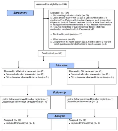
From the 244 CL patients screened from July 2007 and August 2008, 154 subjects met the inclusion criteria, 90 were included in the trial and completed the treatment, and 87 were followed for the entire 6 months after therapy, up to March 2009 (Figure 1). The patients ranged in age from 4 to 65 years with the mean age of 22615 years, which did not differ significantly between the two treatment groups (p = 0.85). Overall, there was a predominance of male patients (67.8% vs. 32.2%), and there were statistically more males in the miltefosine group than in the Sbvgroup (75.0% vs. 53.3%, p = 0.04). The majority presented with a single lesion (76.7%), and significantly more patients in the miltefosine group had $2 lesions (30% vs. 10%, p = 0.04). The lesion with the greatest area at presentation was considered to be the main lesion. The main lesion area was not different between treatment groups (410.6 vs. 461.2 mm2, p = 0.47). The clinical characteristics of patients in the two treatment groups and age groups (2–12 years and 13–65 years) are shown in Table 1. A positive culture for leishmania promastigotes was obtained in 52 out of 90 patients (57.7%).L braziliensiswas identified in 41 obtained samples from biopsy by PCR. In 26 patients (28.8%) neither PCR nor culture identified leishmania.
Efficacy
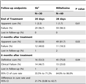
Two months after the end of the treatment, 60% (18/30) of patients in the Sbvgroup had cured lesions, compared with 81.7% (49/60) in the miltefosine group. The cure rates at 6 months of follow-up were 53.3% (16/30) in the Sbvgroup and 75% (45/60) in the miltefosine group (p = 0.04) (Table 2). The absolute difference was 21.7% (95% CI 0.08% to 42.7%). The mean time to cure was 75615 days in the Sbvgroup and 73617 days in the miltefosine group (p = 0.63).
In age groups 2–12 years-old and 13–65 years-old cure rates at 6 months for miltefosine and Sbvwere 68.2% (15/22)versus70% (7/10) (p = 1.0), and 78.9% (30/38)versus45% (9/20) (p = 0.02), respectively.
Two patients (one from miltefosine group and the other from Sbvgroup) were lost for follow-up after the end of the treatment. One patient in the miltefosine group was excluded by irregular use of the medication.
Relapses occurred in both groups (two patients treated with Sbv and four patients treated with miltefosine) between the 2 months and 6 months after the end of the treatment. In four patients (miltefosine = 3 and Sbv= 1) there was a reactivation of the ulcer and the two others (miltefosine = 1 and Sbv= 1) had reactivation of the infiltration in the borders of the healed ulcer.
Figure 1. Trial flowchart.
doi:10.1371/journal.pntd.0000912.g001
Table 1.Baseline characteristics of the 90 patients with Cutaneous Leishmaniasis included in the trial and treated with Sbvor miltefosine.
Characteristics
Sbv (n = 30)
Miltefosine
(n = 60) P value
Age group in years Age2–12 y Age13–65 y
2–12 y (n = 10)
13–65 y (n = 20)
2–12 y (n = 22)
13–65 y (n = 38)
Male/female 16/14 45/15 0.04
Number of male/female per age group 5/5 11/9 15/7 30/8 0.44 0.07
Age (years)6SD (range) 22.7614.7 (7–65) 22.0615.2 (4–59) 0.85
No. of lesions (%) 1
$2
27 (90.0) 3 (10.0)
42 (70.0)
18 (30.0) 0.04
No. of lesions per age group NA NA
1 8 19 15 27
2 0 0 6 7
3 1 1 0 4
4 1 0 1 0
5 0 0 0 0
Area of lesion (mm2)
6SD 410.6 461.2 0.47
Toxicity and Tolerability
The frequency of AE was similar in the Sbv and miltefosine groups (76.7% vs. 78.3%, p = 0.86) but they were reported more commonly in patients $13 years-old than those ,13 years-old
(88.3% vs. 66.7%, p = 0.07). Although the detection of AE was similar, the type of side effects varied widely between the treatment arms (Table 3). The AE that were significantly more frequent in the miltefosine group were vomiting (41.7%), nausea (40%) and abdominal pain (23.3%). In the Sbvgroup, arthralgias (20.7%), mialgias (20.7%) and fever (23.3%) were significantly more frequent than in the miltefosine group (Table 3). Others common AE were diarrhea (10% of miltefosine patients) and headache (43% of Sbvpatients). None of the reported AE required complete discontinuation of therapy in any patient. In the miltefosine group, one patient presented CTC grade 3 vomiting (6 episodes in 24 hrs but did not require IV fluids) and other patient grade 3 diarrhea (increase of$7 stools per day over baseline and interfering with activities of daily living, but did not require hospitalization). Both were able to continue the treatment with miltefosine after a period of 3 to 5 days of interruption and oral fluid supplementation. One patient in the Sbvgroup presented one episode of CTC grade 3 urticaria at the end of therapy. This patient needed to use oral antihistaminic for 7 days with total regression of urticaria.
Mild and transient raised liver enzymes was detected in less than 5% of patients in both groups. No raised levels of urea and creatinine or sodium and potassium abnormalities were detected (data not shown).
Discussion
The treatment of CL caused byL. braziliensiswith Sbvin rural endemic areas in Brazil has been associated with decreasing cure rates, in a setting where the parenteral route can be implicated with unsatisfactory adherence. Additionally, the monotherapy of CL with Sbv after several decades may induce Leishmania resistance. It also has been shown that CL caused byL. braziliensis
Table 2.Follow-up endpoints results in patients with CL treated with Sbvor miltefosine.
Follow-up endpoints Sbv Miltefosine P value
N = 30 N = 60
End of Treatment 20 days 28 days
Apparent cure (%) 1 (3.3) 1 (1.7) 0.61
Failure (%) 29 (96.7) 59 (98.3)
Lost in follow-up (%) 0 0
2 months after treatment
Apparent cure (%) 18 (60.0) 49 (81.7) 0.03
Failure (%) 12 (40.0) 11 (18.3)
Lost in follow-up (%) 1 2
6 months after treatment
Definitive cure (%) 16 (53.3) 45 (75.0) 0.04 Clinical Failure (%) 14 (46.7) 15 (25.0)
Lost in follow-up (%) 1 2
95% CI of cure rate 35.5% to 71.2% 64.0% to 86.0% Difference in cure rate and
95% CI 21.7% (0.08 to 42.7)
doi:10.1371/journal.pntd.0000912.t002
Table 3.Clinical Toxicity Data in patients with CL treated with Sbvor miltefosine.
Side effect symptoms (%)
Sbv
(N = 30) CTC Grade
Miltefosine
(N = 60) CTC Grade P value**
Vomiting 1 (3.3) 2 25 (41.7)
1 (17)* 2 (7)
3 (1) 0.0001
Nausea 3 (10.0) 1 24 (40.0) 1 (18)
2 (6)
0.003
Abdominal pain 0 (0) _ 14 (23.3) 1 (14) 0.004
Diarrhea
1 (3.3) 1 6 (10.3)
1 (4) 2 (1) 3 (1)
0.42
Epigastralgia 2 (6.7) 1 5 (8.6) 1 (5) 1.0
Headache 13 (43.3) 1 (9)
2 (4)
17 (28.3) 1 (15)
2 (2)
0.17
Dizziness 4 (13.3) 1 (3)
2 (1)
8 (13.3) 1 (7)
2 (1)
1.0
Arthralgias 6 (20.7) 1 (3)
2 (3)
0 (0) _ 0.001
Mialgias 6 (20.7) 1 (3)
2 (3)
0 (0) _ 0.001
Fever 7 (23.3) 1 (5)
2 (2)
2 (3.3) 1 (2) 0.006
Anorexia 5 (16.7) 1 7 (11.7) 1 (7) 0.5
Urticaria 3 (10.0) 1 (2)
3 (1)
1 (1.7) 2 (1) 0.1
*In parenthesis, the number absolute of patients for each CTC grade.
has higher therapeutic failure when compared to CL due to other Leishmania species [15]. Therefore the development of new therapeutic strategies associated to a better patient compliance and higher efficacy is needed to a better control of CL.
Our study is the first to evaluate the efficacy of miltefosine in CL caused byL. braziliensisin adults and children in Brazil. Our data shows that a better therapeutic outcome is found after miltefosine treatment with 75% of cure, compared to 53% in the Sbvgroup irrespective of the age group. While we found no difference between Sbv(70%) and miltefosine (68%) cure rates in children, we observed that in patients above 12 years-old the superiority of miltefosine is higher, with 79% of cure compared to 45% with Sbv treatment. The reasons for miltefosine be more effective than antimony on adults and have similar efficacy in children is not completely understood. A decrease in the efficacy of miltefosine in children could be explained by the difficulty to ingest the capsules and also by differences in the biodisposition of the drug in children. However, the efficacy of miltefosine in children and adults was similar in the present study, and the difference found in cure rates of miltefosine and Sbv was due to the high rate of antimony failure in the adult population. It can not be ruled out that no difference between the two drugs was documented in the children population due to the small number of children in the study, and consequently lack of power to detect the differences. Alternatively the genetic polymorphism of L. braziliensis could explain the decreased susceptibility to Sbvin isolates infecting the adult population. We have shown thatL. braziliensisis polymorphic in the endemic region of Corte de Pedra [14], and severe forms of the disease with lower response to antimony therapy are observed predominantly in the adult population with agricultural labor [3,4,16]. For instance, in disseminated leishmaniasis less than 40%
of patients are cured with one Sbvcourse [4], and atypical CL failure to Sbv treatment is documented in up to 95% of the patients [16].
Although the use of miltefosine is associated with several AE (as well as Sbv therapy), we reported no SAE or biochemical abnormalities that caused treatment to be abandoned. Miltefosine is a safe and effective oral treatment of CL caused byL. braziliensis in Bahia, Brazil and should be regarded as an option for CL therapy in rural endemic areas ofL. braziliensis transmission in Brazil.
Supporting Information
Checklist S1 Miltefosine consort - checklist.
Found at: doi:10.1371/journal.pntd.0000912.s001 (0.06 MB PDF)
Protocol S1 Miltefosine protocol.
Found at: doi:10.1371/journal.pntd.0000912.s002 (0.11 MB PDF)
Acknowledgments
We thank Marlete Silva Santos, Ednaldo Lago and Maria Neuza Souza for their dedication and clinical work in the endemic area; Elbe Silva for secretarial assistance and Drs Sine´sio Talhari and Reynaldo Dietze for their support during the planning and development of the trial.
Author Contributions
Conceived and designed the experiments: PRM AT GP EMC. Performed the experiments: PRM JA LHG LV. Analyzed the data: ATR. Contributed reagents/materials/analysis tools: JA AS RSS. Wrote the paper: PRM EMC. Assignment of interventions: PRM. Collected the data: PRM JA. Evaluated clinical outcome: LHG EMC.
References
1. Barral-Netto M, Machado P, Bittencourt A, Barral A (1997) Recent Advances in the Pathophysiology and Treatment of Human Cutaneous Leishmaniasis. Curr Op Dermatol 4: 51–58.
2. Jones TC, Johnson WDJ, Barretto AC, Lago E, Marsden PD (1987) Epidemiology of American cutaneous leishmaniasis due to Leishmania braziliensis. J Infect Dis 156: 73–83.
3. Lessa MM, Lessa HA, Castro TW, Oliveira A, Scherifer A, et al. (2007) Mucosal leishmaniasis: epidemiological and clinical aspects. Braz J Otorhinolaryngol 73: 843–847.
4. Turetz M, Machado PRL, Ko A, Alves F, Bittencourt A, et al. (2002) Disseminated leishmaniasis: a new and emerging form of leishmaniasis observed in northeastern Brazil. J Infect Dis 186: 1829–1834.
5. Romero GAS, Guerra MVF, Paes MG, Maceˆdo VO (2001) Comparison of cutaneous leishmaniasis due to Leishmania (Viannia) braziliensis and L. (V.) guyanensis in Brazil: Therapeutic response to meglumine antimonate. Am J Trop Med Hyg 65: 456–465.
6. O’Neal SE, Guimara˜es LH, Machado PR, Alcaˆntara L, Morgan DJ, et al. (2007) Influence of helminth infections on the clinical course of and immune response to Leishmania braziliensis cutaneous leishmaniasis. J Infect Dis 195: 142–148. 7. Unger A, O’Neal S, Machado PR, Guimara˜es LH, Morgan D, et al. (2009)
Association of Treatment of American Cutaneous Leishmaniasis Prior to Ulcer Development with High Rate of Failure in Northeastern Brazil. Am J Trop Med Hyg 80: 574–579.
8. Rojas R, Valderrama L, Valderrama M, Varona MX, Ouellette M, et al. (2006) Resistance to antimony and treatment failure in human Leishmania (Viannia) infection. J Infect Dis 193: 1375–1383.
9. Sundar S, Jha TK, Thakur CP, Engel J, Sindermann H, et al. (2002) Oral miltefosine for Indian visceral leishmaniasis. N Engl J Med 347: 1739–1746. 10. Soto J, Arana BA, Toledo J, Rizzo N, Vega JC, et al. (2004) Miltefosine for New
World Cutaneous Leishmaniasis. Clin Infect Dis 38: 1266–1272.
11. Soto J, Rea J, Balderrama M, Toledo J, Soto P, et al. (2008) Short Report: Efficacy of Miltefosine for Bolivian Cutaneous Leishmaniasis. Am J Trop Med Hyg 78: 210–211.
12. Oliveira-Neto MP, Schubach A, Mattos M, Gonc¸alves-Costa SC, Pirmez C (1997) A low-dose antimony treatment in 159 patients with American cutaneous leishmaniasis: extensive follow-up studies (up to 10 years). Am J Trop Med Hyg 57: 651–655.
13. Manual MS Ministe´rio da Sau´de, Fundac¸a˜o Nacional de Sau´de, Centro Nacional de Epidemiologia (Brazil) (2007) Manual de Vigilaˆncia da Leishmaniose Tegumentar Americana, 2nd edition. Brası´lia: Ministe´rio da Sau´de. Available at http://portal.saude.gov.br/portal/arquivos/pdf/manual_lta_2ed.pdf. 14. Schriefer A, Schriefer AL, Go´es-Neto A, Guimara˜es LH, Carvalho LP, et al.
(2004) Multiclonal Leishmania braziliensis population structure and its clinical implication in a region of endemicity for American tegumentary leishmaniasis. Infect Immun 72: 508–514.
15. Arevalo J, Ramirez L, Adaui V, Zimic M, Tulliano G, et al. (2007) Influence of Leishmania (Viannia) species on the response to antimonial treatment in patients with American tegumentary leishmaniasis. J Infect Dis 195: 1846–1851. 16. Guimara˜es LH, Machado PR, Lago EL, Morgan DJ, Schriefer A, et al. (2009)