520
Revista da Sociedade Brasileira de Medicina Tropical 44(4):520-521, jul-ago, 2011
1. Laboratório de Imunologia, Departamento de Ciências Biológicas, Universidade Federal do Triângulo Mineiro, Uberaba, MG. 2. Escola Técnica de Saúde, Universidade Federal da Paraíba, João Pessoa, PB. 3.Disciplina de Doenças Infecciosas, Departamento de Clínica Médica, Universidade Federal do Triângulo Mineiro e Bolsista Produtividade em Pesquisa do CNPq, Uberaba, MG.
Address to: Prof. Lúcio Roberto Cançado Castellano. Escola Técnica Saúde/UFPB. Cidade Universitária s/n, Campus I, 58051-900 João Pessoa, PB, Brasil.
Fax: 55 83 3216-7189
e-mail: [email protected], [email protected] Received in 07/10/2010
Accepted in 01/02/2011
Case Report/Relato de Caso
INTRODUCTION
CASE REPORT
Immunophenotyping of circulating T cells in a mucosal leishmaniasis
patient coinfected with HIV
Immunofenotipagem de células T circulantes em um paciente com leishmaniose mucosa
co-infectado com HIV
Lúcio Roberto Castellano
1,2, Mauricio Llaguno
1, Marcos Vinícius Silva
1, Juliana Reis Machado
1,
Dalmo Correia
3, Mario León Silva-Vergara
3and Virmondes Rodrigues
1ABSTACT
HIV coinfection modiies the clinical course of leishmaniasis by promoting a h2 patern of cytokine production. However, litle information is available regarding the lymphocytic response in untreated coinfected patients. his work presents the immunophenotyping of Leishmania-stimulated T cells from a treatment-naïve HIV+ patient with ML. Leishmania braziliensis antigens induced CD69 expression on CD3+CD4+ and CD3+CD8+ cells. It also increased IL-4 intracellular staining on CD3+CD4+GATA3- population and decreased the percentage of CD3+CD4+IL-17+ cells. his suggests that modulations in the IL-4R/STAT6 pathway and the h17 population may serve as parasitic evasion mechanisms in HIV/ML. Further studies are required to conirm these results.
Keywords: Mucosal leishmaniasis. Leishmania braziliensis. HIV coinfection.
RESUMO
A co-infecção por HIV modiica o curso clínico da leishmaniose ao promover aumento no peril h2 de produção de citocinas. No entanto, há pouca informação a respeito da resposta linfocitária em pacientes co-infectados sem tratamento. Neste trabalho, foi realizada a imunofenotipagem de células T estimuladas com antígenos de Leishmaniabraziliensis em paciente não tratado HIV+ e com leishmaniose mucosa. Os resultados mostraram aumento na expressão de CD69 em células CD3+CD4+ e CD3+CD8+. Além disso, foi observado aumento de IL-4 na população de linfócitos CD3+CD4+GATA3 -e diminuição no p-erc-entual d-e células CD3+CD4+IL-17+. Estes resultados sugerem que a modulação da via IL-4R/STAT6 e da população de células h17 funcione como mecanismo de evasão parasitária em HIV/LM. Estudos futuros são necessários para conirmar estes resultados.
Palavras-chaves: Leishmaniose mucosa. Leishmania braziliensis. Co-infecção por HIV.
Human protection against localized cutaneous leishmaniasis (LCL) due to Leishmania (Viannia) braziliensis (Lb) is dependent on an eicient T helper lymphocyte 1 (h1) response, whereas susceptibility is associated with an increased h2 and T regulatory (Treg) profile1,2. Mucosal leishmaniasis (ML) is defined as an
uncontrolled h1-type inlammation of the oropharyngeal region, presenting a disiguring facial lesion and can occur ater unsuccessful healing of a previous LCL3. Recent data demonstrate that HIV
coinfection is crucial to unbalancing the immune response and could favour the occurrence of reactivated leishmanial lesions4,5. he report
discusses some data concerning the anti-Leishmania speciic cellular immune response of an HIV+ ML patient.
A 36-year-old male from Itaporã City (State of Mato Grosso do Sul, Brazil) was admited to the Hospital das Clínicas of the Triângulo Mineiro Federal University on December 2009, with a 5-years progressive mucosal lesion on his right septum. At the time of admission, physical examination revealed a septal perforation and a roundish scar on his right ankle originating from an untreated clinically resolved ulcerative lesion 15 years previously. Chest radiography and electrocardiography (ECG) were normal, with no historical record of altered blood pressure or diabetes events among close relatives. he patient reported past illicit drug use. Laboratory analysis revealed negative results for Mycobacterium leprae bacilloscopy, fungi and mycobacteria cultures, serology for Trypanosoma cruzi, hepatitis B and C viruses and Treponema pallidum (FTA-Abs). Serology for HIV was repeatedly positive (ELISA), with a viral load of 25560 RNA copies/ml, a CD4+/CD8+ ratio of 0.87 (377 CD4+ cells/mm3; 432
CD8+ cells/mm3) and 1430 CD45+ cells/mm3. A tissue fragment
collected from the scar on the right ankle revealed negative histology for fungi and mycobacteria, while a fragment from the nasal septum showed chronic granulomatous inlammation and amastigote forms of Leishmaniasp. Computerized tomography revealed a concentric mucosal hypertrophy of the ethmoidal and maxillary sinuses and the presence of luidic discharge in the inferoventral portion of the nasal septa and the inferior turbinate.
Venous blood was collected before any treatment regimen began. Peripheral blood mononuclear cells (PBMC) were separated using Ficoll-PaqueTM Plus gradient (GE Health Care, Uppsala,
Sweden) and cultured in RPMI 1640 (GIBCO, Grand Island, NY, USA) medium alone or in the presence of Leishmania (V.) braziliensis antigens (AgLb) in a 5% CO2 atmosphere at 37ºC for 24h, as described elsewhere1. Cells were then harvested, suspended
in 100µl Hanks’ balanced salt solution (Sigma, St. Louis, MO, USA) at a inal concentration of 5x10⁵ cells/mL and proceeded to
521
Castellano LR et al - T cell response in a ML/HIV coinfected patient
DISCUSSION
FINANCIAL SUPPORT
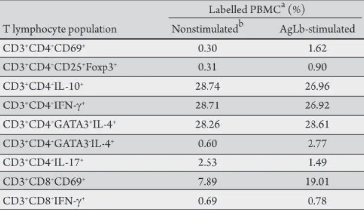
REFERENCES TABLE 1 - Frequency of T cell populations in peripheral blood of a mucosal
leishmaniasis patient coinfected with HIV+.
Labelled PBMCa (%) T lymphocyte population Nonstimulatedb AgLb-stimulated
CD3+CD4+CD69+ 0.30 1.62
CD3+CD4+CD25+Foxp3+ 0.31 0.90
CD3+CD4+IL-10+ 28.74 26.96
CD3+CD4+IFN-g+ 28.71 26.92
CD3+CD4+GATA3+IL-4+ 28.26 28.61
CD3+CD4+GATA3-IL-4+ 0.60 2.77
CD3+CD4+IL-17+ 2.53 1.49
CD3+CD8+CD69+ 7.89 19.01
CD3+CD8+IFN-g+ 0.69 0.78
PBMC: peripheral blood mononuclear cells, aater 24h in vitro cell culture, bmedium alone.
luorochrome-conjugated antibodies (BD Pharmingen, San Diego, CA, USA) against the following surface markers: CD3-PE (clone S4.1), CD3-APC (clone HIT3a), CD4-PE-Cy7 (clone RPA-T4), CD8-FITC (clone HIT8a), CD8-PE-Cy5 (clone HIT8a), CD69-PE (clone FN50) and CD25-FITC (clone PC61). For intracellular staining, cells were permeabilized with BD Cytoix/Cytoperm™ Plus (BD Biosciences) and then incubated with 10µl of the following antibodies: FoxP3-PE (clone 259D/C7), IL-17-Alexa 488 (clone N49-653), IFN-
γ
-FITC (clone 4S.B3), IL-10-PE (clone JES3-9D7), IL-4-FITC (clone MP4-25D2) and GATA-3-PE (clone L50-823). Multiparameter low cytometry was performed using a FACScalibur low cytometer (Becton Dickinson, Mountain View, CA, USA) compensated with single luorochromes. Data was analyzed using Cell Quest Pro sotware (Becton Dickinson) and the results were ploted (Table 1). Dead cells were omited by side scater/forward scater (SSC/FSC) gating, and isotype-matched control antibodies were used to determine background levels of staining.After blood sampling, a delayed-type hypersensitivity test (Montenegro skin test) was performed and resulted in a 12mm positive induration. The patient was then followed and treated in accordance with standard Brazilian Ministry of Health clinical practice.
Very little data is available regarding the immunological response in HIV+ ML patients. It has been demonstrated that cells
from HIV- ML patients present a strong anti-Leishmania speciic
TNF-α and IFN-γ production, with a concomitant decrease in IL-10 and TGF-β levels3. In a late stage AIDS-associated ML patient,
the low lymphocyte proliferative response and IFN-γ production were restored ater the irst speciic immunochemotherapy course. Apparently, this restoration was dependent on predominating CD8+ rather than CD4+ responding T cells6. Another HIV+/ML
case series showed that circulating CD4+ T cell count was < 150
cells/mm3 and that the leishmaniasis clinical outcome showed
strong variability among the patients5. To our knowledge, no data is
available regarding the efects of early HIV infection on the cytokine proile of ML patients. Here, observation veriied that the ML patient responded to AgLb and that this response was dependent on both CD4+ and CD8+ T cells, with increased expression of the CD69+
cellular activating surface marker (Table 1). he activation state
observed on the CD4+ cells was associated with an increase in the
expression of transcription factor Foxp3, characteristic of a Treg cell phenotype, and in intracellular staining of the h2 cytokine IL-4 in the GATA-3- subpopulation, which indicates up-regulation in the
IL-4R/STAT6 pathway. Concomitantly, a decrease in CD4+IL-17A+
cells was observed. he exact role of the h17 response in human parasitic diseases remains unclear, but seems to be related to the
in situ inlammatory milieu observed in ML patients7 and could be
down-modulated during the course of HIV, favoring parasite evasion and the establishment of infection. Studies by Botrel et al showed that the CD8+ cell population revealed only a slight increase in IFN-γ
intracellular staining following stimulation with Lb antigens, which suggests a secondary role of this cell population in host protection and infection clearance8.
hese results show the existence of some modulating mechanism in this HIV coinfected ML patient and brings to light some new aspects concerning effector T helper cell involvement during
Leishmania (V.) braziliensis infection. Further case-control studies are required to conirm these results.
Postgraduatetrainning fellowships were provided to L.R.C. and J.R.M. by Coordenação de Aperfeiçoamento de Pessoal de Nível Superior
(CAPES) and to M.L. and M.V.S. by Fundação de Amparo à Pesquisa do Estado de Minas Gerais (FAPEMIG).
1. Castellano LR, Filho DC, Argiro L, Dessein H, Prata A, Dessein A, et al. h1/h2 immune responses are associated with active cutaneous leishmaniasis and clinical cure is associated with strong interferon-gamma production. Hum Immunol 2009; 70:383-390.
2. Salhi A, Rodrigues Jr V, Santoro F, Dessein H, Romano A, Castellano LR, et al. Immunological and genetic evidence for a crucial role of IL-10 in cutaneous lesions in humans infected with Leishmania braziliensis. J Immunol 2008; 180:6139-6148.
3. Bacellar O, Lessa H, Schriefer A, Machado P, Ribeiro de Jesus A, Dutra WO, et al. Up-regulation of h1-type responses in mucosal leishmaniasis patients. Infect Immun 2002; 70: 6734-6740.
4. Alvar J, Aparicio P, Aseffa A, Den Boer M, Canavate C, Dedet JP, et al. The relationship between leishmaniasis and AIDS: the second 10 years. Clin Microbiol Rev 2008; 21:334-359.
5. Lindoso JA, Barbosa RN, Posada-Vergara MP, Duarte MI, Oyafuso LK, Amato VS, et al. Unusual manifestations of tegumentary leishmaniasis in AIDS patients from the New World. Br J Dermatol 2009; 160:311-318. 6. Da-Cruz AM, Matos M, Oliveira-Neto MP, Coutinho Z, Machado ES, Coutinho
SG. Cellular immune responses to Leishmania braziliensis in patients with AIDS-associated American cutaneous leishmaniasis. Trans R Soc Trop Med Hyg 2000; 94:569-571.
7. Bacellar O, Faria D, Nascimento M, Cardoso TM, Gollob KJ, Dutra WO, et al. Interleukin 17 production among patients with American cutaneous leishmaniasis. J Infect Dis 2009; 200:75-78.