UNIVERSIDADE FEDERAL DO CEARÁ
FACULDADE DE FARMÁCIA, ODONTOLOGIA E ENFERMAGEM
CURSO DE ODONTOLOGIA
PROGRAMA DE PÓS-GRADUAÇÃO EM ODONTOLOGIA
RAMILLE ARAÚJO LIMA
ESTUDOS DA AÇÃO DA TERAPIA FOTODINÂMICA
ANTIMICROBIANA EM BIOFILMES DE Streptococcus mutans -
EFEITO NA VIABILIDADE BACTERIANA E NA MATRIZ DE
POLISSACARÍDEOS.
ESTUDOS DA AÇÃO DA TERAPIA FOTODINÂMICA ANTIMICROBIANA EM BIOFILMES DE Streptococcus mutans – EFEITO NA CÉLULA E NA MATRIZ DE
POLISSACARÍDEOS
Tese apresentada ao Programa de Pós-graduação em Odontologia da Faculdade de Farmácia, Odontologia e Enfermagem da Universidade Federal do Ceará, como requisito parcial para obtenção do título de doutor em Odontologia.
Área de concentração: Clínica Odontológica
Orientadora: Profa. Dra. Iriana Carla Junqueira Zanin dos Santos
Dados Internacionais de Catalogação na Publicação Universidade Federal do Ceará
Biblioteca de Ciências da Saúde
L71e Lima, Ramille Araújo.
Estudos da ação da terapia fotodinâmica antimicrobiana em biofilmes de streptococcus mutans – efeito na célula e na matriz de polissacarídeos./ Ramille Araújo Lima. – 2014.
63 f.: il. color., enc.; 30 cm.
Tese (doutorado) – Universidade Federal do Ceará; Faculdade de Farmácia, Odontologia e Enfermagem; Departamento de Odontologia; Programa de Pós-Graduação em Odontologia; Doutorado em Odontologia, Fortaleza, 2014.
Área de Concentração: Clínica Odontológica.
Orientação: Profa. Dra. Iriana Carla Junqueira Zanin dos Santos. 1. Cárie Dentária. 2. Placa Dentária. 3. Fotoquimioterapia. I. Título.
RAMILLE ARAÚJO LIMA
ESTUDOS DA AÇÃO DA TERAPIA FOTODINÂMICA ANTIMICROBIANA EM BIOFILMES DE Streptococcus mutans – EFEITO NA CÉLULA E NA MATRIZ DE
POLISSACARÍDEOS
Tese apresentada ao Programa de Pós-graduação em
Odontologia da Faculdade de Farmácia, Odontologia e
Enfermagem da Universidade Federal do Ceará, como
requisito parcial para obtenção do título de doutor em
Odontologia.
Aprovada em: 07/08/2014.
novo mundo dе possibilidades. Que mostrou sua misericórdia nos momentos difíceis e me abençoou com sua divina providência nos momentos que mais precisei.
À minha FAMÍLIA, pelo apoio e por sempre acreditarem no meu sucesso desde criança. E em especial aos meus pais, ARNALDO ARAÚJO LIMA e MARIA NEVES LIMA, e ao irmão, ARNALDO ARAÚJO LIMA JÚNIOR.
À minha orientadora, Dra. IRIANA CARLA JUNQUEIRA ZANIN, pelo apoio durante esses
anos, por ter me oferecido a oportunidade de realizer um doutorado sanduíche, pela ajuda no processo do doutorado sanduíche e principalmente por ter me integrado ao seu grupo de pesquisa. Obrigada!
À professora Dra. LIDIANY KARLA AZEVEDO RODRIGUES, por ser acima de tudo um exemplo de como ser um pesquisador de sucesso em nosso país e por ter contribuído para a minha formação profissional e científica durante o período do doutorado.
À Dra. SIMONE DUARTE, que me recebeu na Universidade de Nova York. Agradeço por ter feito eu me sentir tão feliz e confortável em um ambiente tão desafiador. Obrigada pelos ensinamentos valiosos, pelas conversas, pela companhia, pelas festas e por ter me abrigado em seu lar diversas vezes. Palavras não são suficientes para agradecer tudo que fez por mim.
Às professoras NÁDIA ACCIOLY PINTO NOGUEIRA E GISLAINE CRISTINA PADOVANI, pelas contribuições fundamentais durante o exame de qualificação
Ao meu namorado FERNANDO AUGUSTO FERNANDES TÁVORA, pelo apoio incondicional, companherismo, pela presença constante mesmo no período de distância física, e por não ter permitido que eu desistisse quando as dificuldades surgiram.
À todos os colegas do Programa de Pós-graduação em Odontologia, em especial às colegas BEATRIZ NEVES, DANIELLA BEZERRA, SARAH GUEDES, VANARA PASSOS, MARY ANNE MELO, JULIANA LIMA, obrigada pelas risadas, pela companhia e pela amizade.
À amiga DENISE LINS DE SOUSA, pela ajuda indispensável na realização dos experimentos e pela amizade desenvolvida ao longo de 9 meses. Obrigada pela companhia em Nova York, nos momentos alegres e difíceis.
Às novas amizades em Nova York, XI WEI, MEERA DIVYA RATHI e SMRUTI PUSHALKAR e as amizades nacionais-internacionais, THEREZA CRISTINA BOTELHO DANTAS e CAROLINE BRANDI SALES, obrigada pela companhia e pelos momentos de descontração durante o doutorado sanduíche.
À Universidade Federal do Ceará – UFC, na pessoa do reitor Dr. JESUALDO PEREIRA DA SILVA.
À Faculdade de Farmácia, Odontologia e Enfermagem – FFOE, na pessoa de sua diretora Dra. MARIA GORETTI RODRIGUES DE QUEIROZ.
Ao Curso de Odontologia da FFOE, na pessoa do seu coordenador Dr. FABRÍCIO BITU SOUSA.
Ao Programa de Pós-graduação em Odontologia – PPGO, na pessoa da sua coordenadora, Dra. LIDIANY KARLA AZEVEDO RODRIGUES.
À Fundação Cearense de Apoio ao Desenvolvimento Científico e Tecnológico – FUNCAP, pela concessão da bolsa de estudo durante o doutorado, no Brasil.
À Coordenação de Aperfeiçoamento de Nível Superior – CAPES, pela concessão de bolsa de
antimicrobianos tradicionais diante da emergência de cepas resistentes a estes agentes. Este estudo est́ dividido em tr̂s caṕtulos, cujos objetivos foram: Caṕtulo 1) Comparar o efeito antimicrobiano da TFDA realizada com o fotossensibilizador azul de orto-toluidina (TBO) e duas diferentes fontes de luz vermelha {Laserbeam® (LB) ou LumaCare® (LC)} em biofilmes maduros
de Streptpcoccus mutans; Caṕtulo 2) Quantificar as espécies reativas de oxigênio (ERO) encontradas a nível intracelular em biofilmes de S. mutans como resultado da TFDA realizada com TBO e a fonte de luz vermelha LC; e Caṕtulo 3) Avaliar os efeitos da TFDA realizada com TBO e a fonte de luz vermelha LC na viabilidade microbiana, na matriz de polissacarídeos extracelulares (PEC) e na topografia de biofilmes de S. mutans maduros e em formação. Biofilmes foram crescidos em discos de hidroxiapatita imersos em caldo de peptona caséna soja e extrato de levedura com 1% de sacarose por cinco dias. No capítulo 1, biofilmes maduros foram submetidos à TFDA utilizando o fotossensibilizador TBO (100 g/mL) e duas fontes de luz
diferentes: LB(56,6 J/cm2) e LC (56,6 J/cm2; 158,5 J/cm2; 317,0 J/cm2; 475,6 J/cm2). No capítulo 2, os biofilmes foram expostos a terapia usando o LC nas densidades de energia 211.37 J/cm2 e 422.74 J/cm,e a produção de ERO como consequência da terapia foi determinada utilizando o marcador dihidrorodamina 123. No capítulo 3, as mesmas densidades de energia do capítulo 2 (211.37 J/cm2 e 422.74 J/cm2) foram testadas em biofilmes maduros e em formação. Verificamos que não existe diferença na redução da viabilidade bacteriana quando LBeLC são utilizados na mesma dose de energia, porém LC é superior nas exposições 317,0 J/cm2 e 475,6 J/cm2 (capítulo 1). A TFDA gerou 3x mais ERO que o grupo controle negativo. A TFDA nas densidades de energia 211.37 J/cm2 e 422.74 J/cm2 obteve reduções da viabilidade bacteriana de 2 a 5 logs, respectivamente, em biofilmes maduros, e de 6 a 6,5 logs em biofilmes em formação. A TFDA aplicada sobre biofilmes em formação, além de promover redução da viabilidade bacteriana, também reduziu a formação de PEC e o número de conglomerados bacterianos, além de promover a perda de estruturas de conexão dos PEC. (capítulo 3). Conclui-se que TFDA, nos parâmetros utilizados, apresenta-se como uma promissora abordagem terapêutica na inibição da formação de biofilmes de S. mutans e na inativação de biofilmes já estabelecidos.
antimicrobial chemotherapy (PACT) arises as an alternative treatment to these agents. This study was divided into three chapters, whose objectives were: Chapter 1) compare the antimicrobial effect of PACT performed with the photosensitizer TBO and two different sources of red lights {Laserbeam® (LB) or LumaCare® (LC)} in S. mutans mature biofilms, Chapter 2) verify the
amount of intracellular reactive oxygen species (ROS) found in S. mutans biofilms as a result of PACT performed with TBO and light source LC and Chapter 3) evaluate the effects of PACT performed with TBO and red LED LC on microbial viability, polysaccharide matrix and topography of S. mutans mature biofilm and S. mutans biofilm formation. Biofilms were formed on saliva-coated hydroxyapatite discs using batch culture method at 37oC, 5% CO2.
Tryptone-yeast extract broth containing 1% sucrose was changed daily. In chapter 1, the mature biofilms were subjected to PACT using TBO (100μg/mL) and two different light sources: LB (56.6 J/cm2) and LC (56.6 J/cm2; 158.5 J/cm2; 317.0 J/cm2; 475.6 J/cm2). The results were expressed as colony forming units (CFU) per mg of biofilm. In chapter 2, the biofilms were exposed to PACT using TBO and LC energy densities of 211.37 J/cm2 and 422.74 J/cm2 and the reactive oxygen species (ROS) production, as a result of the therapy, was determined using dihidrorhodamine 123 dye. On chapter 3, these same energy densities (211.37 J/cm2 and 422.74 J/cm2) were tested in mature biofilms and forming-biofilms. We verified that there is no difference in the reduction of bacterial viability when LB and LC were used in the same energy dose, however LC was superior when the exposures 317.0 J/cm2 and 475.6 J/cm2 were tested (Chapter 1). PACT generated 3-times more ROS than negative control group. PACT using energy densities 211.37 J/cm2 and
422.74 J/cm2 achieved reductions in bacterial viability up to 2-5 logs in mature biofilms,
respectively, and 6 to 6.5 logs in forming-biofilms. Therapy in forming-biofilms also promoted a reduction in the PEC formation, while reducing the number of bacterial clusters and promote the
lost of PEC connection structures (Chapter 3). We conclude that photodynamic therapy, in the parameters used, comes as a promising therapeutic approach for inhibiting the cariogenic
biofilms formation and inactivation of biofilms already established.
2 PROPOSIÇÃO...15
3 CAPÍTULOS...16
3.1 CAPÍTULO 1...17
3.2 CAPÍTULO 2 ...31
3.3 CAPÍTULO 3...41
4 DISCUSSÃO GERAL ...57
5 CONCLUSÃO GERAL ...61
1 INTRODŨ̧O GERAL
A cárie dentária continua sendo um grande problema na Odontologia e deve receber muita atenção na prática diária. É uma das doenças crônicas mais prevalentes no mundo, atingindo cerca de 60% a 90% das crianças em idade escolar e quase 100% da população adulta (PETERSEN et al., 2005). Apesar do declínio da prevalência de cárie entre crianças, os índices de cárie aumentam com a idade e permanece um problema na população adulta (BERNABÉ e
SHEIHAM, 2014).
A produção de ácidos pelas bactérias acidogênicas e acidúricas no biofilme dental é um pré-requisito essencial para o desenvolvimento das lesões de cárie. Os biofilmes bacterianos são formados quando micro-organismos unicelulares se tornam irreversivelmente aderidos a uma superfície sólida e envolvida por uma matriz de polissacarídeos extracelulares, podendo haver a formação de biofilmes a partir de uma ou múltiplas espécies bacterianas (SPRATT e PRATTEN, 2003). A diversidade anatômica existente na cavidade oral e a interdependência entre as suas estruturas tornam o desenvolvimento dos biofilmes particularmente interessante. Contrariamente ao que ocorre nas superfícies mucosas, onde há uma constante descamação do epitélio e consequente renovação dos micro-organismos aderidos, os dentes constituem superfícies duras favoráveis à formação e maturação do biofilme bacteriano tanto na região supragengival como na região subgengival (MARSH, 1994). A placa dental é um biofilme bacteriano encontrado naturalmente na superfície dos dentes, apresentando composição bacteriana e bioquímica que pode variar em decorrência de fatores intrínsecos e extrínsecos ao hospedeiro (KOLENBRANDER, 2000).
Nos últimos anos, tem sido observado que bactérias inseridas nos biofilmes passam a exibir características fisiológicas distintas que resultam no aumento de resistência dos biofilmes aos agentes antimicrobianos quando comparadas às mesmas bactérias crescidas de forma planctônica (SVENSATER et al., 2001). Aparentemente, o crescimento dos biofilmes de forma
organizada sobre a superfície dos dentes pode funcionar como uma proteção contra as forças mecânicas da mastigação, contra os mecanismos de defesa inata e adquirida do hospedeiro, bem
interior dos biofilmes, a ativação de mecanismos gerais de resposta ao estresse, bem como a expressão de fenótipos específicos para microrganismos organizados na forma de biofilmes
(MAH e O’TOOLE, 2001).
Apesar da complexidade da comunidade bacteriana que coloniza o biofilme dental, existem evidências consideráveis de que a presença de Streptococcus mutans esteja diretamente relacionada aos estágios iniciais da formação das lesões de cárie em humanos. Isso se dá devido a sua presença em altos níveis imediatamente antes do surgimento das lesões; a sua habilidade em rapidamente degradar carboidratos fermentáveis promovendo a formação abundante de ácido; além da sua capacidade de tolerar ambientes com baixo pH (SVENSATER et al., 2001). Em adição, S. mutans tem alta afinidade à superfície dentária mediada pela presença de adesinas e polissacarídeos extracelulares que contribuem para a sua patogenicidade (SENADHEERA et al., 2005).
Encontra-se estabelecido na comunidade científica o conceito de que o surgimento das duas doenças mais prevalentes na cavidade bucal de humanos, a cárie dental (LOESCHE, 1986) e a doença periodontal (SOCRANSKY & HAFFAJEE, 1991), estejam intimamente relacionadas à presença dos biofilmes organizados sobre a superfície dos dentes. O tratamento das doenças relacionadas à presença dos biofilmes bacterianos envolve basicamente a remoção mecânica desses biofilmes e o uso de antibióticos e/ou agentes anti-sépticos. Entretanto, diante da emergência de cepas resistentes aos agentes antimicrobianos tradicionais, têm aumentado o interesse da comunidade científica em desenvolver terapias antimicrobianas alternativas que tornem o surgimento de cepas geneticamente resistentes improvável (HAMBLIN; HASAN,
2004).
dessas reações fotoquímicas podem então danificar componentes essenciais das células ou alterar as atividades metabólicas de maneira irreversível resultando na morte bacteriana.
Como a maioria das espécies bacterianas não apresenta componentes fotossensíveis, a utilização de um agente cromóforo que atraia para si a luz e inicie a formação de radicais livres é importante para a efetividade da terapia fotodinâmica. No que diz respeito à interação entre fotossensibilizador e bactéria, a efetividade da terapia está relacionada a três principais aspectos: (1) capacidade do fotossensibilizador em interagir com a membrana bacteriana; (2) capacidade do fotossensibilizador em penetrar e agir dentro da célula; e (3) formação de oxigênio singleto ao redor da célula bacteriana (NAGATA et al., 2012). Assim, células desprovidas de componentes fotossensíveis endógenos podem se tornar sensíveis à luz se forem coradas com fotossensibilizadores exógenos como derivados das porfirinas, azul de orto-toluidina (TBO) ou compostos de cloro conjugado (ROLIM et al., 2012). Dentre esses, o uso do azul de orto-toluidina torna-se interessante devida sua natureza catiônica e, portanto, capaz de inativar tanto bactérias Gram-positivo quanto Gram- negativo (NAGATA et al., 2012).
Com relação à fonte de luz utilizada, a muitos dos estudos sobre o efeito antimicrobiano da terapia fotodinâmica utiliza lasers de baixa potência com diversos meios ativos e comprimentos de onda. O uso de dispositivos semicondutores que quando polarizados adequadamente emitem luz na faixa visível ou invisível, os chamados diodos emissores de luz (LED) surgiram como fontes alternativas de luz para essa terapia (ZANIN et al., 2005; ZANIN et al., 2006). Embora tanto os lasers quanto os LED produzam luzes monocromáticas, os LED apresentam colimação e coerência menos eficientes, resultando em bandas de emissão mais
largas que acabam por favorecer a obtenção de efeito antimicrobiano, uma vez que a luz é emitida em todo o espectro de absorção dos fotossensibilizadores durante a realização da terapia fotodinâmica (NAGATA et al., 2012).
antibiótico-resistentes, e repetidas aplicações não resultam em seleção de cepas resistentes (GURSOY et al., 2013). Finalmente, o uso do fotossensibilizador ou da luz sozinhos não
apresentam efeito significativo sobre a viabilidade das bactérias, de modo que a terapia pode ser confinada à área da lesão pela aplicação tópica cuidadosa do corante e restrição da irradiação por meio do uso de fibra ótica (WILSON, 2004).
Estudos prévios têm demonstrado o efeito antimicrobiano da terapia fotodinâmica em bactérias orais em culturas planctônicas (WILLIAMS et al., 2003; ROLIM et al. 2012), biofilmes desorganizados (O’NEILL et al., 2002) e organizados (ZANIN et al., 2005; ZANIN et al., 2006; TEIXEIRA et al., 2012) e em lesões de cárie dentinária formadas in vitro (MELO et al., 2010) e in situ (LIMA et al., 2009) quando a combinação apropriada de fotossensibilizador e luz é utilizada. No entanto, a inativação total das bactérias presentes nos biofilmes e lesões de cárie não tem sido possível e a presença da matriz de polissacarídeos extracelulares produzida por S. mutans tem sido apontada como o principal responsável pela efetividade parcial da terapia (ZANIN et al., 2005). Dessa forma, este estudo teve por objetivo avaliar o efeito antibacteriano da terapia fotodinâmica realizada com a combinação do fotossensibilizador TBO e LED vermelho, e analisar os efeitos dessa terapia tanto na viabilidade bacteriana quanto na matriz de polissacarídeos de biofilmes de S. mutans.
2 PROPOSĨ̧O
Esta tese de doutorado ́ apresentada em tr̂s caṕtulos, tendo como objetivos:
Caṕtulo 1: Comparar o efeito antimicrobiano da terapia fotodinâmica realizada com o fotossensibilizador TBO e duas diferentes fontes de luz vermelha (Laserbeam® ou LumaCare®) em biofilmes maduros de S. mutans.
Caṕtulo 2: Verificar a quantidade de espécies reativas de oxigênio encontradas a nível intracelular em biofilmes de S. mutans como resultado da terapia fotodinâmica antimicrobiana realizada com TBO e a fonte de luz vermelha LumaCare®, e assim verificar o stress oxidativo promovido por essa terapia.
3 CAṔTULOS
Esta tese est́ baseada no artigo 46 do regimento Interno do Programa de Ṕs -graduã̧o em Odontologia da Universidade Federal do Ceaŕ, que regulamenta o formato alternativo para dissertã̧es de Mestrado e teses de Doutorado, e permite a inseŗ̃o de artigos cient́ficos de autoria ou co-autoria do candidato. Dessa forma, esta tese ́ composta por tr̂s caṕtulos, contendo artigos a serem submetidos para publicã̧o em revistas cient́ficas, conforme descrito abaixo:
Caṕtulo 1
“Photodynamic antimicrobial chemotherapy on S. mutans mature biofilms: comparative effect of two red lights sources”. Iriana Carla Junqueira Zanin, Ramille Araújo Lima, Denise Lins de Sousa, Simone Duarte. Este artigo seŕ submetido ̀ publicã̧o no perídico “Caries Research”.
Caṕtulo 2
“Intracellular Reactive Oxygen Species in S. mutans biofilm after Photodynamic Antimicrobial Chemotherapy –PACT”. Ramille Araújo Lima, Juliana Aparecida Delben, Iriana Carla Junqueira Zanin, Simone Duarte. Este artigo seŕ submetido ̀ publicã̧o no perídico “Lasers in Medical Science”.
Caṕtulo 3
3.1 Capítulo 1
Title: Photodynamic antimicrobial chemotherapy on S. mutans mature biofilms: comparative effect of two red light sources.
Short Title: Effect of PACT using different lights on S. mutans biofilms
Iriana Carla Junqueira Zanin1, Ramille Araújo Lima2, Denise Lins de Sousa2, Simone
Duarte3
1 Department of Microbiology, College of Dentistry, Federal University of Ceara, Sobral, CE,
Brazil
2Department of Dental Clinics, School of Pharmacy, Dentistry and Nursing, Federal University of
Ceara, Fortaleza, Ceara, Brazil.
3 Department of Basic Science and Craniofacial Biology, College of Dentistry, New York
University, New York, USA.
Key-words: Photodynamic therapies; Streptococcus mutans; biofilm; extracellular polysaccharides
# Corresponding Author:
Iriana Carla Junqueira Zanin, DDs, Ms, PhD
College of Dentistry, Federal University of Ceara, Sobral, CE, Brazil. Coronel Estanislau Frota Street, s/n
Sobral – CE- Brazil Zip code: 62010-560
Phone/fax: ++ 55 88 36132603 Mobile: ++ 55 88 97159496
E-mail: irianaz@yahoo.com.br, iriana.zanin@pq.cnpq.br
Este artigo está escrito segundo às normas de publicação de artigos da revista Caries Research
Declaration of interests
Abstract
Photodynamic antimicrobial therapy (PACT) promotes bacterial death as a result of the photosensitization of microbial components. This study evaluated the comparative antimicrobial effect of PACT performed with different red light sources on in vitroS. mutans maturebiofilms. For that, S. mutans UA159 biofilms were formed on saliva-coated hydroxyapatite discs using batch culture method at 37oC, 5%CO2. Tryptone-yeast extract broth containing 1% sucrose was
changed daily. The antimicrobial effect of toluidine blue O (TBO) (100ug/ml), associated with
two different red light sources, LB (Laserbeam®, λ ≅638.8nm) or LC (LumaCare®, λ ≅630nm) was evaluated. On the 5th day, biofilms were treated with sensitizer-S (TBO) and/or light-L in the
test (S+L+) and control (S-L-, S+L-, S-L+) groups. LB was tested using 900 sec irradiation time
(56.6 Jcm-2) while LC was tested using 22 sec (56.6 Jcm-2); 60 sec (158.5 Jcm-2), 120 sec (317.0 Jcm-2); and 180 sec (475.6 Jcm-2) irradiation time. After treatments, biofilms were colleted, weighted and disrupted using ultrasonic pulses. Ten-fold serial dilutions were carried out and aliquots were plated onto 5% Blood agar which were then incubated at 37°C, 5% CO2 for 48
hours before enumerating the viable microorganisms. The results were expressed as colony forming units (CFU) mg-1 of biofilm and the transformed to log10 of CFU mg-1 of biofilm. The
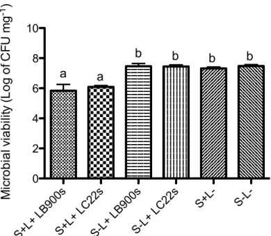
results demonstrated that groups receiving the PACT (S+L+) were statistically different from all tested controls (S-L-, S+L-, S-L+) for both red lights used (p <0.05). Also, there were no statistical difference among all tested control groups (p>0.05). Comparing microbial counts after PACT for the pair LB 900 sec (5.84±0.41) and LC 22 sec (6.09±0.20), both using energy density of 56.6 Jcm-2, there was no difference between them (p>0.05). Also LC 120 sec (4.88±0.18) and LC180 sec (4.98±0.35) presented similar results (p>0.05). Furthermore, considering the clinical
Introduction
Streptococcus mutans is the major etiological agent in dental caries [Bowen, 1999] and it has a great influence on the formation and composition of pathogenic biofilms mainly due its ability to quickly degrade fermentable carbohydrates and synthesize extracellular polysaccharides (EPS) [Paes Leme et al., 2006]. Bacteria in biofilms show distinct physiological characteristics that result in increased resistance to antimicrobial agents when compared to the planktonic forms [Svensater et al., 2001]. Defense mechanisms of biofilm microorganisms may include the formation of a physical barrier (polysaccharides), difficulty dissemination of antimicrobial agents within the biofilm, as well as the expression of specific phenotypes [Mah and O'Toole, 2001].
Front the emergence of strains resistant to traditional antimicrobial agents, the photodynamic antimicrobial chemotherapy (PACT) appears as an alternative therapy to killing oral bacteria. It is based on the use of extrinsic photosensitizers (PS), light-absorbing molecules that initiate a photochemical reaction when exposed to light of a specific wavelength. This photochemistry process leads to reactive oxygen species (ROS) formation, which may cause irreversible damage to essential bacterial cell compounds and may change cell metabolism resulting in bacterial death [Dougherty et al., 1998]. The use of ortho-toluidine blue (TBO) as PS seems to be interesting due its cationic nature, and therefore able to inactivate both gram-positive as gram-negative bacteria [Nagata et al. 2012; Rolim et al. 2012].
ranging from 10 to 20 minutes, which precluded the possibility of clinical use of this therapy. Thus, there is the need of alternatives protocols more effective and less time-demanding.
Therefore, the aim of this study was to compare the antimicrobial effect of PACT performed with TBO and two different red light sources (Laserbeam® 900 sec; or LumaCare® 22sec, 60sec, 120 sec or 180 sec) on S. mutans mature biofilms.
Materials and methods
Experimental Design For this in vitro experiment, 108 hydroxyapatite (HA) sterile discs (0.74
cm2) were randomly allocated into 12 groups, with 3 experimental units per set of group. Biofilms of Streptococcus mutans UA159 were grown on HA discs immersed in batch culture and the mature biofilm was submitted to PACT after 5 days [Duarte et al. 2008]. To minimize the inherent bias related to microbiological procedures, three independent experiments were performed at different time points. The treatment conditions in which biofilms were exposed are as follows: biofilms were exposed to both TBO and light under different conditions (S+L+), biofilms were not exposed to sensitizer or light (S-L-), biofilms were exposed only to sensitizer (S+L-) or biofilms were exposed only to light (S-L+) using different light sources and energy densities (figure 1).
Figure 1. Experimental design. LB represents the Led device Laserbeam® and LC represents the non-coherent light
Light Sources and Parameters: A red light-emitting diode (LED, Laserbeam®, Rio de Janeiro, RJ, Brazil) with spectrum of emission ranging from 620 to 660 nm and predominant wavelength
of 638.8 nm, fixed output power of 40 mW and optic fiber spot size of 0.69 cm2 was used as one of the light sources. Biofilms were exposed to a 56.6 Jcm-2 energy density after 900 seconds of irradiation. For comparing purpose, a non-coherent red light LumaCare® (Model LC-122, Medical Group, Newport Beach, CA) with spectrum of emission ranging from 570 to 690 nm and predominant wavelength of 630 nm, fixed output power of 1.68W, and optic fiber spot size of 1.13 cm2 was also tested. Biofilms were exposed to four different energy densities and respectively energies doses as follows: 56.6 Jcm2, 35.9 J(22sec); 158.5 Jcm-2, 100.64 J (60sec); 317.05 Jcm-2, 201.32 J (120 sec); 475.58 Jcm-2, 3012.99 J (180 sec). Irradiation was performed in a noncontact mode with a focused beam at 10 mm of working distance.
Photosensitizer: The photosensitizer (S) used was toluidine blue ortho (TBO, CI 52040, Sigma Chemicals, Poole, UK), dissolved in deionized water (100 μgmL-1) and stored at room temperature in the dark. The pre-irradiation time was 5 minutes [Zanin et al., 2005].
Inoculum and biofilm model: Streptococcus mutans UA159 (ATCC 700610) was obtained from single colonies isolated on 5% blood agar plates, inoculated in tryptone yeast-extract broth containing 1 % glucose (w/v) and incubated for 18–24 h at 37 °C under microaerophilic conditions in partial atmosphere of 5 % of CO2. Biofilms of S. mutans UA159 were formed on
saliva-coated hydroxyapatite discs (HA) (0.635cm2) placed in batch cultures at 37 °C in 5 % CO 2
for 5 days. The biofilms were grown in tryptone yeast-extract broth containing 1 % sucrose (w/v) and were kept undisturbed for 24 h to allow initial biofilm formation. After that, the culture medium was replaced once daily [Duarte et al., 2006].
maintained at room temperature during the same period (groups S+L- and S-L-). The biofilms were then scrapped with a sterile spatula and transferred to a pre-weighed microtube containing 1
mL of NaCl 0.89% solution. To disperse the biofilm, 2 pulses of 10 seconds with 1 min of interval at an output of 7W were performed (Branson Sonifier 150; Branson Ultrassonics, Danbury, CT). Ten-fold serial dilutions were carried out and aliquots were plated onto Blood agar which were then incubated at 37°C, 5% CO2 for 48 hours before enumerating the viable
microorganisms. The results were expressed as colony forming units (CFU) mg-1 of biofilm.
Statistical analysis: The normality distribution of data was checked using the Kolmogorov– Smirnov statistical test. The mean and the standard deviation of numbers of surviving microorganisms for each treatment were calculated. Colony forming units (CFU) were transformed in log10 CFU in order to reduce variance heterogeneity. One-way analysis of variance test (ANOVA) was used to detect differences, followed by a Tukey–Kramer test for pairwise comparisons. Significance level was set at 5% (p< 0.05) and the confidence interval was set at 95%. Prism 5.0 (GraphPad Software, Inc.; La Jolla, CA, USA) was used to perform the analyses.
Results
The results indicated that neither incubation with TBO alone (S+L-), nor treatment with light alone (S-L+), had a significant effect on the viability of S. mutans (p 0.05). Significant decreases in the viability of monospecies biofilms were only observed when biofilms were exposed to both TBO and irradiation at the same time. At same energy density (56.6 Jcm-2), Laserbeam® (900 sec) and LumaCare® (22 sec) showed the same performance, with no difference between the groups, with median viable counts of 9.54 105 ± 6.21 105 (SD)in the group submitted to LB(S+L+) and 1.37 106 ± 6.35 105 (SD)in the group submitted to LC(S+L+); contrasting with the control groups: 2.43 107 ± 5.53 106 (S-L-), 2.36 107 ± 6.45 106 (S+L-),
S+L+ LB9 00s S+L+ LC 22s S-L + LB
900s
S-L + LC
22s S+L- S-L
-0 2 4 6 8 10 a a
b b b b
M ic ro b ia l v ia b il ity ( L o g o f C F U m g -1 )
Figure 2. Microbial viability (Log of CFU mg-1) after photodynamic therapy performed with LB and LC (energy density 56.6 Jcm-2 ) + TBO (S+L+) compared with negative controls (S-L+ LB900s; S-L+ LC22s, S+L-, S-L-). Vertical bars denote standard deviation and different letters represent statistically significant differences by Tukey test (p<0.05).
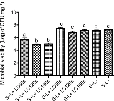
Following, the remaining energy densities of LC (60 sec, 120 sec and 180 sec) also achieved a statistically significant reduction in microbial viability. When compared, 120 sec and 180 sec irradiation time presented similar results (p> 0.05), and both showed better results than 60 sec irradiation time (p <0.05) (figure 3). The log reduction results were calculated by subtraction of log10 counts in the negative control (S-L-), from log10 counts in the other groups
(S+L-, S-L+, S+L+), and log10 reductions of up to 2.38 and 2.27 were observed when S-L- and S+L+
S+L+ LC 60s S+L+ LC 120s S+L+ LC 180s S-L+ LC 60s S-L + LC
120s
S-L + LC
180s S+L- S-L -0 2 4 6 8 10 a b b c
c c c c
M ic ro b ia l v ia b ili ty ( L o g o f C F U m g -1 )
Figure 3. Microbial viability (Log of CFU mg-1) after photodynamic therapy performed with LumaCare® 60, 120 and 180 seconds (energy densities 149 Jcm-2, 317.05 Jcm-2, 475.58 Jcm-2 ) compared with negative controls (S-L+ LC60s; S-L+ LC120s, S-L+ LC180s, S+L-, S-L-). Vertical bars denote standard deviation and different letters represent statistically significant differences by Tukey test (p<0.05).
Discussion
that TBO may bind to the polyphosphates of the membrane and produce molecular damage to lipids and proteins. Besides, Rolim et al., (2012) demonstrated that TBO, associated to a LED,
was the most effective photosensitizer against planktonic cultures of S. mutans out of other five tested at the same molar concentration and irradiation conditions.
Our results confirmed the antimicrobial activity of photodynamic therapy performed with TBO and red LED in S. mutans biofilms, as observed in other studies [Zanin et al., 2005; Zanin et al., 2006; Teixeira et al., 2012]. Although we observed a statistical reduction of microbial viability, the total inactivation was not achieved, in contrast with Rolim et al. (2012) and Paschoal et al. (2013), who tested similar parameters in S. mutans planktonic cultures. It has been know that bacteria in biofilm are less sensitive to antibacterial agents than planktonic bacteria, manly due differences in cell wall composition, rate of growth, metabolic activity and gene expression. Furthermore, bacteria in biofilms are embedded in a polysaccharide matrix, which protects them from external threats, including antimicrobial agents [Anwar et al., 1992; Costerton et al., 1999; Svensater et al., 2001].
Zanin et al (2006) conducted a study performing PACT in S. mutans biofilms using the red LED of 638.8 nm and output power 40 mW (energy density 85.7 J.cm2) and TBO at
the same concentration used in this study, also observed a 2-log reduction in microbial viability, similar results to that obtained in our study using a lower energy density (55 J.cm2). Similar
results obtained with different energies doses can be explained by the difference in substrates. The previously cited study had used enamel slabs as substrate and our study used hydroxyapatite discs. Li et al. (2010) reported that the nature of a surface influences biofilm characteristics
including biomass accumulation and susceptibility to antimicrobial treatments. Paschoal et al. (2013) used the red probe LC, energy densities 18 J.cm2, 35 J.cm2 and 53 J.cm2, in combination with TBO at 25 M, 50 M and 100 M concentrations, and achieved total inactivation of microbial viability. However, this study used planktonic suspensions of S. mutans, unlike our
study using mature biofilms of S. mutans. To our knowledge, this is the first study that had used this non-coherent red light with (630 nm, 1.68W) against mature biofilms.
to increased reduction of the bacterial viability. This may have occurred due photosensitizer saturation, or photobleaching process [Metcalf et al., 2006]. On the other hand, both were
superior to all other experimental groups of this study. In addition to its greater output power, another advantage of the LC is the possibility to produce the entire spectrum of visible light by changing the specific wavelenghts probes. Thus photosensitizers can be activated using the same device, only changing the probe. The most relevant aspect of this study is that the therapy performed with LB irradiation time 15 minutes had the same performance of LC irradiation time 22 seconds, both reaching a energy density of 56.6 Jcm-2. Although this result was expected considering that both treatments used the same density energy, its represent a substantial advance in PACT therapy if we consider a clinical application. The huge difference of irradiation time between the two devices is due to the difference in the output power (LB 40mW and LC 1.68W). Also, the LC device allows the increase of power without significant increase of temperature (data not shown).
As conclusion, PACT significantly decreased the microbial viability of mature S. mutans biofilms when compared to the control groups. PACT performed with LB and LC at same energy density, did not statistically differ from each other, demostrating the LC be a better alternative to PACT, due to its reduced irradiation time – from 15 min to 22 sec. Also, PACT using LC irradiation time of 120 sec, seems to be the most suitable for reducing mature S. mutans in vitro biofilms when the photosensitizer TBO is used.
Acknowledgment
This research was supported by Brazilian Government Agencies as follows:
References
Anwar H, Strap JL, Costerton JW. Establishment of aging biofilms: Possible mechanism of bacterial resistance to antimicrobial chemotherapy. Antimicrob Agents Chemotherapy 1992; 36:1347-1351.
Araújo PV, Teixeira K, Lanza LD, Cortes ME, Poletto LT: In vitro lethal photosensitization of S. mutans using methylene blue and toluidine blue O as photosensitizers. Acta Odontol Latinoam.
2009; 22(2): 93-7.
Bevilacqua IM, Nicolau RA, Khouri S, Brugnera A Jr, Teodoro GR, Zângaro RA, Pacheco MT: The impact of photodynamic therapy on the viability of Streptococcus mutans in a planktonic culture. Photomed Laser Surg. 2007 Dec;25(6):513-8.
Bowen WH: Wither or whither caries research. Caries Res 1999; 33(1): 1-3.
Costerton JW, Stewart PS, Geenberg EP. Bacterial biofilms: A common cause of persistent infections. Science 1999; 284:1318-1322.
Dougherty TJ, Gomer CJ, Henderson BW, Jori G, Kessel D, Korbelik M, Moan J, Peng Q: Photodynamic therapy. J Natl Cancer Inst 1998; 90(12):889-905.
Duarte S, Klein MI, Aires CP, Cury JA, Bowen WH, Koo H: Influences of starch and sucrose on
Streptococcus mutans biofilms. Oral Microbiol Immunol 2008; 23:206–212.
Li L, Finnegan MB, Özkan S, Kim Y, Lillehoj PB, Ho CM, Lux R, Mito R, Loewy Z, Shi W: In vitro study of biofilm formation and effectiveness of antimicrobial treatment on various dental material surfaces. Mol Oral Microbiol. 2010 Dec; 25(6): 384-90.
Mah TC, O´Toole, GA: Mechanisms of biofilms resistance to antimicrobial agents. Trends Microbiol. 2001; 9(1): 34-9.
Matevski D, Weersink R, Tenenbaum HC, Wilson B, Ellen RP, Lépine G: Lethal photosensitization of periodontal pathogens by a red-filtered Xenon lamp in vitro. J Periodontal
Res. 2003 Aug; 38(4): 428-35.
Nagata JY, Hioka N, Kimura E, Batistela VR, Terada RS, Graciano AX, Baesso ML, Hayacibara MF: Antibacterial photodynamic therapy for dental caries: evaluation of the photosensitizers used
and light source properties. Photodiagnosis Photodyn. Ther. 2012; 9(2): 122-131.
O’Neill JF, Hope C, Wilson M: Oral bacteria in multi-species biofilms can be killed by red light in the presence of toluidine blue. Lasers Surg Med 2002;3:86–90.
Paes Leme AF, Bellato CM, Bedi G, Cury JA: The role of sucrose in cariogenic dental biofilm formation— New insight. J. Dent. Res. 2006; 85(10): 878-887.
Paschoal MA, Lin M, Santos-Pinto L, Duarte S: Photodynamic antimicrobial chemotherapy on Streptococcus mutans using curcumin and toluidine blue activated by a novel LED device. Lasers Med Sci. 2013 Nov 19. [Epub ahead of print]
Peterson P, Bourgeois D, Ogawa H, Estupinan-Day S, Ndiaye C: The global burden of oral diseases and risks to oral health. Bull World Healh Organ 2005; 83: 661-9.
Rios A, He J, Glickman GN, Spears R, Schneiderman ED, Honeyman AL: Evaluation of photodynamic therapy using a light-emitting diode lamp against Enterococcus faecalis in extracted human teeth. J Endod. 2011 Jun;37(6):856-9.
Rolim JP, de-Melo MA, Guedes SF, Albuquerque-Filho FB, de Souza JR, Nogueira NA, Zanin IC, Rodrigues LK: The antimicrobial activity of photodynamic therapy against Streptococcus mutans using different photosensitizers. J Photochem Photobiol B 2012; 106:40–46.
Senadheera MD, Guggenheim B, Spatafora GA, Huang YC, Choi J, Hung DC, Treglown JS, Goodman SD, Ellen RP, Cvitkovitch DG: A VicRK signal transduction system in Streptococcus mutans affects gtfBCD, gbpB, and ftf expression, biofilm formation, and genetic competence development. J Bacteriol. 2005 Jun; 187(12): 4064-76.
Shemesh M, Tam A, Steinberg D: Differential gene expression profiling of Streptococcus mutans cultured under biofilm and plank- tonic conditions. Microbiology 2007;153: 1307–1317.
Svensäter G, Welin J, Wilkins JC, Beighton D, Hamilton IR: Protein expression by planktonic and biofilm cells of Streptococcus mutans. FEMS Microbiology Letters. 2001; 205:139-46.
2012;46:549–554.
Tseng SP, Teng LJ, Chen CT, Lo TH, Hung WC, Chen HJ, Hsueh PR, Tsai JC. Toluidine blue O photodynamic inactivation on multidrug-resistant Pseudomonas aeruginosa: Lasers Surg Med 2009 Jul;41(5):391-7.
Usacheva, M.N., Teichert, M.C., and Biel, M.A: Comparison of the methylene blue and toluidine blue O bacterial efficacy against gram-positive and gram-negative microorganisms. Laser Surg. Med. 2001; 29: 165–173.
Zanin IC, Goncalves RB, Junior AB, Hope CK, Pratten J: Susceptibility of Streptococcus mutans
biofilms to photodynamic therapy: an in vitro study. J. Antimicrob. Chemother. 2005; 56(2): 324-330.
Zanin ICJ, Rodrigues LKA, Pimenta LAF, Ho ̈fling JF, Gonçalves RB: Photosensitization of in
vitro biofilms by toluidine blue O combined with a light-emitting diode. Eur. J. Oral Sci. 2006; 114: 64-69.
3.2 Capítulo 2
Intracellular Reactive Oxygen Species in S. mutans biofilm after Photodynamic Antimicrobial Chemotherapy.
Ramille Araújo Lima1, Juliana Aparecida Delben 2, Iriana Carla Junqueira Zanin3, Simone
Duarte3
1 Department of Dental Clinics, School of Pharmacy, Dentistry and Nursing, Federal University
of Ceara, Fortaleza, Ceara, Brazil.
2 Department of Dental Materials and Prosthodontics, College of Dentistry, State University of
São Paulo, Araraquara, São Paulo, Brasil
3 Departmentof Microbiology, College of Dentistry, Federal University of Ceara, Sobral, CE,
Brazil
4 Department of Basic Science and Craniofacial Biology, College of Dentistry, New York
University, New York, USA.
Correspondence author: Simone Duarte
Department of Basic Science and Craniofacial Biology, College of Dentistry, New York
University, NYU, 345 East 24th Street, New York, NY 10010, USA
e-mail: sduarte@nyu.edu
Abstract
Photodynamic Antimicrobial Chemotherapy (PACT) appears as an alternative treatment to dental plaque-related diseases. Products of these photochemical reactions such as reactive oxygen species (ROS) can damage essential components of cells or change their metabolic activities resulting in bacterial death. However, more studies are needed to determine whether reactive oxygen species (ROS) produced are able to penetrate into bacterial cells organized in biofilms
and protected by a matrix of polysaccharides. Therefore, the aim of this study was to measure the amount of intracellular ROS found in S. mutans biofilms after PACT performed with orto-toluidine blue (TBO) and a non-coherent red light (LumaCare®; 570-690 nm). Biofilms of
Streptococcus mutans UA159 were formed on saliva-coated hydroxyapatite discs during 5 days and after that the biofilms were exposed to PACT using the energy density of 211.37 Jcm-2 (1-minute irradiation) and 422.74 Jcm-2 (2-minutes irradiation). Production of ROS in S. mutans
cells as consequence of PACT was determined by using the oxidative-stress-sensitive probe dihydrorhodamine - DHR 123. The 2-minutes PACT group showed the highest levels of ROS (p <0.0001). PACT performed for 1 minute, despite having a lower production of ROS compared to the 2-minutes group, it was also superior to control groups (p<0.05). In conclusion, PACT under tested conditions generates significant levels of intracellular oxidative species in S. mutans
biofilms.
Introduction
Dental caries remains as a major worldwide problem. Although it was observed a decline in the prevalence of this condition, it is one of the most prevalent chronic diseases in the world, reaching about 60% to 90% of school children and almost 100% of the adult population [1] (Petersen et al, 2005). Streptococcus mutans is acknowledged as the major etiological agent in dental caries [2] and it has a great influence on the formation and composition of pathogenic biofilms. The production of acids by acidogenic and aciduric bacteria in the biofilm is essential for the development of carious lesions and it has been observed that bacteria in biofilms show distinct physiological characteristics that result in increased resistance of biofilms to antimicrobial agents when compared to the same bacteria grown in planktonic form [3].
The current treatment for plaque-related diseases involves the use of traditional antimicrobials in conjunction with the mechanical removal of the biofilm. However, due to the emergence of strains resistant to traditional antimicrobial agents, it has increased the interest of the scientific community to develop alternative antimicrobial therapies that make the emergence of genetically resistant strains unlikely [4].
In this regard, photodynamic antimicrobial chemotherapy (PACT) emerges as an alternative treatment to the use of traditional antimicrobials. During this process, photosensitive cell compounds pass into an excited state when exposed to a light-length complementary wave, which is characterized by the passage of electrons to higher energy levels. In this excited state, the photosensitizer can interact with the molecular oxygen, initiating the formation of highly reactive singlet oxygen (Type II photoprocess) or to interact with other molecules, as electron acceptors, resulting in the production of hydroxyls and other organic radicals (Type I photoprocess) [5]. Products of these photochemical reactions, the reactive oxygen species (ROS), can damage essential components of cells or change their metabolic activities resulting in bacterial death.
intracellular ROS found in S. mutans biofilms as result of PACT treatments using the association of ortho-toluidine blue O (TBO) and a non-coherent red light source, and thus verify the
oxidative stress promoted by this therapy, and evaluate the use of dihydrorhodamine 123 dye in biofilms. Our hypothesis was that S. mutans biofilms submitted to PACT will present the highest levels of ROS.
Materials and methods
Experimental design: Hydroxyapatite (HA) sterile discs (63.58mm2) were randomly allocated
into 5 groups, with 3 experimental units per set of group (45 discs). To minimize the inherent bias
related to microbiological procedures, three independent experiments were performed at different time points. The treatment conditions in which biofilms were exposed are as follows: biofilms were exposed to both TBO and light under different conditions (S+L+), biofilms were not exposed to sensitizer or light (S-L-), biofilms were exposed only to sensitizer (S+L-) or biofilms were exposed only to light (S-L+) using different irradiation times (energy density) as follows (Figure 1).
Group Description
S-L- Milli-Q Water (5 min) followed by a 2 min-period in the dark
S+L- TBO Sensitization (5 min) followed by a 2 min-period in the dark
S-L+ 2min Milli-Q Water (5 min) followed by a 2 min LED irradiation (422.74 J/cm2)
S+L+ 1min TBO Sensitization (5 min) followed by 1 min LED irradiation (211.37cm2)
S+L+ 2min TBO Sensitization (5 min) followed by a 2 min LED irradiation (422.74cm2)
Figure 1. Experimental group description
Inoculum and biofilm model: Streptococcus mutans UA159 (ATCC 700610) was obtained from single colonies isolated on blood agar plates, inoculated in tryptone yeast-extract broth containing
1 % (w/v) glucose and incubated for 18–24 h at 37 °C under microaerophilic conditions (5 % of CO2). S. mutans UA159 biofilms were formed on saliva-coated hydroxyapatite discs (HA) placed
yeast-extract broth containing 1 % sucrose (w/v) and were kept undisturbed for 24 h to allow initial biofilm formation. The culture medium was replaced once daily.
Light Sources and Parameters: The non-coherent red light LumaCare® (Model LC-122, Medical Group, Newport Beach, CA) with predominant wavelength of 630 nm (570 nm – 690 nm), spot size of 113.1 mm2, fixed output power of 2.24W was used and 2 energy densities were tested: 211.37 J/cm2 (1 minute irradiation time, energy dose 134.21 J) and 422.74 J/cm2 (2 minutes irradiation time, energy dose 268.43 J). The distance between the light source tip and the exposed sample was 3mm.
Photosensitizer: The photosensitizer (PS) used was toluidine blue ortho (TBO, CI 52040, Sigma
Chemicals, Poole, UK), dissolved in deionized water (100μg mL-1) and stored at room
temperature in the dark. The pre-irradiation time used was 5 minutes.
PACT treatment and measurement ROS: Production of ROS in S. mutans cells as consequence of PACT was determined by using the oxidative-stress-sensitive probe dihydrorhodamine 123 (DHR 123) (Life Technologies®, Carlsbad, CA, USA). At 5th day, the HA discs with S. mutans
mature biofilms were transferred to 24-well plates containing 1mL of 15 DHR 123. The discs remained at this solution for 30 minutes in the dark. Thereafter, the PACT was performed using different treatments as described in figure 1. Immediately after treatment, the biofilms were dip-washed three times in NaCl 0.89%, in order to remove loosely bound biofilm, placed in 5 ml sterile of the same solution and the HA discs surfaces were gently scraped with a sterile spatula to harvest adherent cells. The removed biofilms were subjected to sonication using three 15-s pulses at an output of 7 W (Fisher Scientific, Sonic Dismembrator model 100; USA). Following, the homogenized suspensions were distributed in 96-well black plates and the oxidation of DHR 123
was measured by fluorimetry (SpectraMax M5, Molecular Devices, USA) using excitation and emission wavelengths of 507 nm and 529 nm respectively. This experiment was performed in
triplicates.
Statistical analyses: The normality distribution was checked using the Kolmogorov–Smirnov
between the groups, followed by a Tukey–Kramer test for pairwise comparisons. Significance
level was set at 5% (p< 0.05) and the confidence interval at 95%. Prism 5.0 (GraphPad Software,
Inc.; La Jolla, CA, USA) was used to perform the analyses.
Results
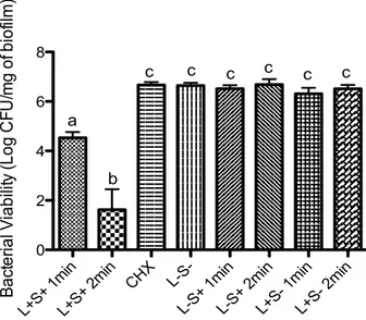
The results were expressed as units of fluorescence, since this means the ROS amount detected. All groups tested emitted some level of fluorescence (Figure 1). Overall, the results showed that ROS production seems to be light dose-dependent once S+L+ 2min group showed the highest levels of ROS (p <0.0001) followed by S+L+ 1min, (p<0.05). The presence of sensitizer in absence of light do not to affect ROS production on S. mutans biofilm. However, the presence of 2 min light irradiation increase the ROS production (p> 0.05) indicating the stress of environment in this tested condition (Figure 2).
S+L + 1m
in
S+L + 2m
in
S-L + 2m
in
S+L
-S-L - 0 500 1000 1500 2000 2500 a b c c,d d F lu o re s c e n c e U n its
Figure 2. Fluorescence units (arbitrary units) representative of
intracellular amount of ROS in S. mutans biofilm cells after exposed
to experimental groups. Data represent mean values (n = 3) and
Discussion
The present study analyzed the production of intracellular ROS in S. mutans biofilms exposed to PACT. Since the antimicrobial mechanism of photodynamic therapy is based on the production of reactive oxygen species, it is essential to check whether the proposed therapy produced significant levels of ROS and if these are able to penetrate into bacterial cells organized in biofilms. It is known that S. mutans biofilm presents a thick layer of extracellular polysaccharide [8], which could hinder the penetration of these species. Although previous studies have analyzed the ROS [6] and singlet oxygen [7] production generated by a number of photosensitizers and light sources combinations, as far as we know, this is the first one that analyzed the presence of intracellular ROS in S. mutans biofilm after PACT exposure.
Dihydrorhodamine 123 (DHR 123) was used as an indicator of intracellular ROS presence. Previous researchers have reported that DRH is no fluorescent, uncharged, and readily taken up by cells, whereas DHR-123, the product of DHR oxidation, exhibits green fluorescence
[9,10]. Farrell et al. [11] using the same dye, analyzed the production of intracellular ROS in
Candida albicans after exposed to pulsed light. In Farrell’s study, the light exposure was not directly proportional to ROS production, with fluorescence peaks observed after 20 and 150 pulses. This result contrasts with those observed in our study, where the irradiation time (energy density) was directly proportional to the levels of fluorescence obtained.
All experimental groups showed some levels of ROS production. Oxidative stress is an unavoidable consequence of life in an oxygen-rich atmosphere. Oxygen radicals and other activated oxygen species are generated as products of aerobic metabolism [11]. In addition, biofilms were removed from the culture medium and then exposed to experimental groups. Removal of the discs from their nutritional medium can itself generate a degree of oxidative stress. However, only elevated levels of intracellular ROS can be biologically deleterious, potentially damaging a wide range of macromolecules including nucleic acids, proteins and lipids [11].
The intracelular ROS amount found to S+L+ 2 min group it was almost 3 times higher
than that observed to negative control S-L- group. This result was expected since the photodynamic therapy mechanism is based on the production of these species. We emphasize that 2-minutes irradiation (422.74 J/cm2) increased by 69% the ROS production compares to 1-minute
LED (energy densities of between 49 and 294 J/cm2) on S. mutans biofilms and they verified that the bactericidal effect was light dose-dependent, which may be a reflection of the production of
ROS.
Biofilms exposed only to 2 min red light irradiation, had higher levels of intracellular ROS than those exposed only to the water in the dark in negative control group (p<0.05). Despite this difference, that amount of ROS detected may not mean reduction of microbial viability. As mentioned above, only elevated ROS levels may be harmful to the cell. Previous studies have demonstrated that only the exposure to red light, or only the exposure to TBO, shows no bactericidal effect in S. mutans planktonic cultures [7,13] and biofilms [12, 14-15]. However, the energy density used in this study was 2 to 23 times greater than used in these studies previously cited. The high energy density used in this study was only possible due to the great output power provided by LumaCare® device (2.24W), allowing higher energies doses in shorter irradiation times, without considerable heating (data not shown).
The results of this study demonstrate that photodynamic therapy may be considered an approach to the treatment of dental plaque-related diseases, since it generates significant levels of intracellular oxidative stress by S. mutans into biofilms. More studies should be conducted in order to verify the relationship between the levels of intracellular ROS and microbial viability of
S. mutans embedded in biofilm. The DHR-123, already used as dye for intracellular ROS in planktonic suspensions, can also be used for bacteria in biofilms. We accept the hypothesis since biofilms receiving therapy showed the highest levels of intracellular ROS.
Acknowledgements
This research was supported by Brazilian Government Agency—Coordenação de Apoio de Pessoal de Nivel Superior— CAPES Foundation grant BEX #7715/13-7 (scholarship to Ramille Araújo Lima).
Conflict of interest
References
1. Peterson P, Bourgeois D, Ogawa H, Estupinan-Day S, Ndiaye C (2005) The global burden of oral diseases and risks to oral health. Bull World Healh Organ. 83: 661-9.
2. Bowen WH (1999) Wither or whither caries research. Caries Res. 33(1): 1-3
3. Svensäter G, Welin J, Wilkins JC, Beighton D, Hamilton IR (2001) Protein expression by planktonic and biofilm cells of Streptococcus mutans. FEMS Microbiology Letters. 205:139-46.
4. Hamblin MR, Hasan T (2004) Photodynamic therapy: a new antimicrobial approach to
infectious disease? Photochem Photobiol Sci. 3(5):436-50.
5. Dougherty TJ, Gomer CJ, Henderson BW, Jori G, Kessel D, Korbelik M, Moan J, Peng Q (1998) Photodynamic therapy. J Natl Cancer Inst. 90(12):889-905.
6. Bouillaguet S, Wataha JC, Zapata O, Campo M, Lange N, Schrenzel J (2010) Production of reactive oxygen species from photosensitizers activated with visible light sources available in dental offices. Photomed Laser Surg. 28(4):519-25.
7. Rolim JP, de-Melo MA, Guedes SF, Albuquerque-Filho FB, de Souza JR, Nogueira NA, Zanin IC, Rodrigues LK (2012) The antimicrobial activity of photodynamic therapy against
Streptococcus mutans using different photosensitizers. J Photochem Photobiol B.106:40–46.
8. Bowen WH, Koo H (2011) Biology of Streptococcus mutans-derived glucosyltransferases: role in extracellular matrix formation of cariogenic biofilms. Caries Res. 45(1): 69-86.
9. Royall JA, Ischiropoulos H (1993) Evaluation of 2',7'-dichlorofluorescin and dihydrorhodamine 123 as fluorescent probes for intracellular H2O2 in cultured endothelial cells. Arch Biochem Biophys. 302(2):348-55.
10. Qin, Y., Lu, M., Gong, X. (2008) Dihydrorhodamine 123 is superior to 2, 7- dichlorodihydorfluorescein diacetate and dihydrorhodamine 6G in detecting intracellular hydrogen peroxide in tumor cells. Cell Biol. Intern. 32, 224–228.
clinically-relevant Candida albicans. Journal of Microbiological Methods 84: 317–326
12. Zanin IC, Goncalves RB, Junior AB, Hope CK, Pratten J (2005) Susceptibility of
Streptococcus mutans biofilms to photodynamic therapy: an in vitro study. J. Antimicrob. Chemother. 56(2): 324-330.
13. Paschoal MA1, Lin M, Santos-Pinto L, Duarte S (2013) Photodynamic antimicrobial chemotherapy on Streptococcus mutans using curcumin and toluidine blue activated by a novel LED device. Lasers Med Sci. Nov 19. [Epub ahead of print]
14. Zanin ICJ, Rodrigues LKA, Pimenta LAF, Ho ̈fling JF, Gonçalves RB (2006)
Photosensitization of in vitro biofilms by toluidine blue O combined with a light-emitting diode. Eur. J. Oral Sci. 114: 64-69.
3.3 Capítulo 3
Photodynamic Antimicrobial Chemotherapy on S. mutans Mature and Forming Biofilms
Ramille Araújo Lima1, Denise Lins de Sousa1, Lidiany Karla Azevedo Rodrigues1, Simone
Duarte2, Iriana Carla Junqueira Zanin3#
1 Department of Dental Clinics, School of Pharmacy, Dentistry and Nursing, Federal University
of Ceara, Fortaleza, Ceara, Brazil. Email: lins.denise@yahoo.com.br
2 Department of Basic Science and Craniofacial Biology, College of Dentistry, New York
University, New York, USA. Email: sduarte@nyu.edu
3 Department of Microbiology, College of Dentistry, Federal University of Ceara, Sobral, CE,
Brazil. Email: irianaz@yahoo.com.br
# Corresponding Author:
Iriana Carla Junqueira Zanin, DDs, Ms, PhD
College of Dentistry, Federal University of Ceara, Sobral, CE, Brazil. Coronel Estanislau Frota Street, s/n
Sobral – CE- Brazil Zip code: 62010-560
Phone/fax: ++ 55 88 36132603 Mobile: ++ 55 88 97159496
E-mail: irianaz@yahoo.com.br, iriana.zanin@pq.cnpq.br
Abstract
Front the emergence of strains resistant to traditional antimicrobial agents, the photodynamic antimicrobial chemotherapy (PACT) appears as an alternative therapy to killing oral bacteria. The aim of this study was to evaluate the effect of PACT, using the association of sensitizer toluidine blue-ortho (TBO) and non-coherent red light LumaCare® (energy densities 211.37 and 422.74
Introduction
The formation of acid ending-products through the metabolism of carbohydrates by
acidogenic microorganisms in biofilms is an important factor in the development of dental caries
(Svensater et al., 2003). Bacteria in biofilms show distinct physiological characteristics that result in increased resistance to antimicrobial agents when compared to the planktonic forms (Svensater
et al., 2001). Defense mechanisms of biofilm microorganisms may include the formation of a physical barrier (polysaccharides), difficulty in dissemination of antimicrobial agents within the biofilm, as well as the expression of specific phenotypes (Mah and O'Toole, 2001). Streptococcus mutans is a key contributor to the formation and composition of cariogenic biofilms mainly due its ability to quickly degrade fermentable carbohydrates and synthesize extracellular polysaccharides (EPS) (Paes Leme et al., 2006). The analyses of EPS matrix formation could advance the current understanding of the development process and structural organization of oral biofilms, which would be essential for designing novel and effective anti-biofilm therapies. (Xiao
and Koo, 2010)
Front the emergence of strains resistant to traditional antimicrobial agents, the photodynamic antimicrobial chemotherapy (PACT) appears as an alternative therapy to killing oral bacteria. It is based on the use of extrinsic photosensitizers (PS), light-absorbing molecules that initiate a photochemical reaction when exposed to light of a specific wavelength. This photochemistry process leads to reactive oxygen species (ROS) formation, which may cause irreversible damage to essential bacterial cell compounds and may change cell metabolism resulting in bacterial death (Dougherty TJ et al. 1998). The use of ortho-toluidine blue (TBO) as PS seems to be interesting due its cationic nature, its ability to inactivate both gram-positive and gram-negative bacteria (Nagata et al. 2012), and also by its capacity of diffuse through the bacterial cell membrane due its high transmembrane permeability coefficient (Usacheva et al. 2001).
Since S. mutans is the major etiological agent in dental caries (Bowen, 1999), and the EPS are essential for the maintenance of cariogenic biofilms (Marsh, 2004), the purposes of this study were to evaluate the effect of PACT, using the association of sensitizer toluidine blue-ortho (TBO) and non-coherent red light (energy densities 211.37 and 422.74 J/cm2), on the microbial
Materials and methods
Light Sources: The light source used in this study was a non-coherent red light (LumaCare® Model LC-122, Medical Group, Newport Beach, CA) with 630 nm predominant wavelength (570-690nm), spot size of 1.13 cm2, and fixed output power of 2.24W. The distance between the light source tip and sample was 3 mm and two energy densities were tested: 211.37 J/cm2 (1 min
irradiation time, energy dose 134.21 J) and 422.74 J/cm2 (2 min irradiation time, energy dose
268.43 J).
Photosensitizer: The PS used is the toluidine blue-ortho (TBO) (Sigma, CI 52040, Steinheim,
Germany) at concentration of 100μg/ml and stored at room temperature in the dark. The pre-irradiation time was 5 minutes.
Inoculum and biofilm model: Streptococcus mutans UA159 (ATCC 700610) was obtained from single colonies isolated on blood agar plates, inoculated in tryptone yeast-extract broth containing 1 % glucose (w/v) and incubated for 18–24 h at 37 °C under microaerophilic conditions in partial atmosphere of 5 % CO2. S. mutans biofilms were formed on saliva-coated hydroxyapatite discs
(HA) placed in batch cultures at 37 °C in 5 % CO2 during 5 days. The biofilms were grown in
tryptone yeast-extract broth containing 1 % sucrose (w/v) and were kept undisturbed for 24 h to allow initial biofilm formation. The culture medium was replaced once daily.
(422.74 J/cm2); CHX - Immersion in 0.12% chlorhexidine digluconate during 1min; L-S- - Milli-Q water (5min) followed by a 1min-period in the dark; L-S+ 1min - TBO sensitization (5min)
followed by a 1min-period in the dark; L-S+ 2min - TBO sensitization (5min) followed by a 2 min-period in the dark; L+S- 1min - Milli-Q water (5min) followed by light irradiation 1min (211.37 J/cm2); L+S- 2min - Milli-Q water (5min) followed by light irradiation 2min (422.74 J/cm2).
After 24h initial biofilm formation, the biofilms were exposed to the different treatments twice daily (10 a.m. and 4 p.m.) until the fifth day of the experimental period to analyze the effects of PACT on the biofilm formation. To evaluate the effect of PACT on mature biofilms, treatments were held only once, on the fifth day.
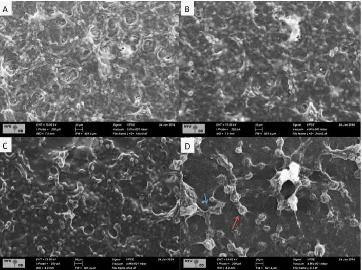
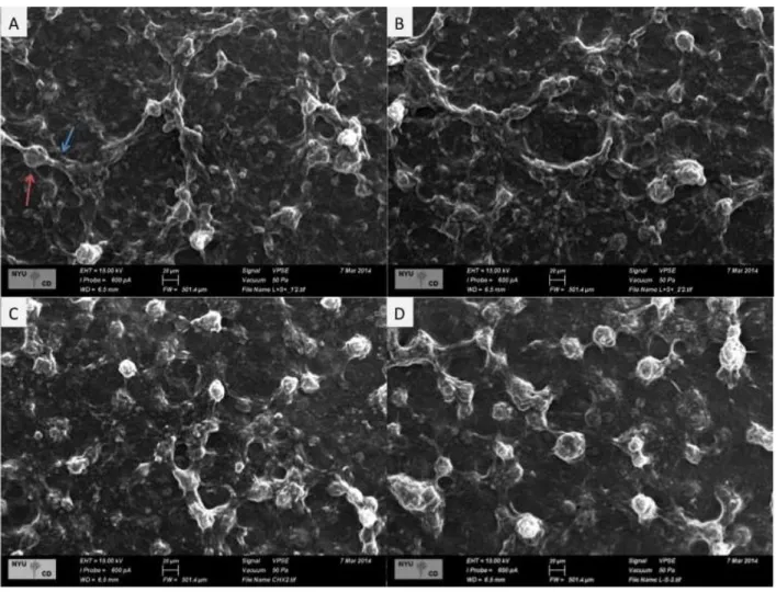
Biofilm analysis: At the end of the experimental period, the biofilms were dip-washed three times in order to remove the loosely bound biofilm, the hydroxyapatite surfaces were gently scraped with a sterile spatula to harvest adherent cells and the biofilm collected was placed in 5 ml sterile saline solution. These biofilms were subjected to sonication using three 15-s pulses at an output of 7 W (Fisher Scientific, Sonic Dismembrator model 100; USA). The homogenized suspension was used for dry weight, bacterial viability (colony-forming units – CFU per miligram of dry weight biofilm), and polysaccharide analyses (EPS-soluble, EPS-insoluble) as described in the previous study (Duarte et al. 2006).
Dry weight: For the dry weight determination, three volumes of cold ethanol (-20°C) were added to 1 ml biofilm suspension, and the resulting precipitate was centrifuged (10,000 g for 10 min at
4°C). The supernatant was discarded, and the pellet was washed with cold ethanol, and then lyophilized and weighed (Duarte et al. 2006)
Bacterial viability: An aliquot (0.1 mL) of the homogenized suspension was serially diluted (1:10; 1:100; 1:1000; 1:10000; 1:100000; 1:1000000) and plated on blood agar. The plates were incubated in 5% CO2 at 37°C for 48 h, and then the number of CFU/mg of biofilm dry weight
was determined.
Polysaccharide analyses: Soluble and insoluble extracellular polysaccharides (EPS-soluble and