Acta Cir. Bras. 2018;33(5):408-414 DOI: http://dx.doi.org/10.1590/s0102-865020180050000002
Adnan GücükI, Gizem SöylerII, Uğur ÜyetürkIII, Burak YılmazIV, İsmail NalbantV, Sebahat GücükVI, Aysel
KüknerVII, Ayhan ÇetinkayaVIII
Does ureteral access sheat usage lead to permanent
damage in the ureter? A placebo controlled trial in a
rabbit model
1Abstract
Purpose: To evaluate the clinical stenosis or precursor histological changes that ureteral access sheaths commonly used in ureteroscopic surgeries may cause in the long term in ureter. Methods: In this study, the animals were divided into 9 groups and according to their groups, ureters of the rabbits were endoscopically fitted with 2F and 3F ureter catheters. The catheters were left in place and withdrawn after a specified period of time. All the ureters were excised and evaluated macroscopically, microscopically and histologically. Ureter diameters were measured and FGF-2 (+) labeled fibroblasts were counted in connective tissue as stenosis precursors.
Results: Macroscopically or microscopically, no stenosis was found in any group. The ureter diameter of the group that were catheterized for the longest time with the catheter that had the widest diameter was significantly lower than the group with the shorter duration and the catheter with the narrower diameter and the control group. When the groups were compared in terms of their FGF values, there was a significant difference in FGF-2 counts at all three ureter levels (p <0.05).
Conclusion: The use of ureteral access sheath may lead to histological changes, as its diameter and duration increase.
Key words: Ureteroscopy. Urinary Catheters. Rabbits.
IAssociate Professor, Department of Urology, Abant Izzet Baysal University, Faculty of Medicine, Bolu, Turkey. Conception and design of the study, manuscript writing, final approval.
IIDoctor, Department of Histology and Embryology, Faculty of Medicine, Near East University, Nicosia, Cyprus. Histopathological examinations.
IIIAssociate Professor, Department of Urology, Abant Izzet Baysal University, Faculty of Medicine, Bolu, Turkey. Conception and design of the study.
IVDoctor, Department of Urology, Abant Izzet Baysal University, Faculty of Medicine, Bolu, Turkey. Scientific and intellectual content of the study, technical procedures.
VAssistant Professor, Department of Urology, Ordu University, Faculty of Medicine, Ordu, Turkey. Scientific and intellectual content of the study, technical procedures.
VIAssistant Professor, Department of Family Medicine, Abant Izzet Baysal University, Faculty of Medicine, Bolu, Turkey. Statistical analysis, technical procedures.
VIIProfessor, Department of Histology and Embryology, Faculty of Medicine, Near East University, Nicosia, Cyprus. Histopathological examinations.
provided by the Experimental Animal Center of the University were used. The animals were housed in a temperature-controlled (19C±2C), humidity-controlled, (40%–70%), and light period controlled (12h/12h light/dark cycle) environment. They were fed a standard rabbit pellet diet and had access to tap water ad
libitum. While starting our study, we could not find a literature with similar endoscopic surgical
procedures. Therefore, in order to familiarize
with the localization of the rabbit ureter
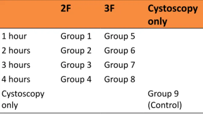
endoscopically and determine the largest diameter of ureteral sheath we can place, we calibrated the ureter via the bladder using open surgery. We determined that a maximum of a 3F ureteral catheter could penetrate in rabbits of a weight of 3-3.5. As seen in Table 1, the animals were divided into 9 groups based on the catheter diameter used and the
duration. Each group contained 8 rabbits. 2F catheter was applied in the first four groups
and 3F catheter was applied in the second four
groups. After the ureteral catheter was placed, it was left there for 1, 2, 3 or 4 hours based on the groups. At the end of these durations,
the catheters were removed. Only cystoscopy
was applied and no catheterization was made
in the 9th group which was the control group.
Table 1 - Groups according to catheter size and
duration.
2F 3F Cystoscopy
only
1 hour Group 1 Group 5
2 hours Group 2 Group 6
3 hours Group 3 Group 7
4 hours Group 4 Group 8
Cystoscopy only
Group 9 (Control)
Before the interventional procedures,
ketamine HCl at a dose of 35mg/kg and xylazine at a dose of 10mg/kg were administered intramuscularly for general
■
Introducti
on
Ureteral access sheath (UAS) is used increasing popularity among urologists as it
makes it easier to enter the collective tubules
in minimally invasive treatments of complex upper urinary system diseases, and minimizes the damage to the ureter causes by repeated entries and exits1. However, its disadvantages
include damage to the ureter due to various mechanisms in this prevalent usage2. Various
studies have shown that excessive distension
of the ureters by access sheaths affects ureteral blood flow and causes ureteral ischemia in
following stages3. It was reported that the
reperfusion occurring as a result of removing the access sheath may harm the ureter via free radicals4. The literature stated that access
sheaths may have chronic effects in the ureter
in the long-run, and recommended usage of
sheaths with suitable diameters in different
cases3. To the best of our knowledge, there
has been no study that investigated the effects of the duration of ischemia due to usage of
sheaths and their diameter in the form that is
applicable to the current clinical practice.
In the study that we planned with the reasons stated above, we placed catheters
equivalent to different diameters of ureteral
sheaths into rabbit ureters endoscopically for
different durations. Then, we analyzed the possible permanent effects in the ureter via both visual and histopathological examination.
Therefore, we aimed to describe an issue that has not been clearly explained in the literature
by simulating a practice that is identical in
humans on rabbits.
■
Methods
This study was approval from the Ethics Board for Animal Studies of Abant Izzet Baysal University.
anesthesia. Briefly, the animals were placed
in a supine position, and their genitalia were scrubbed with povidone iodine solution. An 8F pediatric Cysto-Urethroscope was used for the endoscopic operation. Based on the groups
they belonged to, 2F and 3F ureteral catheters were placed only into the right ureters of the rabbits in correspondence to ureteral access sheaths used in humans, the catheters were
left in their places for a suitable duration based
on the groups, and then removed.
In order to determine the long-term
effects of access sheaths, the rabbits were fed for 1 month and then sacrificed. At the end of 1
month, all ureters were excised by open surgery.
In the meantime, visual evaluation was made
to see whether the ureters were narrowed or whether there was an indicator of macroscopic
dilatations as an indicator of such a case.
They were then taken into histopathological
examination for detection and comparison
of issues such as long-term narrowing in the
ureters in terms of the permanent effects of ischemic damage or potential pathological
issues that are precursors for it. The ureters
of the subject were taken and fixed in 10%
neutralized formalin. The ureters were passed through alcohol gradients and xylol by
separation of upper (renal), medial and lower (bladder) pieces, and embedded in paraffin. The cross-sections with the thickness of 5μm taken
from the blocks were immunohistochemically stained using hematoxylin eosin, Masson’s
3-color stain and FGF-2 (rabbit polyclonal anti
FGF-2, sc-7911).
In the tissue cross-sections of each
subject, ureter diameters were measured with
x4 magnification in units of micrometer from 4 different points, and averages of values were taken. In all cross-sections, the fibroblasts in the connective tissues (lamina propria)
stained with FGF-2 (+) were counted under x40
magnification.
The collected data were analyzed with
the SPSS 20 package software. Due to the
number of the units, normal distribution of the variables was tested using the Shapiro Wilk test. When the variables were not found to be normally distributed, the Kruskal Wallis-H test
was utilized to analyze the differences among the groups. In the case there was significant differences in the Kruskal Wallis-H test, the groups with differences were determined using the Post-Hoc Multiple Comparisons test. While interpreting the results, the level of significance
was determined as 0.05.
■
Results
During the experiment, only one
animal died in the 3rd postoperative day. The
experiment was repeated for this animal.
There was no complication seen other than
this. During the removal of the ureters, the groups were evaluated both macroscopically, and microscopically for histopathological
characteristics.
Macroscopic
During the excision of the ureters, all ureters were macroscopically evaluated
for potential narrowing and dilatation in the proximal. However, narrowing or dilatation of
the proximal as an indirect indicator of it was not seen in any groups including the groups
with statistically significant differences.
Microscopic
The ureteral diameter measurements did not show narrowing in the microscopic
evaluation either. In the in-group comparison
of ureteral diameters, the medial and lower
ureter diameters were significantly lower than
the upper ureter diameter in Group 6, and the
lower ureter diameter was significantly lower than the upper ureter diameter in Group 8
(p<0.05). In the other groups including the
control group, no significant differences were
When the upper ureter sections
were compared in the inter-group comparisons considering the average of four measurements, the diameters in the Groups
4, 6 and 8 were found significantly lower than
the control group (p <0.05). There were no
significant differences in the medial ureter
comparisons. In the comparison of the lower ureter diameters, the ureter diameter in
Group 8 was found significantly lower than
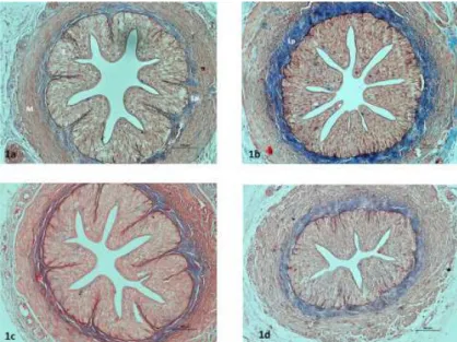
those in the Groups 1, 2 and 3, as well as the control group (Figure 1). Table 2 shows the inter-group comparison values in terms of ureter diameter.
Figure 1 - Light-microscopic images of the ureters in the groups. Control (1a), Group 1 (1b), Group 2 (1c),
Group 8 (1d), Epithelium (E), lamina propria (Lp), muscle layer (M). Masson’s 3-color staining. Bar 100μm.
Table 2 - The results of the Kruskal Wallis H test
regarding the difference among the groups in
terms of ureter diameter (micrometer).
Upper Ureter
Medial Ureter
Lower Ureter
Mean
Group 1 893.55 812.19 850.14
Group 2 884.77 796.43 864.57
Group 3 893.03 854.1 855.59
Group 4 851.3 807.15 843.85
Group 5 908.55 803.38 872.85
Group 6 854.73 760.63 765.25
Group 7 892.43 801.34 733.87
Group 8 837.46 754.21 661.3
Control 1080.1 862.84 916.37
H 19.465 7.682 28.216
p 0.013 0.465 0.001
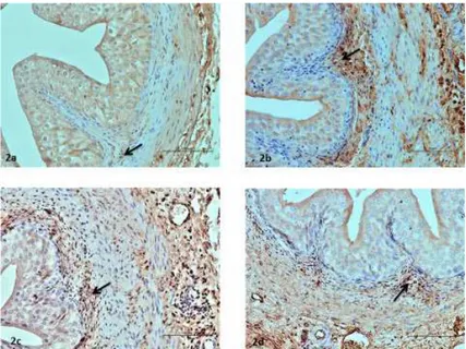
No significant differences were found in
the in-group comparison of upper, medial and
lower sections in terms of FGF values. In the intergroup comparisons, significant differences
were found in terms of FGF-2 numbers for all three levels of ureters (p<0.05). These values
were higher in Group 8 in the upper level in
comparison to the Groups 1, 2 and 3, as well as the control group. In the medial and lower levels, these values were higher than those of
the control group in the Groups 5, 6, 7 and 8
Figure 2 - Images of the FGF-2 immunohistopathologically stained ureters in the groups. Fibroblasts (→)
stained positively by FGF-2 are seen in the lamina propria of the following groups: Control (2a), Group 6 (2b), Group 7 (2c), and Group 8 (2d). Bar 50 μm.
Table 3 - The Results of the Kruskal Wallis H
test in terms of the differences in FGF (+) values
among the groups.
Upper Ureter
Medial Ureter
Lower Ureter
Mean
Group 1 7 8.63 10.5
Group 2 7.63 10.5 11.13
Group 3 6.75 11.5 14.38
Group 4 10.38 10.5 15.88
Group 5 14.12 16.63 19.12
Group 6 14.5 19.88 21.38
Group 7 15.5 22.13 23.38
Group 8 18.75 22.13 26.13
Control 7.63 7.5 8.25
H 37.067 41.529 42.477
p 0.001 0.001 0.001
■
Discussion
Ureteral access sheaths are routinely
used today, especially in retrograd intrarenal
surgeries. By mechanically dilating the ureter
lumen, UAS improves imaging, makes stone
removal easier, and shortens time of surgery3.
It was shown that UASs increase tension in smooth muscles by stretching ureter lumen during this procedure, and they may lead to ischemia this way3,5. It is known that the
higher the diameter of the UAS used, the
better the image will be and the shorter the time of surgery will become. While UAS
usage has various advantages over non-UAS
ureteroscopy practices, there are concerns
on its usage due to increased risk of ureteral damage6. Additionally, UASs may be used with
an internal diameter of 9.5 French to 14 French,
and an external diameter of 11.5F to 18F.
However, placing thick UASs into non-dilated
ureters has potential risks. Likewise, surgery time is affected by UAS diameter in addition to factors such as stone loads, but the relationship between this time and the potential risk of
damage in the ureter is not clearly known. In this study that we conducted with the purpose
of determining these potential relationships, we think we partially found answers to these questions.
the animals’ ureters after sacrificing them. We had aimed to detect potential narrowing and indirect indicator of it as a dilation in
the proximal this way. However, we did not
encounter such narrowing or dilation in any
ureters. In the histological narrowing analysis,
no pathology that would be reflected to the
clinical results were found.
We found it suitable to make
comparisons among different levels of ureters as their diameters may change on different levels. However, no statistically significant difference was found within the groups, with the exception of the Groups 6 and 8. Theoretically, we expected that the narrowest diameters of ureters would be affected the
most, and therefore, it would be more suitable to compare ureter levels with similar diameters.
While the diameter difference in the control group was not significant, the finding that the medial and lower ureters in the Groups 6 and 8 were affected more, at least partially supported this expectation. Additionally, we believe that comparison of ureter diameters on different
levels was useful in terms of increasing the precision of our analysis.
In the ureter diameter comparison
among the groups, we found significant
decreases in ureter diameters in the Groups 4,
6 and 8. These groups represented either a long duration, or the groups treated with thicker
ureter catheters. Accordingly, we found that
catheter diameter and duration are parameters that may affect narrowness. Delvecciho et al.7
investigated long-term structural formation in patients treated with UAS. As a result of UAS
usage, the occurrence rate of narrowness in
radiological images in the first 3 months was
found as 1.4%. Considering that this rate was
1.2% in patients who were not treated with
UAS, they concluded that UAS usage does not
have an influence. Indeed, the clinical outcome of our study agreed with the findings of their
study. However, considering the results in
terms of the reduction in microscopic ureter
diameters and similar FGF (+) outcomes in these
groups, the duration and the catheter diameter affecter histological results negatively.
In the study where Lallas et al.3 used a
pig model to investigate the ischemic effects
of UAS, they used 12F/14F and 14F/16F UASs, placed the UASs with open surgery, kept them
inside for 70 min, measured blood flow using
doppler ultrasonography, and conducted a
histological examination after sacrificing the
animals at the end of 3 days. Histologically, they
were not able to find evidence of ischemia. In difference to their study, we planned to
imitate surgeries we performed on humans as much as possible. This is why we placed the ureter catheters endoscopically. We tried
to determine the influential parameters by keeping the catheter diameters and durations variable among the groups. Additionally, in
order to be able to measure the long-term
effects of these factors in animals, we sacrificed the animals after 1 month and analyzed the
ureters histologically. Lallas et al.3 reported
that UAS decreased blood flow in their sample, but the ureteral wall was left without harm
due to compensatory mechanisms. Therefore, they reported that they found UAS usage to be safe. However, they stated that the chronic
effects of UAS usage has not yet been cleared,
and therefore, it is needed to use a suitable
size of UAS in each patient. They mentioned the necessity of taking preventive measures against UAS-related damage in risky patients. In their study, the histological results reflect an earlier outcome due to the sacrifice carried out after 72 hours. On the other hand, as we carried out the sacrifice after 1 month, we
analyzed later histological and microscopic
outcomes. Long duration and thick catheter placement appeared as influential parameters
that may lead to narrowing in terms of both
ureter diameter and FGF positivity. The fact that our study did not find clinical narrowing may
be related to the compensatory mechanisms
may reveal potential narrowing.
Limitations of our study include that
the process was carried out in an animal like rabbits, which have thinner ureters than
those found in humans. Conducting this study
on animals with similar ureter sizes, such as pigs, could have created more valid results by
obtaining potentially more realistic data by
using the access sheaths we use in humans. However, we did not have the means to work
with this number of pigs. Additionally, while we represented UAS effects, we were not able
to use real UASs as we conducted the study
on rabbits. Despite all these limitations, we
believe that the study simulated the outcomes
of the practices we use on humans as well as
possible.
■
Conclusions
When an upper-limit diameter such as
3F is used in rabbit ureters for a long duration such as 3 or 4 hours, this causes reduction of
diameter in the ureter and histological changes that may be precursors of this. Therefore, while UASs are a widely used highly useful
instruments, usage time and diameter should
be carefully adjusted. Usage of the shortest
duration and lowest diameter of UAS may be a precaution against potential risks.
■
References
1. Kaplan AG, Lipkin ME, Scales CD, Preminger GM. Use of ureteral access sheaths in
ureteroscopy. Nat Rev Urol. 2016;13(3):135-40. doı: 10.1038/nrurol.2015.271.
2. Zelenko N, Coll D, Rosenfeld AT, Smith RC. Normal ureter size on unenhanced helical
CT. Am J Roentgenol. 2004;182(4):1039-41. doı: 10.2214/ajr.182.4.1821039.
3. Lallas CD, Auge BK, Raj GV, Santa-Cruz R, Madden JF, Preminger GM. Laser Doppler
flowmetric determination of ureteral blood flow after ureteral access sheath placement. J Endourol. 2002;16(8):583-90. doı: 10.1089/089277902320913288.
4. Traxer O, Thomas A. Prospective evaluation and classification of ureteral wall ınjuries resulting from ınsertion of a ureteral access sheath during retrograde ıntrarenal surgery. J Urology. 2013;189(2):580-4. doı: 10.1016/j.juro.2012.08.197.
5. Rose JG, Gillenwater JY. Pathophysiology
of ureteral obstruction. Am J Physiol. 1973;225(4):830-7. doı: 10.1152/ ajplegacy.1973.225.4.830.
6. Rizkala ER, Monga M. Controversies in
ureteroscopy: wire, basket, and sheath. Indian J Urol. 2013;29(3):244-8. doı: 10.4103/0970-1591.117287.
7. Delvecchio FC, Auge BK, Brizuela RM, Weizer AZ, Silverstein AD, Lallas CD, Pietrow PK, Albala DM, Preminger GM. Assessment of
stricture formation with the ureteral access sheath. Urology. 2003;61(3):518-22. PMID:
12639636.
Correspondence: Dr. Adnan Gücük
Abant İzzet Baysal Üniversitesi Tıp Fakültesi
Hastanesi
Üroloji Bölümü, Gölköy Kampusu, Bolu Turkey Phone: +905056748193
Received: Jan 18, 2018 Review: Mar 20, 2018 Accepted: Apr 23, 2018
Conflict of interest: none
Financial source: Abant İzzet Baysal University Scientific Research Projects (n. 2016.08.20.1050)
1Research performed at Abant Izzet Baysal
University, Experimental Animals Application