The S-Nitrosylation Status of PCNA Localized
in Cytosol Impacts the Apoptotic Pathway in
a Parkinson
’
s Disease Paradigm
Liang Yin1,2, Yingying Xie1, Songyue Yin1, Xiaolei Lv3, Jia Zhang3, Zezong Gu4, Haidan Sun1*, Siqi Liu1,2,3*
1Key Laboratory of Genome Sciences and Information, Beijing Institute of Genomics, Chinese Academy of Sciences, Beijing, China,2University of Chinese Academy of Sciences, Beijing, China,3Beijing Protein Innovation, Beijing, China,4Department of Pathology and Anatomical Sciences, University of Missouri School of Medicine, Columbia, Missouri, United States of America
*[email protected](HS);[email protected](SL)
Abstract
It is generally accepted that nitric oxide (NO) or its derivatives, reactive nitrogen species (RNS), are involved in the development of Parkinson’s disease (PD). Recently, emerging evidence in the study of PD has indicated that protein S-nitrosylation triggers the signaling changes in neurons. In this study, SH-SY5Y cells treated with rotenone were used as a model of neuronal death in PD. The treated cells underwent significant apoptosis, which was accompanied by an increase in intracellular NO in a rotenone dose-dependent
manner. The CyDye switch approach was employed to screen for changes in S-nitrosylated (SNO) proteins in response to the rotenone treatment. Seven proteins with increased S-nitrosylation were identified in the treated SH-SY5Y cells, which included proliferating cell nuclear antigen (PCNA). Although PCNA is generally located in the nucleus and partici-pates in DNA replication and repair, significant PCNA was identified in the SH-SY5Y cyto-sol. Using immunoprecipitation and pull-down approaches, PCNA was found to interact with caspase-9; using mass spectrometry, the two cysteine residues PCNA-Cys81 and -Cys162 were identified as candidate S-nitrosylated residues. In addition, the evidence ob-tained from in vitro and the cell model studies indicated that the S-nitrosylation of PCNA-Cys81 affected the interaction between PCNA and caspase-9. Furthermore, the
interaction of PCNA and caspase-9 partially blocked caspase-9 activation, indicating that the S-nitrosylation of cytosolic PCNA may be a mediator of the apoptotic pathway.
Introduction
Parkinson’s disease (PD), the second most common neurodegenerative disorder, is character-ized by a massive and specific loss of dopaminergic neurons in the substantia nigra pars com-pacta (SNpc) [1,2]. The exact causes of this neuronal loss have not yet been fully elucidated, but a large body of evidence has revealed the major contribution of apoptosis [3]. Programmed a11111
OPEN ACCESS
Citation:Yin L, Xie Y, Yin S, Lv X, Zhang J, Gu Z, et al. (2015) The S-Nitrosylation Status of PCNA Localized in Cytosol Impacts the Apoptotic Pathway in a Parkinson’s Disease Paradigm. PLoS ONE 10 (2): e0117546. doi:10.1371/journal.pone.0117546
Academic Editor:Hiroyoshi Ariga, Hokkaido University, JAPAN
Received:July 21, 2014
Accepted:December 27, 2014
Published:February 12, 2015
Copyright:© 2015 Yin et al. This is an open access article distributed under the terms of theCreative Commons Attribution License, which permits unrestricted use, distribution, and reproduction in any medium, provided the original author and source are credited.
Data Availability Statement:All relevant data are within the paper and its Supporting Information files.
Funding:Funding was provided by National Key Basic Research Program of China: 2010CB912703; National High Technology Research and Development Program of China: 2012AA020205; Nature Science Foundation of China: 91131009. The funders had no role in study design, data collection and analysis, decision to publish, or preparation of the manuscript.
cell death is a normal process, whereas apoptosis can trigger several pathological changes, in-cluding neurodegenerative disorders. Interestingly, apoptotic neurons have been identified in the SNpc of PD patients via an in situ end labeling method [4], and the activation of caspases has been detected in the nigral neurons of a PD mice model [5]. Many mechanisms are likely involved in neuronal apoptosis, including oxidative stress, mitochondrial dysfunction, energy imbalance, inflammation, defects in familial genes, and dysfunction of the ubiquitin-proteasome system (UPS). It is difficult to attribute neuronal apoptosis to a single causal factor as hypothesized by Sulzer, the neurodegeneration in PD likely results from“multiple hits”[6]. Therefore, an intensive investigation of PD focusing on the detailed events related to the patho-logical process is urgently required.
Nitric oxide (NO) is a free radical in a highly diffusible gaseous state and is regarded as an important regulator for numerous biological processes, such as vasodilation [7], neurotrans-mission [8], and inflammatory responses [9]. Excessive production of NO and NO-derivative reactive nitrogen species (RNS) has been implicated in neuron damage, particularly neurode-generation in PD [10–12]. Increased expression of iNOS has been identified in PD animal models induced by 6-OHDA and LPS or rotenone, leading to increased NO levels and lipid peroxidation products [13–15]. The overexpression of nNOS and the generation of peroxyni-trite (ONOO-) have been observed in PD patients [16]. It is generally accepted that RNS are highly active and able to react with many macromolecules. S-Nitrosylation, which is a re-versible covalent addition of a NO group to a cysteine residue’s sulfhydryl that forms S-nitrosothiols, is a typical protein modification induced by RNS [17]. Several investigators have focused on the correlation between S-nitrosylation and PD. For example, the cysteine resi-due at 644 in dynamin-related protein 1 (Drp1), a member of the Dynamin family of large GTPases, is likely to be S-nitrosylated (SNO). The S-nitrosylation of Drp1 promotes its multi-merization, leading to mitochondrial fission and neuronal damage [18]. In addition, the cyste-ine residues at 51 and 172 in peroxiredoxin 2 (Prx2), an antioxidant protein, are S-nitrosylated. In neuronal cells, SNO-Prx2 becomes inactivated, sensitizing the cells to oxidative stress-dependent cell death [19]. S-Nitrosylation has a direct role in regulating protein functions and affects protein interactions through structural alterations. The S-nitrosylation of GAPDH en-ables its binding and stabilization of the E3 ubiquitin ligase Siah1, thus facilitating nuclear pro-tein degradation and stimulating apoptotic cell death [20]. In addition, the S-nitrosylation of the BIR domain in X-linked inhibitor of apoptosis protein (XIAP) decreases its binding to cas-pase-3, which is accompanied by the loss of the anti-apoptotic ability of XIAP under nitrosative stress [21]. However, no general mechanism for the functional changes of SNO proteins in PD has been identified to date.
of RNS and protein modifications caused by RNS, whereas the inhibition of nitric oxide synthase attenuates rotenone-induced apoptosis [15,19,25]. Generally, it is believed that rote-none can cause neuronal depolarization, resulting in activation of N-methyl-D-aspartate (NMDA) glutamate receptor, a voltage dependent Ca2+channel. Since neuronal nitric oxide synthase (nNOS) is a Ca2+dependent NOS, increased Ca2+due to rotenone treatment thus en-ables activation of nNOS, leading an increase of NO production. In addition, NO has been shown to S-nitrosylate caspases-3 [26], -8 [27], and-9 [28] at the active site cysteine, thus inhib-iting enzymatic activity and affording neuroprotection. How the S-nitrosylation of caspases in-fluences the apoptotic process remains unclear. Thioredoxin2 (TRX2) has been regarded as a transnitrosylation mediator that selectively denitrosylates the S-nitrosothiols of caspase-3 [29], whereas another study proposed that the neuroprotective activity could be abrogated during nitrosative stress via transnitrosylation between XIAP and caspases [30]. According to that model, transnitrosylation from SNO-caspases to XIAP acts as an apoptotic switch. The ubiqui-tination and target degradation functions of XIAP are regulated by its S-nitrosylation status. Therefore, RNS have dual functions, namely, the direct inhibition of caspase activity by S-nitrosylation and the attenuation of caspase degradation by XIAP via SNO-XIAP formation. The mechanism of RNS involvement in apoptosis of rotenone-treated SH-SY5Y cells
is unclear.
In this research, we used rotenone-treated SH-SY5Y cells as a typical PD model to investi-gate the mechanism between SNO proteins and neuronal apoptosis. The canonical approach of SNO proteins detection is biotin switch technique, from which many proteomic strategies were derived for S-nitrosoproteome analysis [31]. Herein, after carefully estimating the nitrosative stress and apoptosis in SH-SY5Y cells, we utilized fluorescence-tagged CyDye thiol reactive agents to label S-nitrosothiols on SNO proteins, which was combined with two-dimensional difference gel electrophoresis (2D-DIGE) to separate the SNO proteins. Approximate two hun-dred fluorescent spots were detected via image analysis; in these spots, 7 proteins were identi-fied as having increased S-nitrosylation in response to rotenone treatment. We focused on the physiological role of the S-nitrosylation of PCNA because this protein is closely related to the cell cycle and the regulation of apoptosis. Its S-nitrosylation was coincidentally up-regulated in another study of PD neuronal model [32]. Interestingly, we observed that PCNA in SH-SY5Y cells was predominately localized in the cytosol and was able to interact with caspase-9. Our data further demonstrate that the up-regulated S-nitrosylation of PCNA decreases the interac-tion of PCNA and caspase-9, leading to an increase of caspase-9 cleavage, so called caspase-9 activation. The active form of caspase-9 is expected to activate the apoptotic pathway in dopa-minergic neuron in PD. This is a reasonable deduction derived from our observation how PCNA S-nitrosylation performs a significant impact to the functions of SH-SY5Y cells, leading to a regulatory mechanism of cell death in PD.
Materials and Methods
Reagents
Rotenone, NG-Methyl-L-arginine acetate salt (L-NMMA), S-Methyl methanethiosulfonate (MMTS), Sodium L-ascorbate, Neocuproine and DMF were purchased from Sigma-Aldrich. 4-Amino-5-Methylamino-20,70-Difluorofluorescein Diacetate (DAF-FM DA) were obtained
Cell culture and treatments
Human neuroblastoma SH-SY5Y and HeLa cervix carcinoma cell lines were obtained from Type Culture Collection (China Center). SH-SY5Y and HeLa cells were cultured in DMEM/ F12 and DMEM (Gibco), respectively, and supplemented with 10% (v/v) fetal bovine serum, 0.1 U/ml penicillin, and 0.1 mg/ml streptomycin (PAA) at 37°C in a humidified 5%
CO2atmosphere.
The SH-SY5Y cells were transferred to a low serum medium that contained 0.5% FBS for 12 h prior to further treatment. The rotenone solution was freshly prepared prior to each ex-periment in dimethyl sulfoxide (DMSO), and DMSO served as the vehicle control. L-NMMA, a NOS inhibitor, was dissolved in deionized water and added to the culture media at the final concentration of 0.5 mM 4 h prior to rotenone treatment. The final volume ratio of vehicle or drug solution to the media was controlled within 0.05%.
Generation and detection of NO
S-Nitrosocysteine (SNOC) was used to generate NO according to previous methods [33] and was freshly prepared prior to each experiment. DAF-FM DA was used as a fluorescent indica-tor of intracellular NO to moniindica-tor the NO generated in response to cell treatment, according to the protocol provided by Molecular Probes. Following the cell treatment, the SH-SY5Y cells were washed with phosphate-buffered saline (PBS), followed by the addition of 5μM DAF-FM
DA for 20 min at 37°C. The cells were then rinsed and maintained in PBS, and the fluorescence within the cells was detected atλex= 495 nm andλem= 515 nm using a Tecan Safire5
microplate reader.
Western blot
The protein samples were dissolved in lysis buffer that contained 50 mM Tris-HCl, pH 7.4, 2% SDS, 10 mM dithiothreitol (DTT), and 5 mM EDTA with supplements of proteinase inhibitors. The denatured proteins were separated on a 12% SDS—PAGE, and the separated proteins were transferred onto a polyvinylidene difluoride membrane. After blocking and washing, the membrane was incubated with the indicated primary antibody followed by incubation with horseradish-linked secondary antibody. The immuno-recognition signals were detected by chemiluminescence using ImageQuant ECL (GE Healthcare), and the chemiluminescence was quantified using PDQuest software version 8.0.1.
Annexin V-FITC/propidium iodide (PI) dual staining assay
SH-SY5Y cells treated with or without 500nM rotenone for 16h were trypsinized and the cell pellets were collected. The cells were resuspended in 100μL binding buffer at 1 × 10⁶cells/mL,
and incubated with 5μL annexin V-FITC and 10μL PI for 15 min in the dark at room
temper-ature. Then, these cells were added into 400μL binding buffer and measured by FACSCalibur
flow cytometer (Becton Dickinson).
Biotin switch technique
beads. The pellets were washed with neutralization buffer that contained 20 mM Hepes, pH 7.4, 100 mM NaCl, 1 mM EDTA, and 0.5% Triton X-100; the pellets were then eluted by Laemmli sample buffer followed by Western blot analysis.
CyDye switch and 2D-DIGE
The combination of CyDye switch and DIGE was used to enrich and separate the SNO proteins in the SH-SY5Y cells. The cell lysates were incubated with 4X volume blocking buffer that con-tained 250mM HEPES-NaOH, pH7.4, 1mM EDTA, 0.1mM Neocuproine, 2.5% SDS, and 20mM MMTS, and the incubation was performed at 50°C for 30 min in the dark with vortex every 5 min. After the free thiols of the cell lysates were blocked and the S-nitrosothiols were reduced, the excess ascorbate was removed by acetone precipitation, and the precipitated pro-teins were dissolved in CyDye labeling buffer, which contained 30 mM Tris-HCl, 7 M urea, and 4% CHAPS, pH 8.0. CyDye DIGE Fluor dyes were used to label proteins at the final con-centration of 10μM. The labeling reaction was quenched by 2X CyDye labeling buffer that
con-tained 2% v/v IPG buffer for pH 3–10 and 130 mM DTT. In the CyDye switch/2D-DIGE experiment, inverse strategy was adopted. On one 2DE running, the control sample labeled with Cy5 was the SH-SY5Y cell without rotenone treatment, while the internal standard sample labeled with Cy3 was a mixture, in which equal quantities of the protein lysates from the SH-SY5Y cells treated with and without rotenone were well mixed. The pooling of the equal quantities of the control sample and the internal standard sample was loaded to electro-phoresis. Following 2DE, the gel was scanned by a Molecular Imager PharosFX System (Bio-Rad) withλex= 548 nm andλem= 560 nm for the Cy3-labeled sample andλex= 641 nm andλem= 660 nm for the Cy5-labeled sample, and the relative fluorescence intensity for a 2DE
spot was estimated by the ratio of Cy5 against Cy3. Inversely, on the other 2DE running, the ro-tenone sample labeled with Cy5 was the SH-SY5Y cell treated with roro-tenone, while the internal standard sample labeled with Cy3 was a mixture of equal quantities of the control and rotenone sample. The sample loading and spot intensity estimation were the same as above. Since the composition in the internal standard sample was consistent, the fold-change for each spot could be obtained by comparison of the relative fluorescence intensity. The florescence images were analyzed using PDQuest software version 8.0.1. All the CyDye switch/2D-DIGE experi-ment, the related proteomic analysis and protein identification were conducted in three independent experiments.
Tryptic digestion and protein identification by mass spectrometry
The selected 2D-DIGE spots were collected and subjected to in-gel digestion with trypsin fol-lowing the protocol previously described [35]. The peptides generated by the tryptic digestion were identified by a micrOTOF-Q (Bruker Daltonics) mass spectrometer using the following instrument settings: nebulizer gas, nitrogen, 1.6 bar; dry gas, nitrogen, 6 l min−1, 190°C; capil-lary,−5,500 V (+4,000 V); end plate offset,−500 V; funnel 1 RF, 200 Vpp; funnel 2 RF, 200 Vpp; in-source CID energy, 0 V; hexapole RF, 100 Vpp; quadrupole ion energy, 5 eV; collision gas, argon; collision energy, 10 eV; collision RF 200/400 Vpp (timing 50/50); transfer time, 70
μs; prepulse storage, 5μs; pulse frequency, 10 kHz; and spectra rate, 3 Hz. The MS/MS spectra
data were searched against the SwissProt database using Data Analysis software (Bruker Dal-tonics) and the MASCOT in-house search engine (MatrixScience).
5–80% acetonitrile with 0.1% formic acid. Each full MS scan was followed by three MS/MS scans of the three most intense ions, with data-dependent selection using the dynamic exclu-sion option. The mass spectrometer settings and spectra analysis method were the same as those used in the spot identification.
Immunofluorescence staining
PCNA subcellular localization in the SH-SY5Y or HeLa cells was observed with a standard immunofluorescence staining protocol [37]. Cells on glass cover slips were fixed in PBS that contained 4% paraformaldehyde for 20 min on ice and permeabilized with 0.3% Triton X-100 for 5 min at room temperature, followed by incubation with primary antibodies overnight at 4°C. The cells were then treated with FITC or TRITC-labeled secondary antibodies and DAPI (for nuclear staining). The fluorescence images were viewed under an FV 500 confocal micro-scope (Olympus).
Enrichment of nuclear and cytosolic fractions
The cell pellet was washed twice with ice-cold PBS and resuspended in 5X volume of ice-cold isolation buffer containing 20 mM Hepes—KOH, pH 7.5, 10 mM KCl, 1.5 mM MgCl2, 1 mM
sodium EDTA, 1 mM sodium EGTA, 1 mM dithiothreitol, 0.1 mM PMSF, and protease inhibi-tors. The resuspended cells were broken by passing several times through a G26 needle. The homogenate was centrifuged at 1000 g for 15 min to precipitate the nuclear fraction, and the supernatant was further centrifuged at 105g for 30 min in L-100 XP ultracentrifuge (Beckman). The resulting supernatant was used as the cytosol fraction.
Coimmunoprecipitation (CoIP) and pull-down assay
The SH-SY5Y cytosol was kept in isolation buffer for 30 min at 4°C. Following centrifugation at 12,000 g for 20 min, the supernatants were incubated with anti-PCNA antibody to immu-noprecipitate PCNA-interacting proteins. The eluted immuimmu-noprecipitates were identified by LC-MS/MS or loaded onto 12% SDS-PAGE gels followed by Western blotting using anti-cas-pase-9 antibody as the primary antibody.
Using the pET system as indicated, wild-type or mutated recombinant His-PCNA proteins were expressed inEscherichia coliBL21 (DE3) cells. The His-tagged fusion proteins were
puri-fied with nickel-agarose beads (QIAGEN) according to the manufacturer’s instructions. Approximately 50μg His-PCNA protein were incubated overnight at 4°C with the lysates
ob-tained from HeLa. Following incubation, nickel-agarose beads were added to capture the re-combinant protein and the proteins that bound to it. The PCNA-interacting proteins were identified by Western blot.
Generation of wild-type and site-directed mutagenesis recombinant
proteins
double-mutant C81A/C162A plasmid was also constructed with the same mutated PCR primers. The mutated PCNAs were inserted into pET-28a for protein expression.
Caspase-9 activation assay
HeLa cytosol was incubated with cytochrome c (Calbiochem), 1 mM dATP and the indicated exogenous PCNA for 30 min at 37°C. The cleavage of caspase-9 was detected by Western blot using a polyclonal antibody against caspase-9 [38].
Overexpression of PCNA in SH-SY5Y cells
The PCR product of PCNA gene with cloning sites of BamH1 and Xho1 was inserted into pcDNA3.1(+) plasmid (Invitrogen) for generation of transfection vector and the pcDNA3.1-PCNA was transfected to SH-SY5Y cells using Lipofectamine 2000 (Invitrogen) according to the manufacturer’s instructions.
Statistical analysis
All experimental data were generated in triplicate (at minimum). The data are presented as the mean ± SEM, and significant differences among the test groups were determined by Student’s
ttest.
Results
Generation of NO stress in SH-SY5Y cells using rotenone
S-Nitrosoproteome in SH-SY5Y cells analyzed by CyDye
switch/2D-DIGE
CyDye switch/2D-DIGE is a common approach to analyze S-nitrosoproteomes [39–41]; in this approach, biotin tags are replaced by fluorescent tags to label SNO proteins. This method was recently employed to study protein S-nitrosylation in microglial cells [42]. To reduce the back-ground noise, we optimized the approach by setting an internal standard as described in “Methods”. Approximately two hundred fluorescent spots were visualized on the images based on analysis using PDQuest software (Fig. 2A). Of the spots that had an intensity change over 1.5-fold due to rotenone treatment in three independent experiments, 7 spots showed an increased fluorescent signal, indicating these proteins had sensitivity to NO stress and were readily S-nitrosylated. The 7 spots were excised from the gels and transferred to the micrO-TOF-Q mass spectrometer for protein identification following trypsin digestion. The differen-tial S-nitrosylated proteins and their fold-changes are summarized inFig. 2B, and all peptides matched with these proteins are listed inS1 Table.
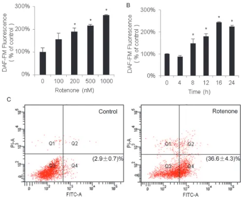
Next, the BST was performed. Its specificity in SH-SY5Y cells was demonstrated (Fig. 2C). Western blot analysis of protein S-nitrosylation in SH-SY5Y showed a strong anti-biotin immunoreactivity for biotinylated proteins only in cells treated with SNOC, a NO-generating Fig 1. Phenotype changes in SH-SY5Y cells in response to rotenone treatment.The dose (A) and time (B) responses of NO generated in SH-SY5Y cells that were treated with rotenone were analyzed. 18h treatment (A) and 500nM rotenone (B) were used for the gradient experiments. The NO contents are represented as the ratio of the intensity of DAF-FM fluorescence in the rotenone-treated group compared with the vehicle-treated group (average ratio±SEM, n = 3,*P<0.05). Apoptosis assessment by flow cytometry for
SH-SY5Y cells treated with or without 500nM rotenone for 16h (C). Dot plot showed annexin V-FITC in x-axis and PI in y-axis. Cells in the fourth quadrant undergoing early stage apoptosis are annexin V-positive/PI negative. And cells at late stage apoptosis or necrotic cells are both annexin V-FITC and PI positive. The left represents the untreated cells as the control. The apoptotic rates are shown as the average ratio±SEM
(n = 3).
reagent, but not in untreated control cells, indicating a complete blocking of free cysteine thiols by MMTS and minimal endogenous S-nitrosylation in untreated cells. The absence of a signal in reaction without ascorbate demonstrates that the modification is SNO-specific. After estab-lishing the efficacy and specificity of the BST in SH-SY5Y cells, three interesting proteins with increased S-nitrosylation, PCNA, PGK1, and TRX, were further examined by BST/Western blot. The lysates from the SH-SY5Y cells in the control or rotenone groups were subjected to BST followed by Western blot using the specific antibodies against the antigens. As shown in
Fig. 2D, the three proteins were readily detectable by the corresponding antibodies in the ly-sates; however, no clearly different immuno-signals were observed between the control and ro-tenone groups. In the lysates that had been incubated with avidin, all immuno-signals in the cells treated with rotenone appeared remarkably augmented compared with the control cells. Thus, the Western blotting results were consistent with the CyDye switch/2D-DIGE results; some proteins in the SH-SY5Y cells were sensitive to NO stress and showed significantly in-creased S-nitrosylation.
Fig 2. Analysis of S-nitrosoproteomes in SH-SY5Y cells with and without rotenone treatment using CyDye switch/2D-DIGE and BST/Western blot.
(A) The representative 2D-DIGE images. The internal standard (Cy3, green) and the target samples (Cy5, red) have been defined in the Methods section. The gel images were acquired by fluorescence scanning atλex= 548 nm andλem= 560 nm for Cy3-labelled samples and atλex= 641 nm andλem= 660 nm
for Cy5-labelled samples. In the merged image, the circled spots represent the spots that differentially responded to rotenone treatment. (B) Comparison of the spot volumes for the seven differential proteins on 2D-DIGE. The spot fold-changes corresponding to the differential spots onFig. 2Awere analyzed using PDQuest software version 8.0.1. The differential spots were defined as changes in spot volume over 1.5-fold in all the cases, and were excised and tryptic digested for protein identification with mass spectrometry (fold-changes of spot volumes±SEM, n = 3,*P<0.05 versus vehicle-treated group). (C)
Specificity and efficiency of biotin switch technique. SH-SY5Y cell lysates were treated with or without 200μM SNOC followed by BST. Protein extract was loaded onto an SDS-PAGE. Western blot analysis was carried out, and the membrane was probed with anti-biotin. Control samples were subjected to PBS or SNOC but not to ascorbate. (D) Verification of the proteins with increased S-nitrosylation in SH-SY5Y cells treated with rotenone treatment. The proteins were extracted from SH-SY5Y cells with and without rotenone treatment and subjected to BST, and the biotinylated proteins were pulled down. The potentially S-nitrosylated proteins were examined by Western blot using the corresponding antibodies. The fold-changes are shown in the bar chart (n = 3, *P<0.05 versus vehicle-treated group).
Localization of PCNA in SH-SY5Y cells
PCNA, a processivity factor for DNA polymeraseεin eukaryotic cells, is predominately located in the nucleus and involved in the apoptotic pathway. Recently, PCNA was also reported to be located in the cytosol of leukocytes [43]. We investigated the localization of PCNA in SH-SY5Y cells using Western blot and confocal microscopy. For the Western blot analysis, SH-SY5Y cells were fractioned into cytosolic and nuclear fractions; the presence of PCNA detected using an antibody specific for PCNA. The Western blot image inFig. 3Aindicates that PCNA in SH-SY5Y cells is dominantly located in the cytosol, not in the nucleus. In the confocal microscopy analysis (Fig. 3B), we stained PCNA and aldose reductase, a cytosolic marker, in SH-SY5Y cells, and observed that aldose reductase was apparently only cytosolic localization but PCNA was detectable in two localizations, cytosol and nuclei. The immuno-signals for PCNA and DAPI staining also do not overlap in SH-SY5Y cells, whereas they strongly overlap in HeLa cells, a positive control for the nuclear localization of PCNA (Fig. 3C). These findings indicate that PCNA in SH-SY5Y cells is primarily distributed in the cytosol and not in the nuclei. We further determined that the location of PCNA in SH-SY5Y cells was unaltered by rotenone treatment (Fig. 3C). Therefore, if PCNA involves apoptosis in SH-SY5Y cells, it is via the apo-ptosis pathways in the cytosol.
Fig 3. Localization of PCNA in SH-SY5Y cells.(A) Localization of PCNA in the cytosolic and nuclear fractions of SH-SY5Y cells was examined by Western blot with antibodies against PCNA, Lamin A (nuclei marker), and aldose reductase (AR, cytosolic marker). (B) Localization of PCNA in SH-SY5Y cells was observed by confocal microscopy. Signal for aldose reductase served as cytosolic marker. (C) Effect to the localization of PCNA in SH-SY5Y cells by rotenone treatment was monitored by confocal microscopy with immunofluorescence using the PCNA antibody. DAPI was used as a nuclei stain.
S-nitrosylation regulation of the interaction of PCNA and caspase-9
How does cytosolic PCNA perform its biological functions in SH-SY5Y cells, especially under NO stress? To answer this question, we searched for proteins that interact with PCNA. CoIP with antibody against PCNA was conducted using the cytosolic fraction of SH-SY5Y cells, and the proteins that interacted with cytosolic PCNA were identified by LC-MS/MS. As shown in
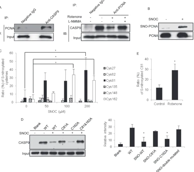
S2 Table, 76 proteins were identified as potential PCNA-interacting candidates, including heat shock protein 90, peroxiredoxin-6, and 14–3–3 protein. Caspase-9, a key caspase that initiates the apoptotic pathways in the cytoplasm, was of particular interest. We subsequently designed experiments to verify the proteomic observations. First, we conducted a diagonal CoIP using two antibodies against PCNA and caspase-9 in SH-SY5Y cells. As depicted inFig. 4A, caspase-9 was found in the precipitate pulled down by the PCNA antibody. Similarly, PCNA was found in the precipitate pulled down by the caspase-9 antibody. These two CoIP results provided con-vincing evidence that the two proteins could physically interact with each other in the cytosol of SH-SY5Y cells. Second, we sought to determine whether the interaction between PCNA and caspase-9 could be affected by NO stress. As shown in the Western blot inFig. 4A, the interac-tion between caspase-9 and PCNA was attenuated by treatment with rotenone. Moreover, the interaction between caspase-9 and PCNA was restored when the NO stress induced by rote-none was reduced by the addition of L-NMMA. Furthermore, the S-nitrosylation status of cas-pase-9 was carefully evaluated, and no change was observed under rotenone treatment of SH-SY5Y cells (S2 Fig.). Thus, the interaction between PCNA and caspase-9 is partially regulated by the S-nitrosylation of PCNA.
There are 6 cysteine residues in PCNA. Which cysteine residue(s) of PCNA is sensitive to NO stress? Considering that the amount of native PCNA immunoprecipitated from SH-SY5Y cells is rather limited, we generated a full-length recombinant PCNA to identify its S-nitrosylation site(s) in vitro. After the recombinant PCNA was incubated with SNOC, its S-nitrosylation status was examined using BST/Western blot.Fig. 4Bshows that the recombi-nant PCNA is clearly S-nitrosylated after exposure to 200μM SNOC for 30 min. For the sake
of which cysteine residues are reactive to NO stress, the recombinant PCNA was treated with various concentrations of SNOC followed by BST labeling. The biotinylated PCNA was di-gested under non-reductive conditions, and the resulting peptides were subjected to LC-MS/ MS. The status of the cysteine modification by MMTS (+46 atomic mass units, free sulfhydryl group) or biotin-HPDP (+428 atomic mass units, S-nitrosylated sulfhydryl group) could be clearly distinguished by MS/MS spectra. With selecting biotin-HPDP(C) and MMTS as the variable modifications in MASCOT, the search results could find the MS/MS spectra corsponding to the biotin-HPDP or MMTS labeled sites on peptides. On the basis of the search re-sults, the spectra from labeled with biotin-HPDP and with MMTS were counted and the labeling efficiency for biotin-HPDP at a certain cysteine site was evaluated, termed as the S-nitrosylation ratio as illustrated inFig. 4C.Fig. 4Cshowed all the cysteine residues on PCNA were possibly S-nitrosylated, however, the S-nitrosylation at the four residues, Cys27, Cys62, Cys135 and Cys148, appear the poor and inconsistent modification signal with the SNOC dose-independent mode. The two PCNA residues, Cys81 and Cys162, exhibited relatively higher S-nitrosylation ratios and a SNOC dose-dependent manner. Therefore, a deduction was logically elicited from the observation, which cysteine Cys81 and Cys162 are potentially reac-tive to NO.S3 Fig. showed the representative MS/MS spectra for the PCNA peptides that con-tained the biotinylated Cys81 and Cys162.
SNOC or control conditions, and then incubated in the dark with a cytosolic fraction of HeLa cells, in which no PCNA was constitutively expressed. The amount of caspase-9 in the pull down was detected by Western blotting. As shown inFig. 4D, wild-type PCNA treated with SNOC led to a decreased interaction between PCNA and caspase-9, while SNOC treatment of the C162A PCNA mutant resulted in the attenuation of the interaction of PCNA and caspase-Fig 4. Effects of the S-nitrosylation status of PCNA on the interactions of PCNA and caspase-9.(A) Diagonal CoIP using two antibodies against PCNA and caspase-9 in SH-SY5Y cells. The interactions of PCNA and caspase-9 were negatively correlated with the NO contents in SH-SY5Y cells. L-NMMA was used for the inhibition of nNOS, and rabbit IgG was used as a negative control for immunoprecipitation. (B) The S-nitrosylation of recombinant PCNA, identified by BST/Western blot, using SNOC as a NO donor. (C) Comparison of the sensitivity of the potential cysteine residues of recombinant PCNA to S-nitrosylation under different NO stress levels. The sensitivity of cysteine residues to NO modification is represented as the ratios of the S-nitrosylated peptides identified by LC-MS/MS to the sum of the corresponding peptides, which include all S-nitrosylated and non-S-nitrosylated peptides at certain sites (n = 3,*P<0.05). (D) Effects of the PCNA mutants under NO stress on the interactions of PCNA and caspase-9. In the pull-down experiment, the wild-type
PCNA and three PCNA mutants, PCNA-C81A,-C162A and-C81A/C162A, were treated with SNOC and incubated with the HeLa cytosol, followed by enrichment with nickel-agarose beads and detection with Western blot using an antibody against caspase-9. The left panel shows the Western blot image, and the right panel presents the interaction of caspase-9 with different SNOC-modified recombinant PCNAs. The relative immune-recognition intensities were estimated based on the ratios of the specific band volume against the total band volumes for caspase-9 in the upper panel (n = 3,*P<0.05 versus WT
PCNA). (E) Comparison of the S-nitrosylated status of PCNA at Cys81 in SH-SY5Y cells with and without rotenone treatment. The S-nitrosylation status of PCNA at Cys81 is represented as the ratios of the S-nitrosylated Cys81 peptide to the sum of the peptides that contained Cys81, which were identified by LC-MS/MS (n = 3,*P<0.05).
9. This interaction was less affected by SNOC treatment of the C81A PCNA mutant. This find-ing implies that compared with Cys162, Cys81 of PCNA is more important for the PCNA in-teraction with caspase-9 and that the S-nitrosylation of PCNA can weaken this inin-teraction.
We next considered whether the S-nitrosylation of Cys81 of PCNA occurs in SH-SY5Y cells. We employed BST in SH-SY5Y cells with and without rotenone treatment and labeled the SNO proteins with biotin-HPDP. The proteins were run on an SDS-PAGE gel, and the band that corresponded to the PCNA molecular mass was excised and in-gel digested by tryp-sin, followed by peptide identification with LC-MS/MS. The spectrum count was used to evalu-ate the protein abundance. Compared with SH-SY5Y cells without rotenone treatment, the percentage of the peptides that contained S-nitrosylated Cys81 was increased following rote-none administration (Fig. 4E). Thus, this PCNA cysteine residue in SH-SY5Y cells acts in a similar manner as in the in vitro response to NO stress.
Regulation of caspase-9 activation by the S-nitrosylation of PCNA
Fig 5. Regulation of caspase-9 activity by the S-nitrosylation status of PCNA.(A) Recombinant wild-type PCNA was immobilized onto nickel-agarose beads followed by incubation with and without SNOC. After the incubated beads were added to the HeLa cytosolic extracts, the activity of caspase-9 cleavage was measured by Western blot using an anti-caspase-9 antibody. As a control, DTT was used to reduce the S-nitrosylation of PCNA induced by SNOC. The relative intensities of cleaved caspase-9 are shown in the bar chart (n = 3,*P<0.05 versus blank group). (B) Western blot analysis to PCNA overexpression and
S-nitrosylation status in SH-SY5Y cells that were transfected with pcDNA3.1-PCNA. (C) Caspase-9 cleavage assay in SH-SY5Y cells with or without PCNA overexpression. The cells were treated with 500 nM rotenone for 16h followed by the cleavage activity assay based upon Western blot using antibody against caspase-9. (D) Annexin V-FITC/PI dual staining assay for rotenone-induced apoptosis in the SH-SY5Y cells which were WT, C81A, C162A, and C81/162A PCNA overexpressed, respectively. The apoptotic rates are shown as the average ratio±SEM (n = 3).
reducing status of cysteine residues in PCNA was another factor to contribute the caspase-9 re-lated apoptosis. These results further supported our hypothesis that rotenone-induced apopto-sis of SH-SY5Y cell was tightly correlated with the S-nitrosylation of PCNA-Cys81 in response to NO stress induced by rotenone, and offered other evidence that PCNA S-nitrosylation had a direct effect on the caspases-9-initiated apoptosis in the PD model.
Discussion
It is generally accepted that a combination of genetic susceptibilities and environmental factors cause PD pathogenesis. In previous decades, several hypotheses for the selective neurodegen-eration upon oxidative stress have been proposed to explain the relevant molecular mecha-nisms. ROS, the main free radicals, likely play an important role in PD, including the abnormalities of iron metabolism and protein modification under oxidative stress in the PD brain. NO is another important free radical species that were widely observed in PD
animal models and PD patients. For instance, in Sprague-Dawley rats injected with rotenone, Yao et al. observed that nitrosative stress was augmented which led to increased parkin S-nitrosylation [44]; in mice injected with 6-OHDA or lipopolysaccharide (LPS), Singh et al. found that an augmentation of nitrite content was detected in both models, whereas the animal pretreated with a NOS inhibitor, N(G)-nitro-L-arginine methyl ester (L-NAME), exhibited protection against the lesions [45]; while in brain tissues of PD patients, Tsang et al. claimed that the S-nitrosylation of XIAP promoted apoptosis, and suggested nitrosative stress as a regu-lator to neuronal survival in PD pathogenesis [21]. As NO is an active free radical, it is general-ly accepted that NO could rapidgeneral-ly modify proteins and form nitrosylated or nitrated adducts. In PD animal models or PD patient brain tissues, several S-nitrosylated proteins were identi-fied, such as PDI, Drp-1, Prx-2, XIAP, and parkin. The detailed mechanisms of NO stress and S-nitrosylated proteins to exert neurotoxicity in PD, nevertheless, remains poorly understood. Moreover, as one of the central molecules responsible for cell proliferation through regulation in DNA replication and repair, PCNA was found that its abnormal interaction with DNA poly-meraseβactivity resulted in neuronal death in vivo, even though that the relevant mechanisms were not clarified yet [46]. What we addressed in the present study is to find the molecular basis that could partially explain the correlation between NO stress and PD neurodegeneration. Although in this study we conducted experiments in a PD cell model, the results gained under NO stress seem to delineate the potential mechanism of apoptosis in PD, which is pivotal in identifying the role of free radicals in PD as well as in designing therapeutic medicines for PD.
the apoptosis of pulmonary artery endothelial cells [50]. Triosephosphate isomerase (TPI) plays an important role in glycolysis by adjusting the rapid equilibrium between dihydroxyace-tone phosphate and glyceraldehyde-3-phosphate. Phosphoglycerate kinase 1 (PGK1) is anoth-er glycolytic enzyme that catalyzes the revanoth-ersible transfanoth-er of a phosphate group from 1, 3-bisphosphoglycerate to ADP and produces 3-phosphoglycerate and ATP. There is evidence indicating that the two glyco-metabolism enzymes are NO sensitive. SNO-TPI was identified in prostate epithelial cells [51], rat cardiac cells [52], and ischemia/reperfusion cardiac cells [53], whereas SNO-PGK1 was identified in myocardium [54]. However, the functions of the modified TPI and PGK1 remain unclear. S-nitrosylation of histone H2A has not been reported to date; in contrast, the S-nitrosylated form of PCNA was identified in prostate epithelial cells in vitro by Lam et al., although no functional analysis was performed in the proteomic screening study.
PCNA was recently identified in the cytosol of several leukocytes and was determined to be an apoptotic regulator through its interaction with caspases [43]. The manner in which cytosol-ic PCNA partcytosol-icipates in the regulation of apoptosis has not been clearly elucidated. PCNA may be regulated by posttranslational modifications. For example, Tyr211 of PCNA was identified as a phosphorylated residue. The phosphorylation at Tyr211 is required to maintain the pro-tein function on chromatin and is dependent on the tyrosine kinase activity of the EGF recep-tor in the nucleus. The increased Tyr211 phosphorylation of PCNA induced by EGFR coincides with pronounced cell proliferation and is closely correlated with the poor survival of breast cancer patients [55]. PCNA can also be acetylated, which affects the subcellular localiza-tion and funclocaliza-tion of the protein. The acetylalocaliza-tion of PCNA at Lys14 induced by UV irradialocaliza-tion results in the dissociation of PCNA and MutT homolog2 (MTH2) [56]. In general, the interac-tion between MTH2 and PCNA in the nucleus increases PCNA stability and facilitates DNA replication and repair. Once cells are exposed to UV light, the interaction between MTH2 and PCNA is disrupted, and PCNA degradation is accelerated. Under exogenous NO stress, SNO-PCNA was first detected in the lysate of prostate epithelial cells [51]. The authors attempted de-termine the biological significance of the modification with regard to the nuclear localization of PCNA, as well as the regulation of DNA replication. For the first time, we demonstrated that PCNA was specifically localized in the cytosolic fraction of SH-SY5Y cells. More importantly, the experimental evidence demonstrated that cytosolic PCNA was sensitive to NO stress in SH-SY5Y cells treated with rotenone. With the evidence from in vitro and the cell model exper-iments, we further confirmed the interaction of PCNA and caspase-9 as a critical step in the regulation of the apoptotic pathway, and provided a potential theoretical model to
investigate the biological functions of PCNA. First, PCNA possesses cysteine residues that are sensitive to NO stress, and the modification status likely results in functional changes. This finding indicates that the oxidative stress induced by drug treatment can cause PCNA signaling cascades. Second, of the several proteins that interacted with PCNA and have been associated with functional regulation in the nucleus, the interaction of PCNA and caspase-9 may be useful for determining the regulation mode of apoptosis. In addition, the function of PCNA com-plexes in the cytosol may differ from those in the nucleus.
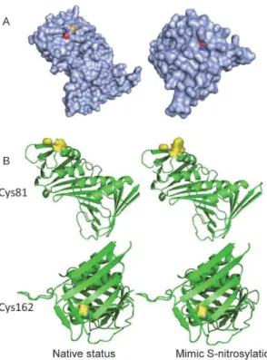
The activation of caspase-9 is an initial event in the intrinsic apoptosis pathway. It is gener-ally accepted that the activation requires binding of its precursor, procaspase-9, to a complex of two proteins, APAF-1 and cytochrome c, and the self-cleavage specific aspartic residues of pro-caspase-9. Cleaved caspase-9 further processes other caspase members, including caspase-3 and caspase-7, which leads to the initiation of the apoptotic pathway. However, the activation of caspase-9 is restricted within the triple protein complex and mediated by several protein components or posttranslational modifications, such as HBXIP [60], TUCAN [61], ERK2 [62], and PKA [63]. XIAP has been reported to directly interact with caspase-9, and the binding of XIAP/caspase-9 has opposing effects on caspase activity and apoptosis [64]. Furthermore, pre-vious studies have found that c-Abl binds directly to caspase-9 and the bound c-Abl phosphor-ylates caspase-9 on Tyr153 and further regulates the caspase-9-related apoptotic response to genotoxic stress [65]. With the exception that they are all kinases, these caspase-9-interacting proteins do not appear to have a specific biochemical function, as TUCAN is located on the ER and HBXIP and XIAP are related to virus proteins. This study contributed new proteins to the list of proteins that interact with caspase-9. Compared with the early observations, the Fig 6. Potential structural consequences of S-nitrosylation for PCNA.(A) The surface accessibility of S-nitrosylation sites. The sulfhydryl group (yellow) of Cys81 was exposed at the protein surface (left), but that of Cys162 was buried inside (right). (B) Molecular simulations of wild-type and SNOC-modified PCNAs. S-nitrosylation at Cys81 clearly affected the local structure of this residue, which may interfere with the interactions, but the Cys162 modification had no significant influence on the structure.
interaction of PCNA and caspase-9 provides two important findings with regards to apoptosis. First, the apoptosome complex contains three key proteins, but it also has multiple associated proteins that interact with the apoptosome proteins based on cell types or biological stimuli. PCNA was shown to interact with caspase-9 in SH-SY5Y cells; however, whether the interac-tion exists globally in many cells must be addressed in future research. Because our data suggest that SNO proteins could exert influences on the apoptotic pathway, NO modification of the apoptosome should be considered in future research.
Supporting Information
S1 Fig. Detection of nNOS expression under the indicated treatments.SH-SY5Y cells in the low-serum media were pretreated with L-NMMA followed by the addition of rotenone. The levels of nNOS under the different treatments were detected by Western blot.
(TIF)
S2 Fig. Evaluation of the potential S-nitrosylation for caspase-9 in SH-SY5Y cells treated with rotenone.The lysate of SH-SY5Y cells with and without rotenone treatment was obtained and subjected to BST. The biotinylated proteins were pulled down with NeutrAvidin beads and detected by Western blot using anti-caspase-9.
(TIF)
S3 Fig. Representative MS/MS spectra for the PCNA peptides that contained the biotiny-lated Cys81 (upper) and Cys162 (lower).δC represents biotin-HPDP derivatized cysteine (+428), which was included in the b- or y-ion series.
(TIF)
S1 Table. The up-regulated SNO-proteins of SH-SY5Y cells in response to rotenone treat-ment.
(DOCX)
S2 Table. The proteins potentially interact with cytosolic PCNA in SH-SY5Y cells. (DOCX)
Author Contributions
Conceived and designed the experiments: LY ZG SL. Performed the experiments: LY XL JZ. Analyzed the data: HS YX. Contributed reagents/materials/analysis tools: LY SY. Wrote the paper: LY SL.
References
1. Michel PP, Ruberg M, Hirsch E (2006) Dopaminergic neurons reduced to silence by oxidative stress: an early step in the death cascade in Parkinson’s disease? Sci STKE 332: pe19.
2. Thomas B, Beal MF (2007) Parkinson’s disease. Hum Mol Genet 16: R183–R194. DOI:10.1093/hmg/ ddm159PMID:17911161
3. Singh S, Dikshit M (2007) Apoptotic neuronal death in Parkinson’s disease: Involvement of nitric oxide. Brain Res Rev 54: 233–250. PMID:17408564
4. Mochizuki H, Goto K, Mori H, Mizuno Y (1996) Histochemical detection of apoptosis in Parkinson’s dis-ease. J Neurol Sci 137: 120–123. PMID:8782165
5. Yang L, Matthews RT, Schulz JB, Klockgether T, Liao AW, et al. (1998) 1-Methyl-4-phenyl-tetrahydro-pyridine neurotoxicity is attenuated in mice over expressing bcl-2. J Neurosci 18: 8145–8152. PMID: 9763461
7. Casey DP, Walker BG, Ranadive SM, Taylor JL, Joyner MJ (2013) Contribution of nitric oxide in the contraction-induced rapid vasodilation in young and older adults. J Appl Physiol 115: 446–455. doi:10. 1152/japplphysiol.00446.2013PMID:23788575
8. Phattanarudee S, Towiwat P, Maher TJ, Ally A (2013) Effects of medullary administration of a nitric oxide precursor on cardiovascular responses and neurotransmission during static exercise following is-chemic stroke. Can J Physiol Pharmacol 91: 510–520. doi:10.1139/cjpp-2013-0066PMID:23826997
9. Kolios G, Valatas V, Ward SG (2004) Nitric oxide in inflammatory bowel disease: a universal messen-ger in an unsolved puzzle. Immunology 113: 427–437. PMID:15554920
10. Tieu K, Ischiropoulos H, Przedborski S (2003) Nitric oxide and reactive oxygen species in Parkinson’s disease. IUBMB Life 55: 329–335. PMID:12938735
11. Tsang AH, Chung KK (2009) Oxidative and nitrosative stress in Parkinson’s disease. Biochim Biophys Acta-Mol Basis Dis 1792: 643–650. doi:10.1016/j.bbadis.2008.12.006PMID:19162179
12. Zhang L, Dawson VL, Dawson TM (2006) Role of nitric oxide in Parkinson’s disease. Pharmacol Thera-peut 109: 33–41.
13. Barthwal MK, Srivastava N, Dikshit M (2001) Role of nitric oxide in a progressive neurodegeneration model of Parkinson’s disease in the rat. Redox Rep 6: 297–302. PMID:11778847
14. Gatto EM, Riobo AN, Carreras CM, Chernavsky A, Rubio A, et al. (2000) Overexpression of neutrophil neuronal nitric oxide synthase in Parkinson’s disease. Nitric Oxide 4: 534–539. PMID:11020342
15. He Y, Imam SZ, Dong Z, Jankovic J, Ali SF, et al. (2003) Role of nitric oxide in rotenone-induced nigros-triatal injury. J Neurochem 86: 1338–1345. PMID:12950443
16. Gatto EM, Carreras MC, Pargament GA, Riobo NA, Reides C et al. (1996) Neutrophil function, nitric oxide, and blood oxidative stress in Parkinson’s disease. Mov Disord 11: 261–267. PMID:8723142
17. Hess DT, Matsumoto A, Kim SO, Marshall HE, Stamler JS (2005) Protein S-nitrosylation: purview and parameters. Nat Rev Mol Cell Biol 6: 150–166. PMID:15688001
18. Cho DH, Nakamura T, Fang J, Cieplak P, Godzik A, et al. (2009) S-Nitrosylation of Drp1 mediates beta-amyloid-related mitochondrial fission and neuronal injury. Science 324: 102–105. doi:10.1126/ science.1171091PMID:19342591
19. Fang J, Nakamura T, Cho DH, Gu Z, Lipton SA (2007) S-Nitrosylation of peroxiredoxin 2 promotes oxi-dative stress-induced neuronal cell death in Parkinson’s disease. Proc Natl Acad Sci USA 104: 18742–18747. PMID:18003920
20. Hara MR, Agrawal N, Kim SF, Cascio MB, Fujimuro M, et al. (2005) S-nitrosylated GAPDH initiates ap-optotic cell death by nuclear translocation following Siah1 binding. Nat Cell Biol 7: 665–674. PMID: 15951807
21. Tsang AH, Lee YI, Ko HS, Savitt JM, Pletnikova O, et al. (2009) S-Nitrosylation of XIAP compromises neuronal survival in Parkinson’s disease. Proc Natl Acad Sci USA 106: 4900–4905. doi:10.1073/pnas. 0810595106PMID:19273858
22. Watabe M, Nakaki T (2004) Rotenone induces apoptosis via activation of bad in human dopaminergic SH-SY5Y cells. J Pharmacol Exp Ther 311: 948–953. PMID:15280438
23. Gandhi S, Wood NW (2005) Molecular pathogenesis of Parkinson’s disease. Hum Mol Genet 14: 2749–2755. PMID:16278972
24. Shaikh SB, Nicholson LF (2009) Effects of chronic low dose rotenone treatment on human microglial cells. Mol Neurodegener 4: 55–68. doi:10.1186/1750-1326-4-55PMID:20042120
25. Ortiz-Ortiz MA, Morán JM, González-Polo RA, Niso-Santano M, Soler G, et al. (2009) Nitric oxide-medi-ated toxicity in paraquat-exposed SH-SY5Y cells: a protective role of 7-nitroindazole. Neurotox Res 16: 160–173. doi:10.1007/s12640-009-9065-6PMID:19526292
26. Jiang ZL, Fletcher NM, Diamond MP, Abu-Soud HM, Saed GM (2009) S-Nitrosylation of caspase-3 is the mechanism by which adhesion fibroblasts manifest lower apoptosis. Wound Repair Regen 17: 224–229. doi:10.1111/j.1524-475X.2009.00459.xPMID:19320891
27. Kim YM, Kim TH, Chung HT, Talanian RV, Yin XM, et al. (2000) Nitric oxide prevents tumor necrosis factor alpha-induced rat hepatocyte apoptosis by the interruption of mitochondrial apoptotic signaling through S-nitrosylation of caspase-8. Hepatology 32: 770–778. PMID:11003621
28. Mannick JB, Schonhoff C, Papeta N, Ghafourifar P, Szibor M, et al. (2001) S-Nitrosylation of mitochon-drial caspases. J Cell Biol 154: 1111–1116. PMID:11551979
30. Nakamura T, Wang L, Wong CC, Scott FL, Eckelman BP, et al. (2010) Transnitrosylation of XIAP regu-lates caspase-dependent neuronal cell death. Mol Cell 39: 184–195. doi:10.1016/j.molcel.2010.07. 002PMID:20670888
31. Torta F, Usuelli V, Malgaroli A, Bachi A (2008) Proteomic analysis of protein S-nitrosylation. Proteomics 8: 4484–4494. doi:10.1002/pmic.200800089PMID:18846506
32. Komatsubara AT, Asano T, Tsumoto H, Shimizu K, Nishiuchi T, et al. (2012) Proteomic analysis of S-nitrosylation induced by 1-methyl-4-phenylpyridinium (MPP+). Proteome Sci 10: 74–81. doi:10.1186/ 1477-5956-10-74PMID:23273257
33. Petit C, Bernardes-Genisson V, Hoffmann P, Souchard J, Labidalle S (1999) Novel donors of nitric oxide derived of S-nitrosocysteine possessing antioxidant activities. Braz J Med Biol Res 32: 1407–1412. PMID:10559842
34. Jaffrey SR, Snyder SH (2001) The biotin switch method for the detection of S-nitrosylated proteins. Sci STKE DOI:10.1126/stke.2001.86.pl1PMID:11865191
35. Camacho-Carvajal MM, Wollscheid B, Aebersold R, Steimle V, Schamel WW (2004) Two-dimensional Blue native/SDS gel electrophoresis of multi-protein complexes from whole cellular lysates: a proteo-mics approach. Mol Cell Proteoproteo-mics 3: 176–182. PMID:14665681
36. Chen JS, Chen KT, Fan CW, Han CL, Chen YJ, et al. (2010) Comparison of membrane fraction proteo-mic profiles of normal and cancerous human colorectal tissues with gel-assisted digestion and iTRAQ labeling mass spectrometry. FEBS J 277: 3028–3038. doi:10.1111/j.1742-4658.2010.07712.xPMID: 20546304
37. Zhang X, Wen Z, Mi X (2014) Expression and anti-apoptotic function of TRAF4 in human breast cancer MCF-7 cells. Oncol Lett 7: 411–414. PMID:24396457
38. McStay GP, Salvesen GS, Green DR (2008) Overlapping cleavage motif selectivity of caspases: impli-cations for analysis of apoptotic pathways. Cell Death Differ 15: 322–331. PMID:17975551
39. Huang B, Chen BC, Wang DL (2009) Shear flow increases S-nitrosylation of proteins in endothelial cells. Cardiovasc Res 83: 536–546. doi:10.1093/cvr/cvp154PMID:19447776
40. Zhang HH, Feng L, Livnat I, Hoh JK, Shim JY, et al. (2010) Estradiol-17βstimulates specific receptor and endogenous nitric oxide-dependent dynamic endothelial protein S-nitrosylation: analysis of endo-thelial nitrosyl-proteome. Endocrinology 151: 3874–3887. doi:10.1210/en.2009-1356PMID: 20519370
41. Zhang HH, Wang YP, Chen DB (2011) Analysis of nitroso-proteomes in normotensive and severe pre-eclamptic Human Placentas. Biol Reprod 84: 966–975. doi:10.1095/biolreprod.110.090688PMID: 21228217
42. Qu Z, Meng F, Zhou H, Li J, Wang Q, et al. (2014) NitroDIGE analysis reveals inhibition of protein S-nitrosylation by epigallocatechin gallates in lipopolysaccharide-stimulated microglial cells. J Neuroin-flammation. doi:10.1186/1742-2094-11-17PMID:24472655
43. Witko-Sarsat V, Mocek J, Bouayad D, Tamassia N, Ribeil JA, et al. (2010) Proliferating cell nuclear anti-gen acts as a cytoplasmic platform controlling human neutrophil survival. J Exp Med 207: 2631–2645. doi:10.1084/jem.20092241PMID:20975039
44. Yao D, Gu Z, Nakamura T, Shi ZQ, Ma Y, et al. (2004) Nitrosative stress linked to sporadic Parkinson’s disease: S-nitrosylation of parkin regulates its E3 ubiquitin ligase activity. Proc Natl Acad Sci U S A 101:10810–10814. PMID:15252205
45. Singh S, Das T, Ravindran A, Chaturvedi RK, Shukla Y, et al. (2005) Involvement of nitric oxide in neu-rodegeneration: a study on the experimental models of Parkinson’s disease. Redox Rep 10:103–109. PMID:15949131
46. Zhang ZT, Zhang ZH, Wang HC, Zhang GX, Hu D, et al. (2014) Proliferating Cell Nuclear Antigen Binds DNA Polymerase-beta and Mediates 1-Methyl-4-Phenyl- pyridinium-Induced Neuronal Death. PLoS One 9(9):e106669. doi:10.1371/journal.pone.0106669PMID:25184665
47. Ito G, Ariga H, Nakagawa Y, Iwatsubo T (2006) Roles of distinct cysteine residues in S-nitrosylation and dimerization of DJ-1. Biochem Biophys Res Commun 339: 667–672. PMID:16316629
48. Hashemy SI, Holmgren A (2008) Regulation of the catalytic activity and structure of human thioredoxin 1 via oxidation and S-nitrosylation of cysteine residues. J Biol Chem 283: 21890–21898. doi:10.1074/ jbc.M801047200PMID:18544525
49. Sumbayev VV (2003) S-nitrosylation of thioredoxin mediates activation of apoptosis signal-regulating kinase 1. Arch Biochem Biophys 415: 133–136. PMID:12801522
51. Lam YW, Yuan Y, Isaac J, Babu CV, Meller J, et al. (2010) Comprehensive identification and modified-Site mapping of S-nitrosylated targets in prostate epithelial cells. PLoS One doi:10.1371/journal.pone. 0014462PMID:21283510
52. Shi Q, Feng JH, Qu HB, Cheng YY (2008) A proteomic study of S-nitrosylation in the rat cardiac pro-teins in vitro. Biol Pharm Bull 31: 1536–1540. PMID:18670085
53. Kohr MJ, Sun J, Aponte A, Wang G, Gucek M, et al. (2011) Simultaneous measurement of protein oxi-dation and S-nitrosylation during preconditioning and ischemia/reperfusion injury with resin-assisted capture. Circ Res 108: 418–426. doi:10.1161/CIRCRESAHA.110.232173PMID:21193739
54. Kohr MJ, Aponte A, Sun J, Gucek M, Steenbergen C, et al. (2012) Measurement of S-nitrosylation oc-cupancy in the myocardium with cysteine-reactive tandem mass tags: short communication. Circ Res 111: 1308–1312. doi:10.1161/CIRCRESAHA.112.271320PMID:22865876
55. Wang SC, Nakajima Y, Yu YL, Xia W, Chen CT, et al. (2006) Tyrosine phosphorylation controls PCNA function through protein stability. Nat Cell Biol 8: 1359–1368. PMID:17115032
56. Yu Y, Cai JP, Tu B, Wu L, Zhao Y, et al. (2009) Proliferating cell nuclear antigen is protected from deg-radation by forming a complex with MutT Homolog2. J Biol Chem 284: 19310–19320. doi:10.1074/jbc. M109.015289PMID:19419956
57. Zhang Z, Zhang S, Lin SH, Wang X, Wu L, et al. (2012) Structure of monoubiquitinated PCNA: implica-tions for DNA polymerase switching and Okazaki fragment maturation. Cell Cycle 11: 2128–2136. doi: 10.4161/cc.20595PMID:22592530
58. Doulias PT, Greene JL, Greco TM, Tenopoulou M, Seeholzer SH, et al. (2010) Structural profiling of en-dogenous S-nitrosocysteine residues reveals unique features that accommodate diverse mechanisms for protein S-nitrosylation. Proc Natl Acad Sci USA 107: 16958–63. doi:10.1073/pnas.1008036107 PMID:20837516
59. Palmer ZJ, Duncan RR, Johnson JR, Lian LY, Mello LV, et al. (2008) S-nitrosylation of syntaxin 1 at Cys(145) is a regulatory switch controlling Munc18–1 binding. Biochem J 413: 479–91. doi:10.1042/ BJ20080069PMID:18452404
60. Marusawa H, Matsuzawa S, Welsh K, Zou H, Armstrong R, et al. (2003) HBXIP functions as a cofactor of survivin in apoptosis suppression. EMBO J 22: 2729–2740. PMID:12773388
61. Pathan N, Marusawa H, Krajewska M, Matsuzawa S, Kim H, et al. (2001) TUCAN, an antiapoptotic cas-pase-associated recruitment domain family protein overexpressed in cancer. J Biol Chem 276: 32220–32229. PMID:11408476
62. Martin MC, Allan LA, Mancini EJ, Clarke PR (2008) The docking interaction of caspase-9 with ERK2 provides a mechanism for the selective inhibitory phosphorylation of caspase-9 at threonine 125. J Biol Chem 283: 3854–3865. PMID:18083711
63. Martin MC, Allan LA, Lickrish M, Sampson C, Morrice N, et al. (2005) Protein kinase A regulates cas-pase-9 activation by Apaf-1 downstream of cytochrome c. J Biol Chem 280: 15449–15455. PMID: 15703181
64. Srinivasula SM, Hegde R, Saleh A, Datta P, Shiozaki E, et al. (2001) A conserved XIAP-interaction motif in caspase-9 and Smac/DIABLO regulates caspase activity and apoptosis. Nature 410: 112–116. PMID:11242052