Prevalence and Impact of Sleep Disordered
Breathing in Patients with Severe Aortic
Stenosis
Markus Linhart*, Jan-Malte Sinning, Alexander Ghanem, Finny J. Kozhuppakalam, Rebecca Fistéra, Christoph Hammerstingl, Carmen Pizarro, Eberhard Grube, Nikos Werner, Georg Nickenig, Dirk Skowasch
Medizinische Klinik und Poliklinik II, Universitätsklinik Bonn, Sigmund-Freud-Str. 25, 53127 Bonn, Germany
Abstract
Background
Unlike the well-established association between sleep disordered breathing (SDB) and chronic heart failure, the relationship between SDB and severe aortic stenosis (AS) is not well investigated. Given the increasing prevalence of AS, and the improving prognosis of high risk AS patients attributable to transcatheter aortic valve implantation (TAVI), the prev-alence and impact of SDB needs to be better understood.
Methods and Results
In this study, 140 patients with severe AS underwent polygraphy prior to TAVI. Clinical and hemodynamic parameters were recorded. Patients were followed for 573±405 days. We
found that 99/140 patients (71%) had SDB with a mean apnoea-hypopnoea-index of 24±17/
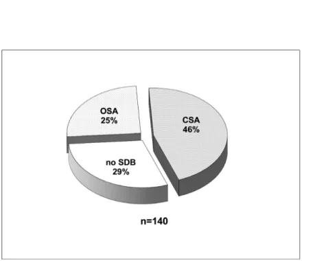
h. SDB was mild in 27%, moderate in 23% and severe in 21% of patients. In addition, 35 patients (25%) had obstructive sleep apnoea (OSA), whereas 64 patients (46%) had central sleep apnoea (CSA). Patients with OSA had predominantly mild SDB (20/38 pts.), and patients with CSA mostly had severe SDB (24/29 pts.). The prevalence and distribution of OSA and CSA were independent of left ventricular function. Overall, 1 and 2 year survival rates (74% and 71%, resp.) did not differ significantly between patients without SDB or those with OSA and CSA (p=0.81).
Conclusions
SDB, with a preponderance of CSA, was found to be highly prevalent in patients with high-grade AS scheduled for TAVI. SDB prevalence was independent of left ventricular function. Mortality after TAVI was not influenced by the type or severity of SDB.
OPEN ACCESS
Citation:Linhart M, Sinning J-M, Ghanem A, Kozhuppakalam FJ, Fistéra R, Hammerstingl C, et al. (2015) Prevalence and Impact of Sleep Disordered Breathing in Patients with Severe Aortic Stenosis. PLoS ONE 10(7): e0133176. doi:10.1371/journal. pone.0133176
Editor:Marc W. Merx, KRH Robert Koch Klinikum Gehrden, GERMANY
Received:January 7, 2015
Accepted:June 23, 2015
Published:July 27, 2015
Copyright:© 2015 Linhart et al. This is an open access article distributed under the terms of the
Creative Commons Attribution License, which permits unrestricted use, distribution, and reproduction in any medium, provided the original author and source are credited.
Data Availability Statement:All relevant data are within the paper.
Funding:The authors received no specific funding for this work.
Introduction
There are many associations between cardiovascular diseases and sleep disordered breathing (SDB). In particular, the obstructive form of SDB (obstructive sleep apnoea, OSA) has been identified as an important risk factor for a variety of cardiovascular diseases such as arterial hypertension, coronary artery disease and atrial fibrillation [1]. On the other hand, central sleep apnoea (CSA) has mainly been associated with chronic congestive heart failure (CHF) and carries a significantly adverse prognosis [2]. Studies have shown that treatment of the asso-ciated cardiac condition can improve the underlying sleep disorder, and vice versa [3–6]. How-ever, preliminary data from the recent SERVE-HF trial showed an increase in cardiovascular mortality in patients with heart failure with reduced ejection fraction and predominant CSA that were treated by adaptive servo-ventilation (http://www.servehf.com) [7]. To date, scarce data exist about the relationship between valvular heart disease and SDB [8–10]. Aortic stenosis is the most frequent valvular heart disease and its prevalence is expected to increase due to aging populations [11]. The aim of this study was to investigate the frequency of SDB in patients with severe aortic stenosis and its impact on mortality after transcatheter aortic valve implantation (TAVI). We found that patients with severe aortic stenosis scheduled for TAVI had a high prevalence of SDB with a preponderance of CSA and that this was not associated with outcomes after TAVI.
Methods
Study Design and Patient Selection
A total of 140 patients with high-grade aortic stenosis who were evaluated for TAVI at our cen-tre between 2010 and 2013 were screened prospectively for our study using polygraphy. The study was approved by the local medical ethics committee (Ethikkommission an der Medizi-nischen Fakultät der RheiMedizi-nischen Friedrich-Wilhelms-Universität Bonn, project approval no. 255/08). Informed written consent was obtained from all patients. All patients underwent our standard preoperative diagnostic tests, which included comprehensive 2D- and 3D-transtho-racic and transesophageal echocardiography (Philips iE 33 ultrasound system, Amsterdam, The Netherlands) as well as coronary angiography with invasive determination of left ventricu-lar end-diastolic pressure (LVEDP) [12,13]. Left ventricular end-diastolic volume, left ventric-ular end-systolic volume, left ventricventric-ular ejection fraction (LVEF) and left atrial volume were determined by 2D echocardiography. Right ventricular systolic pressure was estimated on the basis of the modified Bernoulli equation and was considered equal to the systolic pulmonary artery pressure (sPAP). Body surface area was calculated using the DuBois formula. The 30-day mortality risk was estimated by means of the logistic EuroSCORE (www.euroscore.org) and the Society of Thoracic Surgeons (STS) score ((http://riskcalc.sts.org/) algorithms that are based on the presence of coexisting illnesses.[14] Patients were followed on an outpatient basis 180 and 365 days after the procedure and every year thereafter. Patients who did not come to their follow-up appointments were interviewed by telephone.
Sleep Study and Scoring
defined as a cessation of airflow or a>90% reduction in airflow from baseline for>10 seconds.
Hypopnoea was defined as a reduction in airflow of>30% with an oxygen desaturation of
4%. The apnoea–hypopnoea index (AHI) was defined as the number of episodes of apnoea and hypopnoea per hour. Patients with an AHI>5/h were considered to have SDB [15], which
was subclassified into mild, moderate, and severe forms with 5–14.9, 15–29.9, and30 events/ hour, respectively. Events were scored manually by an experienced sleep laboratory specialist. Events were classified as obstructive when the airflow criteria were met and thoracic or abdom-inal respiratory effort was documented; otherwise they were classified as central. An episode of SDB in which more than 50% of the events were central was defined as CSA whereas those in which50% of the events were obstructive was defined as OSA. Mixed apnoea was defined as an absence of nasal airflow associated with no respiratory effort followed by the resumption of inspiratory effort in the second portion of the event. These events were classified as part of the OSA group [16]. The central apnoea index (CAI) and the obstructive apnoea index (OAI) were calculated as the mean number of episodes of central and obstructive apnoea events per hour, respectively. The oxygen desaturation index (ODI) was defined as the number of oxygen level drops of3% from baseline per hour. The hypopnoea index (HI) was defined as the number of hypopnoeas per hour. Sleep-related symptoms were documented with the German version of the self-rating Epworth Sleepiness Scale questionnaire (ESS) [17]. A result in the 0–9 range was considered normal whereas a result in the 10–24 range indicated daytime sleepiness [18].
Statistical Analysis
Continuous variables are expressed as the mean ± one standard deviation and were analysed by a two-sided unpaired Student’st-test. For values of N-terminal pro–B-type natriuretic
pep-tide with a highly skewed distribution, statistical significance was calculated using the Wil-coxon–Mann–Whitney test. Categorical variables were compared by Fisher’s exact test or the chi-square test for trend, as appropriate. Correlation between measures was calculated using Spearman’s rank correlation. Survival rates were compared with the log-rank and Breslow test. Statistical analyses were performed with SPSS software version 22.0 (IBM Inc., Armonk, New York, USA). A p value<0.05 was considered statistically significant.
Results
Baseline characteristics
140 patients were included in the study. The mean age of the patients was 81±6 years, and 52% of the patients were male. The aortic valve areas were narrowed to an average of 0.39±0.11 cm2/m2with a mean peak-to-peak gradient of 47±24 mmHg. Mean EuroSCORE was 23±16%. Patients were not obese, with a mean BMI of 26±4 kg/m2. On average, renal function was only mildly impaired (eGFR 55±22 ml/min) and mean left ventricular systolic function was only mildly reduced (54±17%).Table 1displays the clinical and hemodynamic parameters of the patients at baseline.
Prevalence of sleep-disordered breathing
The prevalence of SDB as defined by an AHI>5/h was 71% (99/140 pts.). Mean AHI in
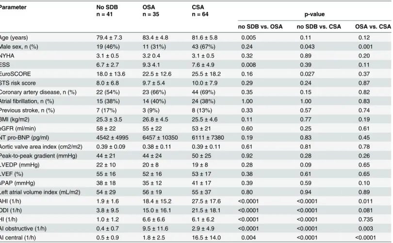
Table 1. Baseline patient characteristics.
n = 140
Age (years) 81.4±6.2
Male sex, n (%) 73 (52%)
NYHA 3.1±0.5
ESS 7.8±4.3
EuroSCORE 22.6±15.8
STS risk score 9.4±7.0
Coronary artery disease, n (%) 89 (64%)
Atrialfibrillation, n (%) 53 (38%)
Previous stroke, n (%) 18 (13%)
BMI (kg/m2) 25.8±4.3
eGFR (ml/min) 55±22
NT pro-BNP (pg/ml) 5763±7704
Aortic valve area index (cm2/m2) 0.39±0.11
Peak-to-peak gradient (mmHg) 47±24
LVEDP (mmHg) 20±9
LVEF (%) 54±17
sPAP (mmHg) 38±16
Left atrial volume index (mL/m2) 55±31
ESS Epworth sleepiness scale, STS Society of Thoracic Surgeons, BMI body mass index, LVEF left ventricular ejection fraction; LVEDP, left ventricular end-diastolic pressure, sPAP systolic pulmonary artery pressure.
doi:10.1371/journal.pone.0133176.t001
Fig 1. Prevalence and type of sleep disordered breathing (SDB) in 140 patients with severe aortic stenosis.
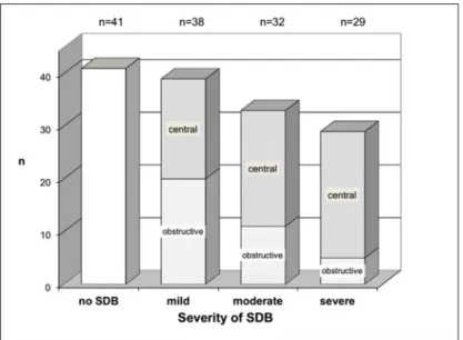
with increasing SDB severity. The ratio of CSA to OSA in mild SDB was approximately 1:1 whereas it reached almost 5:1 in severe SDB (Fig 2).
Relationship between SDB and LVEF as well as other clinical and
hemodynamic parameters
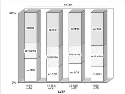
The prevalence of SDB and distribution of its subtypes (OSA and CSA) was independent of the left ventricular ejection fraction. Mean LVEF was not different between the groups (Table 2). When stratified into categories of normal, mildly reduced, moderately reduced and severely reduced LVEF, there was a similar distribution of SDB in each category with no statistically sig-nificant differences (Chi-square test for independence, p = 0.95,Fig 3). Likewise, LVEDP and NT pro-BNP levels were not significantly different between the groups (Table 2). Although renal dysfunction may have been a confounder for the raised NT pro-BNP levels, our findings remained consistent even when patients with renal dysfunction was excluded from the analysis (p = 0.56).
There was a weak but significant positive correlation between AHI and left atrial volume index (r = 0.31, p = 0.02) in the CSA but not the OSA group. No significant correlations were seen between SDB, in terms of the overall group as well as the OSA and CSA subgroups, and multiple clinical parameters including LVEF, LVEDP, NT pro-BNP and sPAP.
Relation of SDB to outcome after TAVI
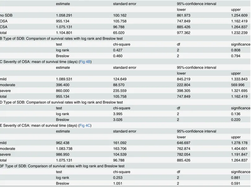
Median patient follow-up was 573±405 days. A total of 11 patients were lost to follow-up. Overall, one and two year survival rates were 74.4% and 71.3%, respectively. The rates of sur-vival were not significantly different in patients without SDB and those with OSA or CSA (p = 0.81,Fig 4A,Table 3A and 3B). There was also no significant difference observed in the survival rates of patients with OSA or CSA with regard to the severity of SDB (p = 0.14 and p = 0.88, resp.,Fig 4B and 4C,Table 3C–3F).
Fig 2. Distribution of sleep disordered breathing.With increasing severity of sleep disordered breathing (SDB), the prevalence of the central subtype increases at the expense of the obstructive subtype of SDB. Severity is indicated by the apnoea-hypopnoea-index: mild 5–14.9/h, moderate 15–29.9/h, severe>30/h.
Discussion
This is the largest study to date investigating SDB in patients with severe aortic stenosis. The main findings are:
1. There is a high prevalence of SDB (71%) in this patient population, with a preponderance of the CSA subtype (46%).
2. With increasing severity of SDB as defined by AHI, the prevalence of CSA increases at the expense of OSA.
3. The prevalence and distribution of SDB is independent of the left ventricular function and not associated with survival after TAVI.
Unlike in CHF, arterial hypertension or atrial fibrillation, the association between valvular heart disease and SDB is not well characterised. Aortic stenosis is the most common valvular heart disease in developed countries and its impact on public health and health care resources is expected to grow due to aging populations [11]. Concurrently, because of the development of TAVI, an increasing number of AS patients with high or excessive surgical risk can be Table 2. Comparison of clinical and hemodynamic parameters between patients without SDB or with OSA or CSA.
Parameter No SDB OSA CSA
n = 41 n = 35 n = 64 p-value
no SDB vs. OSA no SDB vs. CSA OSA vs. CSA
Age (years) 79.4±7.3 83.4±4.8 81.6±5.8 0.005 0.11 0.12
Male sex, n (%) 19 (46%) 11 (31%) 43 (67%) 0.24 0.043 0.001
NYHA 3.1±0.5 3.2 0.4 3.1±0.5 0.32 0.89 0.20
ESS 6.7±2.7 9.3 4.1 7.6±4.9 0.008 0.39 0.11
EuroSCORE 18.0±13.6 22.5±12.6 25.5±18.2 0.16 0.027 0.37
STS risk score 8.0±6.8 9.7±5.4 10.0±7.9 0.29 0.24 0.87
Coronary artery disease, n (%) 22 (54%) 23 (66%) 44 (69%) 0.35 0.15 0.82
Atrialfibrillation, n (%) 15 (38%) 14 (40%) 24 (38%) 1.00 1.00 0.83
Previous stroke, n (%) 7 (17%) 3 (9%) 8 (13%) 0.33 0.57 0.74
BMI (kg/m2) 25.3±3.5 26.8±4.5 25.5±4.6 0.11 0.77 0.19
eGFR (ml/min) 58±22 55±22 53±21 0.60 0.25 0.61
NT pro-BNP (pg/ml) 4542±4995 6457±10350 6111±7380 0.19 0.83 0.45
Aortic valve area index (cm2/m2) 0.39±0.09 0.38±0.11 0.39±0.11 0.61 0.81 0.78
Peak-to-peak gradient (mmHg) 44±21 44±24 50±25 0.92 0.28 0.26
LVEDP (mmHg) 22±10 20±8 19±8 0.28 0.09 0.65
LVEF (%) 55±16 52±16 53±17 0.38 0.61 0.65
sPAP (mmHg) 38±18 35±12 41±17 0.39 0.59 0.10
Left atrial volume index (mL/m2) 54±29 56±19 55±37 0.80 0.94 0.89
AHI (1/h) 1.9±1.6 18.4±15.2 27.5±17.6 <0.0001 <0.0001 0.011
ODI (1/h) 3.8±9.5 15.0±16.1 21.5±18.1 <0.0001 <0.0001 0.081
HI (1/h) 1.0±1.2 6.6±6.6 6.1±6.2 <0.0001 <0.0001 0.735
AI obstructive (1/h) 0.4±0.7 9.5±11.6 2.9±4.9 <0.0001 <0.0001 0.003
AI central (1/h) 0.5±0.9 1.8±2.5 16.5±14.0 0.004 <0.0001 <0.0001
ESS Epworth sleepiness scale, STS Society of Thoracic Surgeons, BMI body mass index, eGFR estimated glomerularfiltration rate, LVEF left ventricular ejection fraction, LVEDP left ventricular end-diastolic pressure, sPAP systolic pulmonary artery pressure, AHI apnea-hypopnea-index, ODI oxygen desaturation index, HI hypopnoea index, AI apnoea index.
effectively treated. TAVI can therefore greatly improve life expectancy in these patients [19]. With ongoing technical improvements, TAVI is expected to become a good alternative to sur-gery for an increasingly broader range of patients [19]. Therefore, the significance of SDB in these patients needs to be evaluated. Such studies would also allow for greater insights into the mechanism of SDB in cardiac disease.
Prevalence of SDB
We found that there is a very high prevalence of SDB in the AS patient population, with an overall prevalence of 71%. This finding confirms a previous, smaller report, where SDB was found in 30/42 patients with severe aortic stenosis [9]. The prevalence of SDB in AS patients is thus similar to the prevalence in SDB in CHF patients as described in previous studies. For instance, a study of 700 patients with CHF, as defined by NYHA classII and LVEF40%, found similar rates of SDB as our study both in terms of the overall prevalence rate as well as Fig 3. Distribution of sleep apnoea, and its central and obstructive subtypes, is independent of left ventricular ejection fraction.
doi:10.1371/journal.pone.0133176.g003
Fig 4. Kaplan-Meier estimate of survival.Kaplan-Meier estimates of survival with regard to the type of sleep disordered breathing (SDB, Panel A) and the severity of obstructive (OSA, Panel B) and central sleep apnoea (CSA, Panel C), respectively, show no significant differences in mortality between the groups. Mild SDB: AHI 5–14.9/h, moderate SDB: AHI 15–29.9/h, severe SDB AHI>30/h.
the distribution of CSA and OSA [20]. In our study, CSA accounts for the majority of moderate and severe cases of SDB (46/61 pts., 75%) whereas OSA prevails in the mild forms (20/38 pts., 53%). By nature, patients eligible for TAVI are of advanced age. In our study cohort, mean age was 81±6 years. It has been suggested that SDB is age-dependent in elderly individuals whereas it seems to be age-related in middle age individuals [21,22]. In a recent study in 298 elderly women with a mean age of 82±3 years, 35% had an AHI>15/h, as compared with 44% of
patients in our study [23]. Taking together the available data, SDB seems to be highly prevalent also in the general elderly population. However, definition and diagnosis of SDB and its sub-types were varying in the studies, and details on cardiovascular comorbidities were not reported. Thus, to date it remains unclear to what proportion of SDB is age-dependent and what is attributable to severe cardiac disease such as CHF or severe aortic stenosis in this popu-lation. Our findings demonstrate that SDB, in particular CSA, constitutes a frequent comorbid-ity in patients with severe aortic stenosis and requires further investigation as part of the clinical workup.
Table 3. Statistics of survival analysis.
A Type of SDB: mean of survival time (days) (Fig 4A)
estimate standard error 95%-confidence interval
lower upper
no SDB 1.058.291 100.162 861.973 1.254.609
OSA 955.134 105.758 747.849 1.162.419
CSA 1.075.131 96.788 885.426 1.264.837
total 1.104.801 65.020 977.362 1.232.239
B Type of SDB: Comparison of survival rates with log rank and Breslow test
test chi-square df significance
log rank 0.427 2 0.808
Breslow 0.460 2 0.794
C Severity of OSA: mean of survival time (days) (Fig 4B)
estimate standard error 95%-confidence interval
lower upper
mild 1.089.531 124.649 845.219 1.333.843
moderate 396.400 88.570 222.804 569.996
severe 860.000 235.559 398.305 1.321.695
total 955.134 105.758 747.849 1.162.419
D Type of SDB: Comparison of survival rates with log rank and Breslow test
test chi-square df significance
log rank 3.995 2 0.136
Breslow 3.026 2 0.220
E Severity of CSA: mean of survival time (days) (Fig 4C)
estimate standard error 95%-confidence interval
lower upper
mild 962.438 161.092 646.697 1.278.178
moderate 1.083.738 163.706 762.874 1.404.601
severe 986.950 104.539 782.054 1.191.847
total 1.075.131 96.788 885.426 1.264.837
3F Type of SDB: Comparison of survival rates with log rank and Breslow test
test chi-square df significance
log rank 0.253 2 0.881
Breslow 1.051 2 0.591
SDB and left ventricular function
Interestingly, the proportion of SDB and its subtypes was found to be independent of left ven-tricular systolic and diastolic function. Mean LVEF was not different between the groups, nor was the distribution of SDB when stratified according to the degree of LVEF impairment
(Table 2,Fig 3). SDB was also present in a considerable number of patients with normal LVEF.
Likewise, NT pro-BNP was strongly elevated in all groups but was not significantly different between the groups (Table 2). This finding held true even if the analysis was limited to only patients with normal or mildly reduced renal function. This is in contrast to a study of chronic heart failure without valvular disease, where men with low levels of the natriuretic peptide BNP were found to have a low risk of CSA [24]. These findings implicate that SDB in severe AS, as opposed to SDB in CHF, is not directly related to systolic heart failure.
Pathophysiology of SDB in cardiac disease
The mechanistic link between SDB and cardiac disease is not yet fully understood. OSA is believed to be a risk factor for many cardiac diseases by inducing increases in afterload as well as elevations in systemic blood pressure secondary to hypoxia and increasing sympathetic ner-vous system activity during sleep disordered breathing events. These factors ultimately exert harmful effects on the cardiovascular system [1]. Such a mechanism may also be the underlying cause of the development of calcific aortic stenosis, which shares the same risk factors with cor-onary artery disease [25,26]. The current theory of cardiovascular pathogenesis in CSA, in brief, involves pulmonary congestion secondary to heart failure that leads to chronic hyperven-tilation from repeated pulmonary vagal irritant receptor stimulation. This, in turn, facilitated by enhanced chemosensitivity to CO2, drives the PaCO2below the apnoea threshold, and
trig-gers episodes of apnoea [27]. Prior studies in patients with CSA have shown a positive correla-tion between pulmonary capillary wedge pressure and the frequency and severity of central apnoea [16]. In our study cohort, pulmonary capillary wedge pressure was not systematically evaluated. We were unable to document a significant correlation between clinical parameters that, within certain limitations, could indicate an increased pressure load in the pulmonary cir-culation (such as echocardiographically estimated systolic pulmonary pressure (sPAP) or inva-sively determined LVEDP). However, we found a weak but significant, positive correlation between AHI and left atrial volume in the CSA group. This suggests that in left atrial dilatation due to left atrial stretch might be involved in the underlying pathophysiology of CSA.
The recent fluid shift hypothesis suggests that nocturnal fluid shifts contribute to the patho-genesis of both OSA and CSA in patients with CHF. This is thought to occur by means of fluid displacement from the legs to the neck during nocturnal recumbency, which causes an obstruc-tion of the upper airways and, in CSA, also to the lungs, where it aggravates the pulmonary congestion [28]. It is conceivable that this mechanism also applies to SDB in severe aortic ste-nosis; however, specific measurements such as changes in leg fluid volume and neck or calf cir-cumference [28] were not evaluated in our study. Moreover, routine hemodynamic parameters such as those discussed above might not be sensitive enough to predict SDB and its subtypes in this patient group.
Outcomes after TAVI and clinical perspective
with fewer comorbidities and cardiovascular risk factors, such as those who may be eligible for open heart surgery.
Interestingly, we found no association between outcome after TAVI and SDB prevalence in our patients. This in accordance with the lack of association between prevalence, type or sever-ity of SDB with systolic heart function, but stands in contrast to the negative impact of SDB on the prognosis of patients with systolic non-valvular CHF [29]. A plethora of clinical factors, such as kidney function, peripheral arterial disease, chronic obstructive pulmonary disease or frailty, as well as intraprocedural parameters have been described as predictors for the outcome after TAVI [13,30–32]. In particular, peri-prosthetic aortic regurgitation, which switches the exposition of the left ventricle from pressure load to volume overload, has an important impact on mortality [30]. It seems that amongst these heterogeneous parameters, SDB does not stick out as a single, powerful predictor of outcome. We and others have recently shown, in small patient populations, that the degree of SDB as indicated by AHI significantly improves in patients after successful TAVI but does so almost exclusively in patients with CSA [10,33]. Future studies must show whether patients with SDB that remains after interventional or surgi-cal valve replacement have a worse prognosis than those patients in whom SDB resolves.
Limitations
The study was conducted by means of an ambulatory polygraphy system, whereas the‘gold standard’for diagnosing SDB is level 1 polysomnography. However, a recent meta-analysis has argued that portable polygraphy has good sensitivity and specificity in diagnosing OSA [34]. Diagnostic accuracy for CSA has been less well investigated. Thus, there might be some degree of inaccuracy in distinguishing between OSA and CSA in our study. The sPAP was estimated only by Doppler echocardiography, the accuracy of which has been questioned [35].
Conclusions
There is a high prevalence of SDB (71%), with a preponderance of the CSA subtype (46%), in patients with severe aortic stenosis scheduled for TAVI. With increasing severity of SDB as indicated by the AHI, the prevalence of CSA increases at the expense of OSA. This finding is independent of the left ventricular function and is not associated with outcomes after TAVI.
Acknowledgments
We thank Mrs. Karin Springmann for her invaluable support in the sleep study laboratory. We are grateful to Dr. rer. nat. R. Fimmers from the Institut für Medizinische Biometrie, Informa-tik und Epidemiologie (IMBIE) of the University Hospital Bonn for his expert advice in statis-tic calculations.
Author Contributions
Conceived and designed the experiments: ML DS. Performed the experiments: ML JMS AG CP EG NW GN. Analyzed the data: ML FJK RF DS. Wrote the paper: ML JMS AG CH GN DS. Performed and analysed polygraphy: FJK RF. Performed and analysed echocardiography: CP CH.
References
1. Bradley TD, Floras JS. Sleep apnea and heart failure: Part I: obstructive sleep apnea. Circulation. 2003; 107(12):1671–8. PMID:12668504.
3. Sinha AM, Skobel EC, Breithardt OA, Norra C, Markus KU, Breuer C, et al. Cardiac resynchronization therapy improves central sleep apnea and Cheyne-Stokes respiration in patients with chronic heart fail-ure. J Am Coll Cardiol. 2004; 44(1):68–71. PMID:15234409.
4. Pepperell JC, Ramdassingh-Dow S, Crosthwaite N, Mullins R, Jenkinson C, Stradling JR, et al. Ambu-latory blood pressure after therapeutic and subtherapeutic nasal continuous positive airway pressure for obstructive sleep apnoea: a randomised parallel trial. Lancet. 2002; 359(9302):204–10. PMID:
11812555.
5. Kaneko Y, Floras JS, Usui K, Plante J, Tkacova R, Kubo T, et al. Cardiovascular effects of continuous positive airway pressure in patients with heart failure and obstructive sleep apnea. N Engl J Med. 2003; 348(13):1233–41. PMID:12660387.
6. Kourouklis SP, Vagiakis E, Paraskevaidis IA, Farmakis D, Kostikas K, Parissis JT, et al. Effective sleep apnoea treatment improves cardiac function in patients with chronic heart failure. Int J Cardiol. 2013; 168(1):157–62. PMID:23041002. doi:10.1016/j.ijcard.2012.09.101
7. Cowie MR, Woehrle H, Wegscheider K, Angermann C, d'Ortho MP, Erdmann E, et al. Rationale and design of the SERVE-HF study: treatment of sleep-disordered breathing with predominant central sleep apnoea with adaptive servo-ventilation in patients with chronic heart failure. Eur J Heart Fail. 2013; 15(8):937–43. PMID:23535165. doi:10.1093/eurjhf/hft051
8. Abe H, Takahashi M, Yaegashi H, Eda S, Kitahara H, Tsunemoto H, et al. Valve repair improves central sleep apnea in heart failure patients with valvular heart diseases. Circ J. 2009; 73(11):2148–53. PMID:
19713650.
9. Prinz C, Bitter T, Oldenburg O, Faber L, Horstkotte D, Piper C. Sleep apnoea in severe aortic stenosis. Postgrad Med J. 2011; 87(1029):458–62. PMID:21441165. doi:10.1136/pgmj.2010.112052
10. Linhart M, Pabst S, Fistera R, Ghanem A, Sinning JM, Hammerstingl C, et al. Transcatheter valve implantation improves central sleep apnoea in severe aortic stenosis. EuroIntervention. 2013; 9 (8):923–8. PMID:23974781.
11. Osnabrugge RL, Mylotte D, Head SJ, Van Mieghem NM, Nkomo VT, LeReun CM, et al. Aortic stenosis in the elderly: disease prevalence and number of candidates for transcatheter aortic valve replacement: a meta-analysis and modeling study. J Am Coll Cardiol. 2013; 62(11):1002–12. PMID:23727214. doi:
10.1016/j.jacc.2013.05.015
12. Ghanem A, Muller A, Nahle CP, Kocurek J, Werner N, Hammerstingl C, et al. Risk and fate of cerebral embolism after transfemoral aortic valve implantation: a prospective pilot study with diffusion-weighted magnetic resonance imaging. J Am Coll Cardiol. 2010; 55(14):1427–32. PMID:20188503. doi:10. 1016/j.jacc.2009.12.026
13. Sinning JM, Ghanem A, Steinhauser H, Adenauer V, Hammerstingl C, Nickenig G, et al. Renal function as predictor of mortality in patients after percutaneous transcatheter aortic valve implantation. JACC Cardiovasc Interv. 2010; 3(11):1141–9. PMID:21087750. doi:10.1016/j.jcin.2010.09.009
14. Smith CR, Leon MB, Mack MJ, Miller DC, Moses JW, Svensson LG, et al. Transcatheter versus surgi-cal aortic-valve replacement in high-risk patients. N Engl J Med. 2011; 364(23):2187–98. PMID:
21639811. doi:10.1056/NEJMoa1103510
15. Iber C A-I S, Chesson AL, Quan SF. Das AASM-Manual zum Scoring von Schlaf und assoziierten Ereignissen: Regeln, Technologie und technische Spezifikationen. American Academy of Sleep Medi-cine. 2008; Westchester, IL.
16. Solin P, Bergin P, Richardson M, Kaye DM, Walters EH, Naughton MT. Influence of pulmonary capillary wedge pressure on central apnea in heart failure. Circulation. 1999; 99(12):1574–9. PMID:10096933.
17. Bloch KE, Schoch OD, Zhang JN, Russi EW. German version of the Epworth Sleepiness Scale. Respi-ration. 1999; 66(5):440–7. PMID:10516541.
18. Johns MW. A new method for measuring daytime sleepiness: the Epworth sleepiness scale. Sleep. 1991; 14(6):540–5. PMID:1798888.
19. Webb JG, Wood DA. Current status of transcatheter aortic valve replacement. J Am Coll Cardiol. 2012; 60(6):483–92. PMID:22749306. doi:10.1016/j.jacc.2012.01.071
20. Oldenburg O, Lamp B, Faber L, Teschler H, Horstkotte D, Topfer V. Sleep-disordered breathing in patients with symptomatic heart failure: a contemporary study of prevalence in and characteristics of 700 patients. Eur J Heart Fail. 2007; 9(3):251–7. PMID:17027333.
21. Bliwise DL. Epidemiology of Age-Dependence in Sleep Disordered Breathing (Sdb) in Old Age: the Bay Area Sleep Cohort (Basc). Sleep Med Clin. 2009; 4(1):57–64. PMID:20161180.
23. Yaffe K, Laffan AM, Harrison SL, Redline S, Spira AP, Ensrud KE, et al. Sleep-disordered breathing, hypoxia, and risk of mild cognitive impairment and dementia in older women. JAMA. 2011; 306(6):613– 9. PMID:21828324. doi:10.1001/jama.2011.1115
24. Calvin AD, Somers VK, van der Walt C, Scott CG, Olson LJ. Relation of natriuretic peptide concentra-tions to central sleep apnea in patients with heart failure. Chest. 2011; 140(6):1517–23. PMID:
21636668. doi:10.1378/chest.10-2472
25. Agmon Y, Khandheria BK, Meissner I, Sicks JR, O'Fallon WM, Wiebers DO, et al. Aortic valve sclerosis and aortic atherosclerosis: different manifestations of the same disease? Insights from a population-based study. J Am Coll Cardiol. 2001; 38(3):827–34. PMID:11527641.
26. Stewart BF, Siscovick D, Lind BK, Gardin JM, Gottdiener JS, Smith VE, et al. Clinical factors associated with calcific aortic valve disease. Cardiovascular Health Study. J Am Coll Cardiol. 1997; 29(3):630–4. PMID:9060903.
27. Yumino D, Bradley TD. Central sleep apnea and Cheyne-Stokes respiration. Proc Am Thorac Soc. 2008; 5(2):226–36. PMID:18250216. doi:10.1513/pats.200708-129MG
28. Yumino D, Redolfi S, Ruttanaumpawan P, Su MC, Smith S, Newton GE, et al. Nocturnal rostral fluid shift: a unifying concept for the pathogenesis of obstructive and central sleep apnea in men with heart failure. Circulation. 2010; 121(14):1598–605. PMID:20351237. doi:10.1161/CIRCULATIONAHA.109. 902452
29. Damy T, Margarit L, Noroc A, Bodez D, Guendouz S, Boyer L, et al. Prognostic impact of sleep-disor-dered breathing and its treatment with nocturnal ventilation for chronic heart failure. Eur J Heart Fail. 2012; 14(9):1009–19. PMID:22730336. doi:10.1093/eurjhf/hfs085
30. Sinning JM, Hammerstingl C, Vasa-Nicotera M, Adenauer V, Lema Cachiguango SJ, Scheer AC, et al. Aortic regurgitation index defines severity of peri-prosthetic regurgitation and predicts outcome in patients after transcatheter aortic valve implantation. J Am Coll Cardiol. 2012; 59(13):1134–41. PMID:
22440213. doi:10.1016/j.jacc.2011.11.048
31. Green P, Woglom AE, Genereux P, Daneault B, Paradis JM, Schnell S, et al. The impact of frailty status on survival after transcatheter aortic valve replacement in older adults with severe aortic stenosis: a sin-gle-center experience. JACC Cardiovasc Interv. 2012; 5(9):974–81. PMID:22995885. doi:10.1016/j.
jcin.2012.06.011
32. Sinning JM, Horack M, Grube E, Gerckens U, Erbel R, Eggebrecht H, et al. The impact of peripheral arterial disease on early outcome after transcatheter aortic valve implantation: results from the German Transcatheter Aortic Valve Interventions Registry. Am Heart J. 2012; 164(1):102–10 e1. PMID:
22795289. doi:10.1016/j.ahj.2012.04.016
33. Dimitriadis Z, Wiemer M, Scholtz W, Faber L, Piper C, Bitter T, et al. Sleep-disordered breathing in patients undergoing transfemoral aortic valve implantation for severe aortic stenosis. Clin Res Cardiol. 2013; 102(12):895–903. PMID:23963651. doi:10.1007/s00392-013-0603-0
34. El Shayeb M, Topfer LA, Stafinski T, Pawluk L, Menon D. Diagnostic accuracy of level 3 portable sleep tests versus level 1 polysomnography for sleep-disordered breathing: a systematic review and meta-analysis. CMAJ. 2014; 186(1):E25–51. PMID:24218531. doi:10.1503/cmaj.130952