against VEGF
165
Harleen Kaur, Lin-Yue Lanry Yung*
Department of Chemical and Biomolecular Engineering, National University of Singapore, Singapore, Singapore
Abstract
Vascular endothelial growth factor (VEGF165) is a potent angiogenic mitogen commonly overexpressed in cancerous cells. It contains two main binding domains, the receptor-binding domain (RBD) and the heparin-binding domain (HBD). This study attempted to identify the specific sequences of the VEa5 DNA aptamer that exhibit high binding affinity towards the VEGF165protein by truncating the original VEa5 aptamer into different segments. Using surface plasmon resonance (SPR) spectroscopy for binding affinity analysis, one of the truncated aptamers showed a.200-fold increase in the binding affinity for HBD. This truncated aptamer also exhibited high specificity to HBD with negligible binding affinity for VEGF121, an isoform of VEGF lacking HBD. Exposing colorectal cancer cells to the truncated aptamer sequence further confirmed the binding affinity and specificity of the aptamer to the target VEGF165protein. Hence, our approach of aptamer truncation can potentially be useful in identifying high affinity aptamer sequences for the biological molecules and targeting them as antagonist for cancer cell detection.
Citation:Kaur H, Yung L-YL (2012) Probing High Affinity Sequences of DNA Aptamer against VEGF165. PLoS ONE 7(2): e31196. doi:10.1371/journal.pone.0031196
Editor:Christina Lynn Addison, Ottawa Hospital Research Institute, Canada
ReceivedSeptember 21, 2011;AcceptedJanuary 3, 2012;PublishedFebruary 16, 2012
Copyright:ß2012 Kaur, Yung. This is an open-access article distributed under the terms of the Creative Commons Attribution License, which permits unrestricted use, distribution, and reproduction in any medium, provided the original author and source are credited.
Funding:This work was supported by research funding from the Singapore Ministry of Education Academic Research Fund Tier 2 grant MOE2008-T2-1-046 and Tier 1 grant R279000282112. The funders had no role in study design, data collection and analysis, decision to publish, or preparation of the manuscript.
Competing Interests:The authors have declared that no competing interests exist.
* E-mail: [email protected]
Introduction
Short single stranded nucleic acids referred to as aptamers are widely being explored as molecules of high affinity and specificity for binding a diverse array of target molecules ranging from high molecular weight proteins to small ions and nucleotides [1–3]. Aptamers contain functional moieties that fold into different secondary conformations, such as hairpin stem and loops, G-quadruplexes, bulges and pseudoknots, and they exhibit substan-tial impact on the conformational stability and target binding affinity of the aptamer. The non-immunogenic property of aptamer provides additional advantage over the prevalent antibodies and makes them a promising candidate for therapeutic and diagnostic application [4]. ‘‘Macugen’’ is the first FDA-approved aptamer-based therapeutic for treating the wet-form of age-related macular degeneration. The successful approval of this 27-mer RNA aptamer as therapeutic drug in 2004 has demonstrated the potential of aptamers as future therapeutics [5]. Currently, 8 aptamers are in various phases of clinical trials for treating different diseases [6–13]. These include NOX-E36 L-RNA aptamer against CCL2 ligand in Type 2 diabetes, G-quadruplex forming AS1411 DNA aptamer against nucleolin in acute myeloid leukemia, and phosphorothioate-modified ARC1779 DNA aptamer against von Willebrand factor (vWF) in carotid artery disease [7,8,10].
Aptamers are commonly screened and obtained by in vitro
selection technique, also termed as systematic evolution of ligands by exponential enrichment (SELEX). SELEX starts with a random pool of oligonucleotide library incubated with the target molecule and involves continuous rounds of affinity and amplification steps to screen for the high affinity sequences
[14,15]. Different selection process has been used for isolation of high binding affinity aptamers in SELEX, such as nitrocellulose membrane filtration, surface plasmon resonance (SPR), capillary electrophoresis and bead-based methods [15–18]. Binding affinity and specificity are the crucial criteria for the therapeutic use of aptamers. Generally, not all nucleotide domains of the post-screened aptamer play an important role in target binding. The non-binding domain may actually interfere with the interaction between the aptamer and target protein by formation of complex secondary structures, and eventually prevents the binding domain to fold into the desired conformation for binding to the target [4]. This may result in reduction or complete loss of the binding affinity as well as higher synthesis cost. Therefore, identifying the high binding affinity domains in the post-screened aptamer is a key step to perform for producing potent aptamers with higher affinity/specificity for various biomedical applications.
Different strategies have been adopted to enhance the aptamer binding affinity for its target and to make them suitable for different biological applications. One of the commonly used strategies includes chemical modification of the aptamer structure at 59- or 39-terminus, nucleobase, sugar, and phosphate backbone. The modifications include (i) the addition of functional groups, such as amino (-NH2), fluoro (-F), O-methyl (-OCH3), locked
nucleic acids (LNAs) or phosphorothioate linkages (PS-linkages) to make aptamers nuclease resistant, and (ii) conjugation with high molecular weight polyethylene glycol (PEG) to enhance in vivo
structure [27,28]. Strategies such as partial fragmentation, enzymatic footprinting, and recently microarray based binding sequence determination have also been employed for probing the high affinity binding sequences [13,29,30].
Vascular endothelial growth factor (VEGF) is a mitogenic protein secreted by both endothelial and tumor cells and induces physiological and pathological angiogenesis inside the body. Through alternate exon splicing of single human VEGF gene, several isoforms of this growth factor, including VEGF165,
VEGF121, VEGF189and VEGF206, are generated [31]. Of these,
VEGF165and VEGF121are the predominant isoforms. VEGF165,
which contains both heparin-binding domain (HBD) and receptor-binding domain (RBD), has been shown to be a more potent mitogen in inducing angiogenesis compared with VEGF121, which
contains only RBD [32–34].
A previous work by Ikebukuro and co-workers has identified a 66mer DNA aptamer (VEa5) that binds to HBD of VEGF165
protein with Kd value of 130 nM [35]. Since increasing the
binding affinity of this aptamer is important for further therapeutic development, they later adopted the dimerization method to reduce the Kdof VEa5 aptamer to 6 nM, which is 20 times better
than the original monomeric VEa5 [36]. In the present study, we adopted a different strategy to improve the binding affinity of the VEa5 aptamer. We attempted to identify the high affinity binding sequences within the 66mer VEa5 by truncating its stem-loop regions and investigated the impact on the binding affinity against HBD of VEGF165protein using surface plasmon resonance (SPR)
spectroscopy. Our results demonstrated that the truncation of the stem-loop regions can locate the key binding domain in the aptamer and the truncated sequence exhibits substantially higher binding affinity compared with the entire VEa5 aptamer.In vitro
binding study with colorectal cancer cells overexpressed with VEGF protein further confirmed the high binding affinity of the truncated aptamer.
Materials and Methods
Materials
The HPLC purified oligonucleotides (both unlabeled and fluorescent-labeled) were purchased from Sigma-Aldrich. The recombinant human carrier free VEGF165 (molecular weight of
38 kDa, pI = 8.25) and VEGF121 (molecular weight of 28 kDa,
pI = 6.4) proteins were purchased from R & D systems. CM5 sensor chips were purchased from GE Healthcare for protein immobilization. 1-ethyl-3-[3-dimethylaminopropyl] carbodiimide hydrochloride (EDC), N-hydroxysuccinimide (NHS), and ethanol-amine-HCl were purchased from Sigma-Aldrich. Sodium acetate (anhydrous) was purchased from Fluka. Human colorectal adenocarcinoma HT-29 cell line was a gift from Dr. Partha Roy’s lab. Normal human fetal lung fibroblast MRC-5 cells were obtained from ATCC. Dulbecco’s modified eagle’s media (DMEM) media, RPMI-1640 and fetal bovine serum (FBS) was purchased from Caisson laboratories. Trypsin-EDTA and 1% penicillin/streptomycin mixture were purchased from PAN biotech. Phosphate buffer saline (PBS) buffer was purchased from 1stBase. Tween-20 was purchased from USB Corporation.
Binding affinity of truncated aptamers via surface plasmon resonance spectroscopy
To elucidate the role of stem-loop regions of the VEa5 aptamer in VEGF binding, the original sequence of the VEa5 was truncated. The corresponding binding affinity of truncated aptamers was investigated using surface plasmon resonance (SPR) spectroscopy, where VEGF165 and VEGF121 acted as
ligands and were directly immobilized on the sensor chip. Briefly, the carboxylic group on the sensor chip was activated by standard amine coupling procedure using freshly prepared EDC/NHS. VEGF165or VEGF121 (25mg/ml) in acetate buffer (pH 6.0) was
then injected into the sensor chip at flow rate 8ml/min to reach ,200RU immobilization level. The deactivation was done by ethanolamine-HCl to block unreacted carboxyl groups. The binding analysis was carried out with truncated aptamers at different concentrations (0.2 to 100 nM) using a BIAcore 2000 instrument (GE Healthcare). The running condition was set at 30ml/min flow rate, 25uC, 3 min association time and 5 min dissociation time. PBS and 0.005% tween-20 solution mixture was used as the running buffer, and 50 mM NaOH as the regeneration buffer. All the buffers were filtered and degassed prior to each experiment. Blank surfaces were used for background subtraction. Upon injection of the truncated aptamers, sensorgrams recording the association/dissociation behavior of the VEGF-aptamer complex were collected. By varying the truncated aptamer concentration, a series of sensorgrams (Figure 1) were obtained and subsequently analyzed using the 1:1 Langmuir model provided in the BIAevaluation software (version 4.1) to calculate the equilibrium dissociation constant Kd. All SPR measurements
were performed in triplicates.
Cellular binding of truncated aptamer via flow cytometry analysis
The HT-29 colorectal cancer and MRC-5 human fetal lung fibroblast cells were plated at a seeding density of 105cells/ml in DMEM and RPMI-1640 media and were allowed to attach for 48 hours in humidified incubator containing 5% CO2, 1% O2and
94% N2(hypoxia) at 37uC. The hypoxia condition was maintained
by culturing the cells in a sealed hypoxia chamber (Billups-Rothenberg). The cells were trypsinized for very short time and incubated with 59-PE-texas red labeled truncated aptamer of different concentrations for 2 hours at 37uC in culture medium. The cells were then centrifuged for 5 min at 1500 rpm and re-suspended in PBS buffer for flow cytometry analysis immediately. Analysis was performed on a Beckman-Coulter CyAn ADP flow cytometer using 488 nm excitation and 613/20 nm emission filter. 20,000 events were collected for each sample. All the experiments for binding assay were repeated at least 3 times. Relative fluorescence was determined using SUMMIT V 4.3.02 software. For calculation of equilibrium dissociation (Kd), the fluorescence
intensity signal was plotted against fluorescently labeled aptamer concentration by fitting in equation Y = Bmax X/(Kd+X) using
SigmaPlot software. Sequence specificity of the SL2-B aptamer was
determined using a scrambled sequence. The Kdvalue of the SL2
-B aptamer was calculated by subtracting the fluorescence intensity signal from the scrambled sequence.
Competitive aptamer binding assay was performed to determine the effect of unlabeled SL2-B aptamer on the binding capability of
59-PE-texas red labeled truncated aptamer. HT-29 colorectal cancer cells were incubated with 20-fold excess concentration of unlabeled aptamer as competitor (10 nM) simultaneously with 59 -PE-texas red labeled aptamer (0.5 nM). All other experimental conditions and procedures were same as described for the flow cytometry analysis.
Fluorescence microscopy imaging
the cells were incubated with 59-PE-texas red labeled truncated aptamer at 37uC for 2 hours and washed with PBS (pH = 7.4) three times to remove unbound aptamer. Sequence specificity of the truncated aptamer was determined using a scrambled sequence as control. Images of aptamer binding to cells were acquired using a Leica DMIL fluorescence microscope.
Statistical analysis
Results from at least 3 independent experiments in flow cytometry experiment were analyzed using Student’s t-test. p-value,0.05 was considered significant. Data are expressed as mean6S.D.
Results and Discussion
Binding analysis of aptamer-VEGF complex by surface plasmon resonance (SPR)
The induction of the aptamer folding was done using predictions by the mfold software [37]. As shown in Figure 2, VEa5 displays complex hairpin stem-loop secondary structure with three stem-loop regions and several unpaired terminal nucleotides. Based on the SPR measurement, the original VEa5 aptamer exhibited a binding constant of Kd= 120 nM to the
surface immobilized VEGF165(Table 1). This value is very close to
the Kdvalue of the VEa5 aptamer binding to the heparin-binding
domain (HBD) of VEGF165 reported in the literature
(Kd= 130 nM) [35].
To better understand the significance of stem-loop (SL) regions towards binding affinity, we conducted a few truncations on the SL regions. By truncating SL3 at the 39 end together with 39
-termini hanging nucleotides and the nucleotides between SL2and
SL3, we obtained SL12(Figure 3A and Table 1) and it exhibited a
Kdvalue of 5 nM, a striking 24-fold increase in the binding affinity
compared with the original VEa5 aptamer. Further truncation of SL2to yield SL1(Figure 3B and Table 1), however, resulted in
complete loss of binding activity towards VEGF165. These two
modifications pointed to the indispensable role of SL2 in the
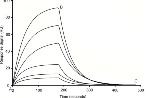
binding process. The increase in the binding affinity could be due to the deletion of non-binding nucleotides that hampers the Figure 1. Typical SPR sensorgrams demonstrating interaction of aptamer with immobilized VEGF165 protein at different
concentrations (bottom to top, 0.2 to 100 nM).Point A to B corresponds to the association phase and point B to C corresponds to the dissociation phase in all the sensorgrams. Shown here is the SL2-B aptamer (Kd= 0.5060.32 nM).
doi:10.1371/journal.pone.0031196.g001
Figure 2. Schematic representation of the secondary structure of original VEa5 aptamer as predicted by the mfold program. Nucleotide 4–20 forms the stem-loop 1 (SL1) region, nucleotide 22–38
forms the stem-loop 2 (SL2) region, and nucleotide 43–50 forms
stem-loop 3 (SL3) region. Nucleotide 28–33 forms internal loop 1 (IL1), and
nucleotide 24–27+34–36 together forms internal loop 2 (IL2) within SL2
region.
binding process and hence, their removal may lead to more desired secondary conformation in the truncated aptamer required for binding to the HBD of VEGF165protein [28].
We next truncated SL1and left with only the SL2sequence for
VEGF165 binding analysis, and surprisingly we obtained lower
binding affinity. With Kdvalue of 49 nM (Figure 3C and Table 1),
the binding affinity of the SL2 sequence was approximately 10
times higher than the Kd of SL12. This prompted us to further
investigate the role of the additional sequence next to the 39and 59 ends of the SL2region. By adding a single nucleotide at both 59
and 39-ends of the SL2aptamer (SL2-A, Figure 3D), we were able
to lower the Kd value by almost 5-fold (Kd= 10 nM, Table 1)
compared to the SL2aptamer. Further addition of nucleotides to
39-end did not yield any improvement in the binding affinity. However, adding another 7 nucleotides at 59-end of the SL2
aptamer (SL2-B, Figure 3E) showed further enhancement in the
binding affinity (Kd= 0.5 nM, Table 1), and this represented a
90-fold increase compared with the SL2aptamer, and more than
200-fold increase compared with the original VEa5 aptamer and 10-fold increase compared with the dimerized VEa5 aptamer [36]. A possible explanation is that the addition of nucleotides to the 59 -end provides more conformational stability to the aptamer which Table 1.Different aptamer sequences along with their equilibrium dissociation constant (Kd) values determined using surface plasmon resonance (SPR) spectroscopy.
Sequences of original and various truncated aptamers (59--- 39)* Kd
VEa5ATACCAGTCTATTCAATTGGGCCCGTCCGTATGGTGGGTGTGCTGGCCAGATAGTATGTGCAATCA 12061.8 nM
SL12ATACCAGTCTATTCAATTGGGCCCGTCCGTATGGTGGGTGTGCTGGCCAGATAGTATGTGCAATCA 560.45 nM
SL1ATACCAGTCTATTCAATTGGGCCCGTCCGTATGGTGGGTGTGCTGGCCAGATAGTATGTGCAATCA No Binding
SL2ATACCAGTCTATTCAATTGGGCCCGTCCGTATGGTGGGTGTGCTGGCCAGATAGTATGTGCAATCA 4962.4 nM
SL2-AATACCAGTCTATTCAATTGGGCCCGTCCGTATGGTGGGTGTGCTGGCCAGATAGTATGTGCAATCA 1061.1 nM
SL2-BATACCAGTCTATTCAATTGGGCCCGTCCGTATGGTGGGTGTGCTGGCCAGATAGTATGTGCAATCA 0.560.32 nM
*The underlined and bold section indicates the aptamer sequence and the non-bold grey section indicates the truncated sequence. In aptamer sequence terminology, the subscript number indicates the presence of the particular stem-loop region.
doi:10.1371/journal.pone.0031196.t001
Figure 3. Schematic representation of the secondary structures of various truncated aptamers as predicted by the mfold program. (A) SL12indicates the presence of SL1and SL2regions together, (B) SL1indicates the presence of SL1region only and (C) SL2indicates the presence of
SL2region only. (D) SL2-A and (E) SL2-B correspond to secondary structures formed after addition of nucleotides to SL2region.
doi:10.1371/journal.pone.0031196.g003
Figure 4. Sensorgrams demonstrating interaction of SL2-B aptamer with immobilized VEGF165and VEGF121proteins using SPR
spectroscopy.Point A to B corresponds to the association phase and point B to C corresponds to the dissociation phase in the sensorgrams. Shown here is SL2-B aptamer binding with VEGF165protein (Kd= 0.5060.32 nM) and VEGF121protein (Kd= 10.261.89mM).
doi:10.1371/journal.pone.0031196.g004
Figure 5. Representative flow cytometry profiles and quantitative analysis of flow cytometry results in (A) HT-29 cells and (B) MRC-5 cells after SL2-B aptamer at different concentrations (red – 0 nM (negative control), green – 0.5 nM, pink – 10 nM, blue – 100 nM),
helps in improving the binding competence of the SL2-B sequence
[38]. We also attempted to truncate the internal loops of SL2(IL1
and IL2, Figure 2) but the removal of either loops reduced the
binding affinity. Therefore, SL2-B is thought to be the minimal
sequence required in VEa5 aptamer to provide high binding affinity to HBD of VEGF165.
To address the selectivity of the truncated SL2-B aptamer to
VEGF165 binding, VEGF121 was selected for the binding
comparison. VEGF121is another spliced form of VEGF mRNA
that constitutes only the receptor-binding domain (RBD) but is devoid of HBD. HBD assists in the binding of VEGF protein to the heparin sulfate (HS) and heparin sulfate proteoglycans (HSPGs) present on the extracellular matrix of the cell membrane [32]. This enhances the interaction of VEGF with its receptors (VEGFR-1/Flt-1 and VEGFR-2/KDR) and the specific co-receptor neuropilins, triggering the cellular angiogenic response in malignant cells [34].
Compared with VEGF165, the binding of VEGF121 to the
membrane receptors is not strong, making it not potent to generate intense angiogenic signal. Using SL2-B for binding analysis with
VEGF121 under same buffer condition (Figure 4), substantial
reduction in the response signal and minimal binding (Kd= 10.2mM) was observed compared to VEGF165. This
confirms the binding selectivity of the truncated SL2-B aptamer
on HBD of the VEGF165protein.
In vitrocellular binding analysis of SL2-B aptamer-VEGF complex
Compared to techniques such as nitrocellulose membrane filtration or capillary electrophoresis, cell-based technique, such as flow cytometry, isolates aptamer sequences that have the ability to bind to their target with high affinity and specificity in a more physiologically relevant environment [39,40]. Thus, it has been considered as a more direct strategy for studying the binding of aptamers to their targets on cellular surface.
To validate the increase in the binding affinity of the truncated SL2-B aptamer and to determine its target specificity at cellular
level, we exposed the fluorescent-labeled SL2-B sequence directly
on the HT-29 colorectal cancer cells and normal MRC-5 fibroblast cells (control) under 1% O2hypoxia condition by flow
cytometry analysis. As shown in Figure 5A, a significant peak shift (increase in the fluorescent signal) was observed for SL2-B aptamer
at different concentrations, compared to control sample (only cells) in response to hypoxia conditions in HT-29 cells (p-value,0.001). The percentage of fluorescent-labeled cells increased with the increase in the concentration of the SL2-B aptamer, indicating the
binding of the SL2-B aptamer to the cell surface. In contrast, when
using normal MRC-5 fibroblast cells, no enhancement in the fluorescent signal was observed after treatment with different SL2
-B aptamer concentrations (Figure 5-B, not significant (n.s.) compared to negative control). Since the VEGF protein is overexpressed in HT-29 cells but not the normal MRC-5 cells, the result showed that SL2-B can specifically form aptamer-VEGF
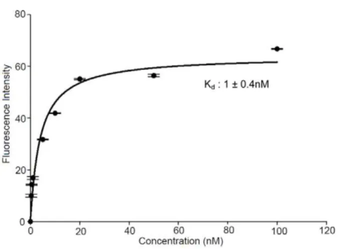
complex on the HT-29 cell membrane. The Kdvalue of the SL2-B
towards VEGF was further evaluated using flow cytometry data (Figure 6). The Kdvalue for the SL2-B aptamer to the cell surface
was found to be 1.0 nM, which is close to the Kdvalue determined
by SPR technique (Figure 6). Repeating the same experiment at Figure 6. Binding curve of SL2-B aptamer with HT-29 cells.Cells
were incubated with different SL2-B aptamer concentrations ranging
from 0 to 100 nM. The fluorescence intensity originating from the scrambled sequence at each concentration was subtracted from the fluorescence intensity of corresponding SL2-B aptamer. The actual
fluorescence intensity was fitted into SigmaPlot software to determine the Kd.
doi:10.1371/journal.pone.0031196.g006
Figure 7. Flow cytometry histogram showing binding competition between the labeled and unlabeled SL2-B aptamer in HT-29 cells
and quantitative analysis of flow cytometry result.Red – 0 nM (negative control), green – aptamer binding without excess of unlabeled SL2-B
aptamer (0.5 nM); pink – aptamer binding in excess of unlabeled SL2-B aptamer (unlabeled aptamer – 10 nM, labeled aptamer – 0.5 nM). *Significant
difference from the negative control sample at p-value,0.05; **Significant difference from the negative control sample at p-value,0.001. doi:10.1371/journal.pone.0031196.g007
normoxia condition (i.e. 21% O2) did not yield any observable
binding of SL2-B aptamer to the cells. The result is in agreement
with the fact that the hypoxia condition enhances production of VEGF protein in HT-29 cells [41–43].
Hypoxia is one of the crucial physiological factors that exert profound impact on the metabolism, invasion and tumor progression, thereby affecting many oncogenic pathways. Inade-quate level of cellular oxygen leads to aggressive phenotypic changes in the tumor cells, and is primarily responsible for their resistance against the therapies and poor prognosis [44]. Exposure to hypoxia milieu upregulates the expression of master regulator Hypoxia-Inducible Factor 1 gene (HIF-1 gene) in the solid tumors, which switch on the transcription of several downstream genes, in particular, VEGF [45]. The expression of VEGF protein and its two tyrosine kinase receptors – VEGFR-1/Flt-1 and VEGFR-2/ KDR is induced by the transcription and stabilization of VEGF
mRNA in response to hypoxia, resulting in increase rate of vascularization in tumor [46–48].
The binding ability of SL2-B aptamer for VEGF protein in
HT-29 cells was evaluated by competing 59-PE-texas red labeled SL2-B
sequence against the unlabeled SL2-B sequence. As shown in
Figure 7, the presence of 20-fold excess of unlabeled aptamer significantly decreased the fluorescent signal from the PE-texas red labeled aptamer (p-value,0.05). This result suggests that both the labeled and unlabeled SL2-B aptamer binds to the same target, the
VEGF protein on HT-29 cell membrane.
In vitrofluorescence imaging of SL2-B aptamer-VEGF complex
The targeting ability and specificity of the high affinity SL2-B
aptamer was further investigated and imaged using live colorectal cancer HT-29 cells and normal MRC-5 fibroblast cells (control) at Figure 8. Binding of PE-texas red-labeled aptamers (SL2-B and scrambled sequence) to HT-29 cells and normal MRC-5 fibroblast
cells under hypoxia condition.(A) & (C) Bright field images of HT-29 cells after exposing to the SL2-B and scrambled sequence respectively. (B) &
(D) Corresponding fluorescence microscopy images of the (A) & (C) bright field images. (E) & (F) Bright field and fluorescence microscopy images of MRC-5 cells after exposing to the SL2-B sequence.
the same hypoxia conditions. By comparing the bright field and the fluorescent images, we observed red fluorescence on HT-29 cells after exposing to PE-texas red labeled SL2-B aptamer
(Figure 8A & B), but no detectable fluorescence was observed with PE-texas red labeled scrambled sequence (Figure 8C & D). This confirms the specific binding of SL2-B aptamer to VEGF165
protein in cancer cells. For MRC-5 fibroblasts, minimal fluorescence was also observed after exposing to the same SL2-B
aptamer (Figure 8E & F). This further demonstrates the specificity and targeting ability of the SL2-B aptamer to cancer cells.
Conclusions
To summarize, this work attempted to identify in the key binding aptamer sequences by truncating based on the stem-loop structure of the original VEa5 aptamer. From the results, we can conclude that the SL2 sequence is important for the binding to
HBD of the VEGF165protein, and the SL2-B aptamer binds HBD
of VEGF165protein strongly and selectively. This newly obtained
SL2-B aptamer sequence can potentially be useful in
oligomer-based cancer therapeutic and diagnostic applications, though further studies are required for better understanding of the SL2-B
aptamer sequence and to elucidate its binding mechanism with HBD of VEGF165 protein. Furthermore, the current stem-loop
modification approaches can be useful in identifying target domains, getting rid of excessive unnecessary nucleotides, and eventually lowering the cost of aptamer synthesis.
Acknowledgments
The authors thank Dr Partha Roy (Division of Bioengineering, National University of Singapore) for providing the HT-29 colorectal cancer cells. We also appreciated the doctoral scholarship (for H.K.) from NUS.
Author Contributions
Conceived and designed the experiments: HK LY. Performed the experiments: HK. Analyzed the data: HK LY. Contributed reagents/ materials/analysis tools: HK LY. Wrote the paper: HK LY.
References
1. Bock LC, Griffin LC, Latham JA, Vermaas EH, Toole JJ (1992) Selection of single-stranded DNA molecules that bind and inhibit human thrombin. Nature 355: 564–566.
2. Wrzesinski J, Ciesiolka J (2005) Characterization of structure and metal ions specificity of Co2+
-binding RNA aptamers. Biochemistry 44: 6257–6268. 3. Davis JH, Szostak JW (2002) Isolation of high-affinity GTP aptamers from
partially structured RNA libraries. Proc Natl Acad Sci USA 99: 11616–11621. 4. Jayasena SD (1999) Aptamers: An emerging class of molecules that rival
antibodies in diagnostics. Clin Chem 45: 1628–1650.
5. Ruckman J, Green LS, Beeson J, Waugh S, Gillette WL, et al. (1998) 29 -fluoropyrimidine RNA-based aptamers to the 165-amino acid form of vascular endothelial growth factor (VEGF165): Inhibition of receptor binding and VEGF-induced vascular permeability through interactions requiring the exon 7-encoded domain. J Biol Chem 273: 20556–20567.
6. Sayyed SG, Ha¨gele H, Kulkarni OP, Endlich K, Segerer S, et al. (2009) Podocytes produce homeostatic chemokine stromal cell-derived factor-1/ CXCL12, which contributes to glomerulosclerosis, podocyte loss and albumin-uria in a mouse model of type 2 diabetes. Diabetologia 52: 2445–2454. 7. Ninichuk V, Clauss S, Kulkarni O, Schmid H, Segerer S, et al. (2008) Late onset of
Ccl2 blockade with the Spiegelmer mNOX-E36-39-PEG prevents glomerulosclerosis and improves glomerular filtration rate in db/db mice. Am J Pathol 172: 628–637. 8. Bates PJ, Laber DA, Miller DM, Thomas SD, Trent JO (2009) Discovery and development of the G-rich oligonucleotide AS1411 as a novel treatment for cancer. Exp Mol Pathol 86: 151–164.
9. Waters EK, Richardson J, Schaub RG, Kurz JC (2009) Effect of NU172 and bivalirudin on ecarin clotting time in human plasma and whole blood. J Thromb Haemost 7: 683.
10. Diener JL, Daniel Lagasse´ HA, Duerschmied D, Merhi Y, Tanguay JF, et al. (2009) Inhibition of von Willebrand factor-mediated platelet activation and thrombosis by the anti-von Willebrand factor A1-domain aptamer ARC1779. J Thromb Haemost 7: 1155–1162.
11. Chan MY, Cohen MG, Dyke CK, Myles SK, Aberle LG, et al. (2008) Phase 1b randomized study of antidote-controlled modulation of factor IXa activity in patients with stable coronary artery disease. Circulation 117: 2865–2874. 12. Biesecker G, Dihel L, Enney K, Bendele RA (1999) Derivation of RNA aptamer
inhibitors of human complement C5. Immunopharmacology 42: 219–230. 13. Green LS, Jellinek D, Jenison R, Ostman A, Heldin CH, et al. (1996) Inhibitory
DNA ligands to platelet-derived growth factor B-chain. Biochemistry 35: 14413–14424.
14. Ellington AD, Szostak JW (1990) In vitro selection of RNA molecules that bind specific ligands. Nature 346: 818–822.
15. Tuerk C, Gold L (1990) Systematic evolution of ligands by exponential enrichment: RNA ligands to bacteriophage T4 DNA polymerase. Science 249: 505–510. 16. Misono TS, Kumar PKR (2005) Selection of RNA aptamers against human
influenza virus hemagglutinin using surface plasmon resonance. Anal Biochem 342: 312–317.
17. Berezovski M, Drabovich A, Krylova SM, Musheev M, Okhonin V, et al. (2005) Nonequilibrium capillary electrophoresis of equilibrium mixtures: A universal tool for development of aptamers. J Am Chem Soc 127: 3165–3171. 18. Tok JB, Fischer NO (2008) Single microbead SELEX for efficient ssDNA
aptamer generation against botulinum neurotoxin. Chem Comm. pp 1883–1885.
19. Jellinek D, Green LS, Bell C, Lynott CK, Gill N, et al. (1995) Potent 29 -amino-29-deoxypyrimidine RNA inhibitors of basic fibroblast growth factor. Biochem-istry 34: 11363–11372.
20. Pagratis NC, Bell C, Chang YF, Jennings S, Fitzwater T, et al. (1997) Potent 29 -amino-, and 29-fluoro-29-deoxyribonucleotide RNA inhibitors of keratinocyte growth factor. Nature Biotechnol 15: 68–73.
21. Chelliserrykattil J, Ellington AD (2004) Evolution of a T7 RNA polymerase variant that transcribes 29-O-methyl RNA. Nature Biotechnol 22: 1155–
1160.
22. Hernandez FJ, Kalra N, Wengel J, Vester B (2009) Aptamers as a model for functional evaluation of LNA and 29-amino LNA. Bioorg Med Chem Lett 19: 6585–6587.
23. Schmidt KS, Borkowski S, Kurreck J, Stephens AW, Bald R, et al. (2004) Application of locked nucleic acids to improve aptamer in vivo stability and targeting function. Nucleic Acids Res 32: 5757–5765.
24. Kawasaki AM, Casper MD, Freier SM, Lesnik EA, Zounes MC, et al. (1993) Uniformly modified 29-deoxy-29-fluoro phosphorothioate oligonucleotides as nuclease-resistant antisense compounds with high affinity and specificity for RNA targets. J Med Chem 36: 831–841.
25. Somasunderam A, Ferguson MR, Rojo DR, Thiviyanathan V, Li X, et al. (2005) Combinatorial selection, inhibition, and antiviral activity of DNA thioaptamers targeting the RNase H domain of HIV-1 reverse transcriptase. Biochemistry 44: 10388–10395.
26. Boomer RM, Lewis SD, Healy JM, Kurz M, Wilson C, et al. (2005) Conjugation to polyethylene glycol polymer promotes aptamer biodistribution to healthy and inflamed tissues. Oligonucleotides 15: 183–195.
27. Kato T, Yano K, Ikebukuro K, Karube I (2000) Interaction of three-way DNA junctions with steroids. Nucleic Acids Res 28: 1963–1968.
28. Shangguan D, Tang Z, Mallikaratchy P, Xiao Z, Tan W (2007) Optimization and modifications of aptamers selected from live cancer cell lines. ChemBio-Chem 8: 603–606.
29. Sayer NM, Cubin M, Rhie A, Bullock M, Tahiri-Alaoui A, et al. (2004) Structural determinants of conformationally selective, prion-binding aptamers. J Biol Chem 279: 13102–13109.
30. Katilius E, Flores C, Woodbury NW (2007) Exploring the sequence space of a DNA aptamer using microarrays. Nucleic Acids Res 35: 7626–7635. 31. Houck KA, Ferrara N, Winer J, Cachianes G, Li B, et al. (1991) The vascular
endothelial growth factor family: Identification of a fourth molecular species and characterization of alternative splicing of RNA. Mol Endocrinol 5: 1806–1814.
32. Park JE, Keller GA, Ferrara N (1993) The vascular endothelial growth factor (VEGF) isoforms: Differential deposition into the subepithelial extracellular matrix and bioactivity of extracellular matrix-bound VEGF. Mol Biol Cell 4: 1317–1326.
33. Cohen T, Gitay-Goren H, Sharon R, Shibuya M, Halaban R, et al. (1995) VEGF121, a vascular endothelial growth factor (VEGF) isoform lacking heparin binding ability, requires cell-surface heparan sulfates for efficient binding to the VEGF receptors of human melanoma cells. J Biol Chem 270: 11322–11326. 34. Keyt BA, Berleau LT, Nguyen HV, Chen H, Heinsohn H, et al. (1996) The
carboxyl-terminal domain (111–165) of vascular endothelial growth factor is critical for its mitogenic potency. J Biol Chem 271: 7788–7795.
35. Hasegawa H, Sode K, Ikebukuro K (2008) Selection of DNA aptamers against VEGF165 using a protein competitor and the aptamer blotting method. Biotechnol Lett 30: 829–834.
36. Hasegawa H, Taira KI, Sode K, Ikebukuro K (2008) Improvement of aptamer affinity by dimerization. Sensors 8: 1090–1098.
37. Zuker M (2003) Mfold web server for nucleic acid folding and hybridization prediction. Nucleic Acids Res 31: 3406–3415.
38. Potty ASR, Kourentzi K, Fang H, Jackson GW, Zhang X, et al. (2009) Biophysical characterization of DNA aptamer interactions with vascular endothelial growth factor. Biopolymers 91: 145–156.
39. Davis KA, Lin Y, Abrams B, Jayasena SD (1998) Staining of cell surface human CD4 with 29-F-pyrimidine-containing RNA aptamers for flow cytometry. Nucleic Acids Res 26: 3915–3924.
40. Daniels DA, Chen H, Hicke BJ, Swiderek KM, Gold L (2003) A tenascin-C aptamer identified by tumor cell SELEX: Systematic evolution of ligands by exponential enrichment. Proc Natl Acad Sci USA 100: 15416–15421. 41. Waleh NS, Brody MD, Knapp MA, Mendonca HL, Lord EM, et al. (1995)
Mapping of the vascular endothelial growth factor-producing hypoxie cells in multicellular tumor spheroids using a hypoxia-specific marker. Cancer Res 55: 6222–6226.
42. Oswald J, Treite F, Haase C, Kampfrath T, Ma¨ding P, et al. (2007) Experimental hypoxia is a potent stimulus for radiotracer uptake in vitro:
Comparison of different tumor cells and primary endothelial cells. Cancer Lett 254: 102–110.
43. Calvani M, Trisciuoglio D, Bergamaschi C, Shoemaker RH, Melillo G (2008) Differential involvement of vascular endothelial growth factor in the survival of hypoxic colon cancer cells. Cancer Res 68: 285–291.
44. Vaupel P, Mayer A (2007) Hypoxia in cancer: Significance and impact on clinical outcome. Cancer Metast Rev 26: 225–239.
45. Minchenko A, Bauer T, Salceda S, Caro J (1994) Hypoxic stimulation of vascular endothelial growth factor expression in vitro and in vivo. Lab Invest 71: 374–379.
46. Neufeld G, Cohen T, Gengrinovitch S, Poltorak Z (1999) Vascular endothelial growth factor (VEGF) and its receptors. FASEB J 13: 9–22.
47. Ikeda E, Achen MG, Breier G, Risau W (1995) Hypoxia-induced transcriptional activation and increased mRNA stability of vascular endothelial growth factor in C6 glioma cells. J Biol Chem 270: 19761–19766.