UNIVERSIDADE FEDERAL DE UBERLÂNDIA
FACULDADE DE MEDICINA
PROGRAMA DE PÓS-GRADUAÇÃO EM CIÊNCIAS DA SAÚDE
Implicações translacionais de uma nova ferramenta de detecção de células
tumorais circulantes no monitoramento do câncer de próstata
Leandro Alves de Oliveira
Leandro Alves de Oliveira
Implicações translacionais de uma nova ferramenta de detecção de células
tumorais circulantes no monitoramento do câncer de próstata
Dissertação apresentada ao Programa
de Pós-Graduação em Ciências da
Saúde da Faculdade de Medicina da
Universidade Federal de Uberlândia,
como requisito parcial para a obtenção
do título de Mestre em Ciências da
Saúde.
Área de concentração:
Ciências da
Saúde.
Orientador:
Prof. Dr. Luiz Ricardo
Goulart
Dados Inrernacionais de Catalogação sa Publicação (CIP) Sistema de Bibliotecas da UFU, MG, Brasil.
0481 1017
Oneutador: Luiz Ricardo Goulart.
Dissertação (mestrado) - Universidade Federal de Uberlândia. Programa de Pôs-Giaduaçào e n C iô id a sd i Siide.
Incln l brblio zrafia.
1. Ciências médica: - Teses. 2. Próstata - Câncer -Teses. 3. Céhrlas Neoplisicas Circulantes - Teses. 4. Citometiia de flucto - Teses. I. Goulart. Luiz Ricardo. II. Universidade Fédéral de Uberlândia. Pioerama de Pós-Graduação em Ciências da Saúde. IEL Titulo
CDU: 61
Oliveira. Leandro AIves de. 1972
Implicações tianslacionais de mua nova ferramenta de detecção de celula: tumorais circulantes no monitoramento do cancer de próstata f
Leandro Aises de Oliveira. - 201”.
FOLHA DE APROVAÇÃO
Leandro Alves de Oliveira
Avaliação do aptâmero A4 selecionado por 3D Cell SELEX no
rastreamento do câncer de próstata
Dissertação apresentada ao Programa de
Pós-Graduação em Ciências da Saúde da
Faculdade de Medicina da Universidade
Federal de Uberlândia, como requisito
parcial para a obtenção do título de Mestre
em Ciências da Saúde.
Área de concentração: Ciências da Saúde.
Banca examinadora
Presidente da banca (orientador):
Prof. Dr. Luiz Ricardo Goulart Filho
Titular:
Prof. Dr. Omar Pacheco Simão.
Instituição: FAMED-UFU.
DEDICATÓRIA
A minha esposa, Silvana, sempre companheira no meu caminho, trazendo amor
e paz, constrói todos os dias nosso lar.
Às minhas filhas, Isabella e Giovanna, nosso presente de Deus, minha maior
realização, são a prova viva da palavra Amor.
Aos meus pais, Hélio e Iêda, que por uma vida de dedicação, amor e bons
exemplos superaram tudo para criarem oportunidades de realização de sonhos e
conquistas dos filhos.
Às minhas irmãs, Ana Beatriz, Ana Paula e Maria, cunhados e sobrinhos,
sempre ao meu lado, uma amizade infinita.
Aos meus sogros, Rubens e Sandra apoio constante e a alegria no convívio
familiar.
AGRADECIMENTOS
Ao Prof. Dr. Luiz Ricardo Goulart Filho, por me orientar nesse trabalho, mas
em especial, pela sua amizade, respeito e dedicação. Muito obrigado.
Ao Prof. Dr. Omar Pacheco Simão, que me incentivou a realização dessa tese, o
convívio contigo transforma-nos em pessoas melhores, vossa liderança e
dedicação é um estímulo e exemplo para as novas gerações de urologistas.
A Dra. Aline Gomes de Souza, incansável pesquisadora, presença constante
contribuiu em todas as etapas deste projeto.
A todos pesquisadores do Laboratório de Nanobiotecnologia, aos funcionáros
do H.C. da UFU pela ajuda constante.
Aos Professores que ajudaram na minha formação de urologistas, Dr. Sebastião
Antônio Borba, Dr. Osvaldo Freitas Filho, Dr. Lindolfo Dumont Prado e Dr.
Eduardo H. Braga.
Aos colegas de residência: Dr. Claiber Vieira Borges (in memorian), Dr.
Ronaldo Manochio Zero, Prof. Dr. Fernando Gonçalves de Almeida e
especialmente ao amigo Dr. Jorge Henrique Moreira Agostinho; vocês
contribuíram para minha formação urológica.
Aos 34 residentes de urologia que auxiliei na formação, meu maior orgulho, a
melhor forma de aprender é ensinar, meus maiores professores.
Epígrafe
“
O objetivo da ciência é substituir as palavras
por fatos e as impressões por demonstrações. ”
RESUMO
Introdução:
O diagnóstico precoce de câncer de próstata (CaP) é essencial para
aumentar a sobrevida dos pacientes, mas os marcadores e métodos atuais não
possuem sensibilidade e especificidade suficientes, tornando o diagnóstico ainda
muito impreciso. Recentemente, as células tumorais circulantes (CTCs) têm
surgido não como método de rastreio do CaP, mas sim como marcadores de
prognóstico utilizando um arsenal de diversos alvos para a captura dessas
células. Contudo, a busca por um método ou marcadores comuns para o rastreio,
diagnóstico, prognóstico e monitoramento da doença ainda se apresenta com um
dos principais objetivos técnico-científicos a ser alcançado.
Objetivo:
de forma exponencial, embora seis pacientes com altos índices de células
circulantes apresentaram PSA<0,02ng/mL, considerados como falha
bioquímica.
Conclusão:
nossos resultados preliminares indicam uma acurácia
elevada de 98% e demonstra um grande potencial de aplicação dessa nova
tecnologia diagnóstica tanto no rastreamento, quanto no monitoramento do
tratamento do CaP, o qual deverá ser melhor investigado em população de risco.
Palavras-chave: Palavras-chave:
Câncer de próstata, diagnóstico, aptâmero,
citometria de fluxo, células tumorais circulantes.
ABSTRACT
the study evaluated 34 PCa patients and 16 health controls. Blood samples were
collected in EDTA tubes, and after erythrocytes lysis, nucleated cells were
incubated with A4 aptamer conjugated with biotin, them the cells were washed
and incubated with streptavidin-FITC for later flow cytometer analysis.
Percentage of CTCs were compared between patient’s groups and correlated
against age, PSA levels, staging and treatment regimen (hormonal blockade,
radiotherapy and surgery). Detection limit above 1% of CTCs was considered
positive, based on the percentage observed on all of the 16 negative controls.
Results:
all patients were positively diagnosed independently of therapy time or
staging, except for one patient undergoing hormonal blockade therapy, which
does not present detectable CTCs. CTCs percentage presented high correlation
against age (R=0.75) and with PSA levels (R=0.80) with exponential behavior,
although, six patients with high CTCs count presented PSA levels <0.02 ng/mL,
and were considered was biochemical errors.
Conclusion:
Our preliminary
results indicated high accuracy (98%) and demonstrate a potential application of
this technology for diagnosis and screening, as well as in the monitoring of PCa
evolution, which should be better investigated in the risk population.
LISTA DE ILUSTRAÇÕES
Figura 1.
Procedimento para a detecção de células tumorais circulantes (CTCs)
com marcação do aptâmero A4 seguido pela análise por citometria de fluxo. A.
Esquema de preparo e processamento da amostra. B. Exemplos de resultados
positivos (paciente) e negativos (controle) da análise realizada após a citometria
de fluxo.
Figura 2.
Percentual de células tumorais circulantes ligantes ao biomarcador
Aptâmero A4 detectado por citometria de fluxo para a detecção do câncer de
próstata e estabelecimento do valor de
cutoff,
conforme resultados obtidos nos
pacientes saudáveis ou com HPB. O
cutoff
considerado positivo para o câncer de
próstata (CaP) foi acima de 1,0%.
Figura 3.
Análise de correlação e equação predita para o comportamento da
tendência entre o percentual de células tumorais circulantes (CTCs) e a idade,
conforme análise por citometria de fluxo da marcação de CTCs com o aptâmero
A4. Com relação às faixas etárias dos grupos de pacientes, o CaP apresentou
idade superior à 55 anos, enquanto que o controle apresentou idades entre 28 a
44 anos.
Figura 5
. Análise de doença residual mínima pela detecção de CTCs em
diferentes esquemas terapêuticos para o câncer de próstata por meio da análise
de citometria de fluxo com a marcação do Aptâmero A4.
Figura 6.
Análise de doença residual mínima pela detecção do percentual CTCs
(A) e dos níveis de PSA sérico (B) em pacientes com câncer de próstata,
conforme o tempo de tratamento.
LISTA DE TABELAS
Tabela 1.
Caracterização clínico-laboratorial dos pacientes com câncer de
próstata e controles investigados quanto ao percentual de CTCs por citometria
de fluxo.
LISTA DE ABREVIATURAS E SÍMBOLOS
CaP
Câncer de Próstata
CEP
Comitê de Ética em Pesquisa
CTC
Célula Tumoral Circulante
DRE
Exame Retal Digital
EDTA
Ácido Etilenodiamino Tetra-Acético
FITC
Isotiocianato de fluoresceína
HPB
Hiperplasia Prostática Benigna
INCA
Instituto Nacional do Câncer
SELEX
Evolução Sistemática de Ligantes por Enriquecimento
Exponencial
PBS
Tampão fosfato salino
PCA3
Gene 3 do Antígeno do Câncer de Próstata
PCR
Reação em cadeia de polimerase
PSA
Antígeno Específico Prostático
SUMÁRIO
1 INTRODUÇÃO...16
2 FUNDAMENTAÇÃO TEÓRICA...17
3 OBJETIVOS...19
3.1 Obj etivo geral...19
3.2 Objetivos específicos...19
4 ARTIGO... 20
1
INTRODUÇÃO
O Câncer de Próstata (CaP) é um grande problema de saúde pública
mundial, devido à sua alta incidência e mortalidade [1, 2]. Quando não tratado o
CaP pode se tornar mais agressivo, possibilitando sua progressão para hormônio
resistente e culminando com a ocorrência de metástases [3, 4]. A sobrevida de
pacientes com CaP está relacionada a muitos fatores, especialmente à extensão
do tumor no momento do diagnóstico [5]. Atualmente, o acompanhamento das
alterações prostáticas é realizado em 4 etapas: toque retal (DRE), dosagem do
antígeno prostático específico (PSA), ultrasonografia transretal e biópsia [6, 7].
Contudo, a complexidade do manejo da doença está cada vez mais aumentado
devido à progressão heterogênea e dos subgrupos com diferentes prognósticos
[8].
2 FUNDAMENTAÇÃO TEÓRICA
Historicamente, a detecção de células tumorais circulantes (CTCs) da
próstata foi primeiramente realizada por meio da RT-PCR com diversos
marcadores e combinações, tais como PSA (prostate specific antigen), PSMA
(prostate specific membrance antigen), PSCA (prostate stem cell antigen), CK19
(cytokeratin 19) e PTHrP (Parathyroid hormone-related protein), dentre os quais
o PSMA e PSCA são altamente expressos em tumores metastáticos [15].
Estima-se que milhares de CTCs são liberadas na circulação sanguínea
diariamente [16] e que elas sobrevivem durante 1-2,5h [17].
Recentemente, a captura de CTCs, usadas como biomarcadores e também
chamadas de biópsias líquidas, tem sido aplicada com sucesso no câncer de
próstata com potencial uso no diagnóstico e rastreamento [18-20]. Uma extensa
revisão recente aborda a importância clínica da detecção e análise de CTCs,
especialmente nos estágios iniciais do câncer [21].
CellSearch® [20]. Este teste baseia-se na seleção positiva de CTCs utilizando
anticorpos contra o antígeno da molécula de adesão celular epitelial (EpCAM)
com detecção complementar com os anticorpos anti-CD45, e anti-citoqueratinas
8, 18, e 19 [24]. Semelhantemente à CellSearch, o AdnaTest também utiliza o
anti-EpCAM para a identificação de CTCs, mas complementa sua detecção com
amplificação de mRNAs dos genes PSA, PSMA e EGFR (epidermal growth
factor receptor), resultando em detecção de CTCs em somente 67% dos
pacientes metastáticos antes do tratamento [25]. Contudo, a grande limitação
desses testes está no uso de marcadores de células mesenquimais em um tumor
altamente heterogêneo, o que restringe a detecção à uma população que
provavelmente esteja associada à apenas alguns fenótipos agressivos do CaP
[26-30]. Nesse contexto, foi desenvolvida uma outra plataforma de imagem com
alto desempenho, a FAST (fiberoptic array scanning technology), em que 3
milhões de células são espalhadas em lâminas de microscopia e detectadas
simultaneamente com DAPI, anti-CD45 e anti-citoqueratinas, resultando em
uma positividade de aproximadamente 93% [31], teste este ainda em estudos de
validação. Duas revisões extensas recentes sobre todos os testes desenvolvidos
atualmente para detecção de CTCs foram apresentados [22], e todas as
tecnologias estão associadas à imunocaptura, imunohistoquímica e/ou a
microfluídica e eletroforese, e apenas um ensaio (Vita-Assay) reivindica o uso
da citometria de fluxo com uma série de marcadores (EpCAM, CK, CD44,
CD34, CD45, vimentin).
virgens de tratamento e em pacientes sob diferentes regimes terapêuticos como
prova-de-conceito, e o potencial uso da tecnologia é discutido nessa pesquisa.
3 OBJETIVOS
3.1 Objetivo geral
Aumentar o desempenho no diagnóstico e monitoramento do CaP através da
biologia molecular.
3.2 Objetivos específicos
Title: Translational Implications of a Novel Circulating Tumor Cells Detection Tool in Prostate Cancer Monitoring
One sentence summary:
A novel aptamer-based flow cytometry tool was developed to detect circulating tumor cells in the peripheral blood of prostate cancer patients, which has been successfully applied for tracking and treatment monitoring.
1 2 1 1 i *
Authors: Leandro A. Oliveira ’ , Aline G. Souza , Vivian A. Goulart , Luiz R. Goulart
Affiliations:
1 Laboratory of Nanobiotechnology, Institute of Genetics and Biochemistry, Federal University of Uberlândia, Uberlândia, MG, Brasil.
2 School of Medicine. Graduate Program in Health Sciences, Federal University of Uberlândia, Uberlândia, MG, Brasil.
Corresponding author
ABSTRACT
Circulating tumor cells (CTCs) have been used as prognostic markers in prostate cancer (PCa). We present a novel aptamer-based flow cytometry tool to detect CTCs in the
peripheral blood of PCa patients with and without treatment and healthy controls. Percentage of CTCs were compared between patients’ groups and correlated with age, serum PSA levels, staging and therapeutic procedures adopted. The cutoff limit >1% of CTCs was considered positive. PCa patients were diagnosed as positive irrespective of time of therapy or staging. CTCs increased exponentially and showed high correlation with age (R = 0.75) and PSA levels (R = 0.80), although six patients with PSA <0.02 ng/mL presented high CTCs%, and were considered biochemical failure. Our results presented an accuracy of 98%, and
demonstrated a potential application of this new diagnostic tool for PCa tracking and treatment monitoring, which should be further investigated in men at risk.
INTRODUCTION
Prostate cancer (PCa) is a major public health problem worldwide because of its high
incidence and mortality (1, 2). When untreated, PCa may become more aggressive, allowing its progression to hormone resistance, culminating with the occurrence of metastases (3, 4). The PCa patients’ survival is related to many factors, especially the tumor extent at the time of diagnosis (5). Currently, monitoring of prostatic changes is performed in four stages: rectal examination (DRE), prostate specific antigen (PSA) dosage, transrectal ultrasonography, and biopsy (6, 7). However, the complexity of the cancer management is increasingly higher due to the heterogeneous progression of the disease and the differential prognoses for patients’ subgroups (8).
The utility of PSA is quite limited in distinguishing between stages of PCa and benign conditions, such as BPH and prostatitis, and their use for PCa screening has not been
recommended (9, 10). Therefore, it is evident the urgent need to identify new tools that may present greater specificity and sensitivity for PCa diagnosis (9, 11). Alhtough several
biological targets have been proposed as diagnostic markers, only the PCA3 non-coding RNA has shown utility, which can detected both in the peripheral blood (12) and in urine (13), as well as in combination with PSA (12). However, the studies present moderate sensitivities and specificities, with approximate accuracy of 70%. Recently, a new aptamer-based diagnostic technology that includes the magnetic capture of the circulating PCA3 RNA followed by amplification through RT-PCR has increased the diagnostic sensitivity to 100% (14). Nevertheless, the sample size was limited, and the new tool still requires purification of total RNA and logistics for sample collection due to greater instability of RNA molecules.
Historically, detection of RNA from circulating tumor cells, namely CTCs of the prostate, was first performed by RT-PCR with various markers and combinations, such as the prostate specific antigen (PSA), prostate specific membrane antigen (PSMA), PSCA (prostate specific stem cell antigen), CK19 (cytokeratin 19) and PTHrP (Parathyroid hormone-related protein), among which PSMA and PSCA are highly expressed in metastatic tumors (15). It is estimated that thousands of CTCs are released into the bloodstream daily (16), which are able to survive for 1-2.5h (17).
Recently, the capture of CTCs as biomarkers, also called liquid biopsies, has been successfully applied to prostate cancer with potential use in diagnosis and screening (18-20).
Although high levels of CTCs suggest an association with disease progression, the predictive value for therapeutic monitoring is still inconclusive (22). More advanced
microfluidic systems associated with immunofluorescence has been subsequently developed to identify the tumor origin by capturing CTCs in the peripheral blood against PSA and PSMA as a proof-of-concept, using healthy blood contaminated with tumor cell lines (DU145, PC3 and LNCaP) (23). However, the same system was tested in five patients with metastases and in 12 controls, but CTCs could be detected in only three patients (60%) and on control individual (8%), without achieving the necessary sensitivity and specificity. On the other hand, although metastases are associated with CTCs and a worse prognosis, 35% of metastatic patients are undetectable by the CellSearch® system (20). This assay is based on the positive selection of CTCs using antibodies against the epithelial cell adhesion molecule antigen (EpCAM) with complementary detection of anti-CD45 antibodies, and anti-
cytokeratins CK-8, CK-18, and CK-19 (24). Similar to CellSearch, AdnaTest also uses anti- EpCAM to identify CTCs, but complements its detection with mRNAs of the PSA, PSMA and EGFR genes, resulting in detection of CTCs in only 67% of the cells in pre-treatment metastatic patients (25). However, the major limitation of these tests is the use of
mesenchymal cell markers in highly heterogeneous tumors, which restricts detection to a population likely to be associated with only a few aggressive PCa phenotypes (26-30). In this context, another high-performance imaging platform, FAST (fiberoptic array scanning technology), was developed in which three million cells are scattered on microscopy slides and detected simultaneously with DAPI, anti-CD45 and anti-cytokeratins, resulting in a positivity of approximately 93% (37), although this test is still under validation. An extensive review of all current CTC detection tests have recently been presented (22), and all
technologies are associated with immunocapture, immunohistochemistry and/or microfluidic and electrophoresis, and only one assay (Vita-Assay) claims the use of flow cytometry with a series of markers (EpCAM, CK, CD44, CD34, CD45, Vimentin).
RESULTS
Establishment of the new aptamer-based flow cytometry for liquid biopsies, and patients’ characteristics.
We have previously selected the A4-aptamer against a tumor cell line by 3D-Cell Selex (32), but its clinical application was not demonstrated. In order to understand its behavior as a CTC marker in clinical settings, we have established a diagnostic methodology based on flow cytometry, which involves a very simple, fast and straightforward procedure, as described in Figure 1.
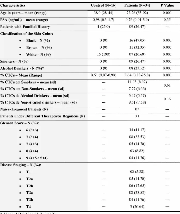
The clinical-laboratory characterization of the groups of patients and controls is presented in Table 1. Regarding the age, PCa patients presented ages greater than 55 years old, whereas the control group presented an age range between 28 to 44 years. Patients with PCa also presented highly significant frequencies for both smoking and alcoholism.
Regarding skin color, 79.4% of PCa patients were classified as black or mulatto, which were significantly different from controls. Among PCa patients, approximately 80% presented Gleason <7, while 38.4% were staged as advanced cancer (T3b and T4). CTCs were counted in both control and patient groups, and the cutoff for CTCs established as negative result was
1% based on results observed for all controls (Figure 2). The distribution of PCa patients according to their disease staging, regardless the treatment status, showed great variability without any correlation with the clinical-laboratory staging.
In a single case, one patient originally classified as T4, and then re-staged to T1, under hormonal blockage had CTC index of less than 1%; however, a reanalysis of the patient. For T2-staged patients, CTC levels were widely distributed, ranging from 1.2% to 25.8%, variability that was also observed in the other stages.
CTC levels are correlated with age and serum PSA levels
In relation to age, there was a high correlation of CTCs with increasing age (R = 0.75) with an exponential behavior as the age advanced, regardless the patients’ staging (Figure 3). The highest levels of CTCs were observed in patients over 65 years of age.
CTC levels are not correlated with different therapeutic interventions or with treatment period
Another important finding was the large variation of CTC levels for different types of treatment (Figure 5). There was no correlation of therapeutic success with CTC levels, since all therapeutic combinations showed highly variable levels of CTCs. However, among the two most frequently used therapeutic options, 6 out of 9 patients undergoing hormonal therapy (66.6%), and 7 out of 14 patients undergoing radical prostatectomy (50%) presented reduced percentages of CTCs (<4.2%). The first therapeutic option was the most effective when staging was considered, since the largest number of patients was in the T4 stage.
Regarding the analysis of minimal residual disease in PCa patients, according to the time of treatment (Figure 6), we have demonstrated that both CTCs% and serum PSA levels showed a low positive correlation with the period of therapy (R=0.34). Therefore, it was not possible to associate both markers with disease progression post-treatment. However, in relation to PSA levels recommended for relapse (<0.2 ng/dL), 15 patients still had lower levels and were considered disease-free, although CTCs ranged from 1.2% to 25.8%. For patients with recurrent disease, the percentages of CTCs ranged from 3.33% to 24.66%. It is important to note that among those 15 patients with PSA <0.2 ng/dL, 9 patients had CTCs% between 1.2% and 3.35% and the treatment time ranged from 9 to 25 months, which led us to establish a cutoff for CTCs% below 5% that coincided with the period of 25 months post treatment as indicative of a stable disease with no progression. The serum PSA levels in most patients in this condition were below 0.2 ng/dL (9/15). It is important to observe that the remaining six patients (40%), with PSA levels below the cutoff for tumor recurrence, presented CTCs% from 14.59 to 25.8, which were considered biochemical failure.
CTC levels detected by the aptamer-based flow cytometry method presents excellent diagnostic parameters
DISCUSSION
This research describes a new methodology based on flow cytometry with a new biomarker that is able to detect circulating tumor cells (CTCs) in the peripheral blood of patients without treatment and under different therapeutic regimens. Although prostate cancer is a heterogeneous, multifocal, and highly complex disease with different stages in its
evolution involving the cascade of testosterone activation, we have demonstrated that the use of CTCs as biomarkers throughout the disease progression was possible due to their capture by a common biological target mediated by the novel A4-aptamer developed by 3D-Cell Selex (32). The new tool allowed the detection of all positive patients in all stages and treatments, except for one patient undergoing hormonal treatment that was re-staged from T4 to T1, who was later characterized as Paget’s disease. Although these may be considered preliminary results due to the small sample size, the importance of these findings is discussed herein.
The ideal characteristics of a excellent biomarker are high specificity and sensitivity, good viability, minimally invasive, reproducibility and low cost, and in the case of PCa, the marker should also be a molecule that may be applied for disease tracking, diagnosis, prognosis, and treatment monitoring (33). PSA was the main tool applied to the PCa screening in the recent past, but it has been shown to be of low specificity, promoting overdiagnosis with unnecessary biopsies, and also leading to overtreatment (5, 10, 34, 35).
However, PSA levels still allow assessment of treatment response, and may help to detect relapse cases following local treatment of the tumor (33, 34, 36). Among various technologies for CTCs detection, the CellSearch capture system, which includes the immunohistochemistry evaluation of several markers, was named liquid biopsy, and its use is restricted to prognosis
(20).
been developed to allow robust formation of CTC clusters without pre-enrichment (39),
which can provide a noninvasive and inexpensive assessment of drug development or personalized therapy. The last three strategies were used to better characterize the molecular aspects of CTCs without any diagnostic pretension.
Our new diagnostic platform based on flow cytometry and aptamer labeling has surprisingly achieved an excellent precision (98%), much higher than those presented by existing epithelial markers (around 65%), such as EpCAM and markers associated with epithelial-mesenchymal transition (CK, CD44, CD34, CD45, vimentin). Interestingly, some technologies are now directed to the analysis of larger blood volumes, particularly in early tumors, where the presence of CTCs is even more infrequent (40). In our investigation, using only 5 mL of peripheral blood, we were able to detect CTCs in all PCa patients, with and without treatment, except in a patient who was later diagnosed as Paget’s disease, with CTCs below 1%. This detection ability may also be associated with labeling of many non-epithelial cell types other than epithelial ones, but with similar size, since no epithelial markers have been included, such as EpCAM. We are currently attempting to use a panel of markers to phenotype all A4+ CTCs by flow cytometry, with greater sample size, mainly to justify the significant correlation of CTCs detected by the A4-aptamer with age and the post-treatment PSA levels, suggesting that this marker may actually represent an advance in the diagnosis.
Interestingly, in addition to improving PCa diagnosis, the marker may also be used for PCa monitoring. The lack of correlation between the positive A4-aptamer detection with different stages and different treatment regimens, and the medium correlation with the time of treatment suggest the need for new therapeutic approaches in order to reduce the minimal residual disease. The various therapeutic regimens in the arsenal against PCa confirmed the current literature regarding the disease relapse after local treatment and putative cure, and also as palliative treatments in patients with systemic disease, since there was great fluctuation of PSA levels (35).
(42). This hypothesis has yet to be substantiated by additional molecular and immunological studies in these CTCs through cell sorting by FACS.
The correlation of post-treatment serum PSA levels with the percentage of CTCs becomes even more significant when taking into account that PSA has been advocated as a marker of response to prostate cancer treatment in both radical prostatectomy and
radiotherapy, and hormone therapy (41). After radical surgery, the lowest PSA value, defined as PSA nadir (PSAn), should be less than 0.2 ng/dL. However, PSA levels above this value after 60 to 90 days post-surgery suggest residual disease, requiring further treatment. Thus, to define prognosis it is used the time it takes to double PSA values, and the time it takes for biochemical relapse (42). For radiotherapy, the most reliable way to monitor PSA after nadir, which can take up to 18 months to achieve, is to observe an increase of at least 2 ng/dL to have a diagnosis of recurrence (43). For PCa monitoring after hormonal blockade and
chemotherapy, there is still no consensus regarding the PSA pattern, because during hormonal refractoriness cells are undifferentiated and do not produce PSA as a naive-treatment tumor cell. Still in relation to PSA levels after therapy in a large study, patients who had
radiotherapy associated with androgenic suppression had a significantly higher chance of being disease-free in 5 years than those who had only radiation therapy, with odds of 80% and 62%, respectively (44). An extensive analysis of 32 studies with therapeutic regimens has been presented in a comprehensive review without reaching a consensus (45). However, radiotherapy remains one of the few therapeutic interventions that offers a potential cure for patients who have PSA-based recurrence after radical prostatectomy. The response rate of PSA to the rescue radiation therapy, defined as any incremental decrease in PSA in response to radiotherapy, has reached up to 90%. A high PSA response rate to radiotherapy suggests that the majority of patients with post-operative PSA relapse may be due either to the local tumor recurrence in the prostate bed or to the distant occult metastasis simultaneously (45). However, despite the different therapeutic options adopted in this investigation, the
percentage of CTCs observed in the various patients indicates that there is still no option defined as more successful, although hormone suppression was more successful than radiotherapy based on the reduced percentage of CTCs.
With regard to prognosis and/or relapse, it was not possible to associate both serum PSA levels and CTC percentage with disease evolution, since both markers showed a low correlation with time after treatment. However, our preliminary data suggest that the
indicative of a stable disease with no progression, since most patients (60%) in this condition had serum PSA levels below 0.2 ng/dL. Therefore, patients’ monitoring data in this
investigation also suggest that the detection and quantification of CTCs can anticipate
treatments or avoid unnecessary procedures by creating new algorithms based on CTC levels. It is important to emphasize that the future of CTC detection lies in the molecular
characterization of these cells, since simple enumeration is not enough. Specific molecular signature is expected to provide critical information about the tumor's biology of the patient, which will allow the identification of new personalized therapies (46).
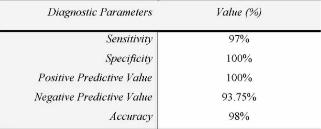
The performance of the A4-aptamer as diagnostic test was excellent, as it provided an accuracy of 98%, with sensitivity of 97% and specificity of 100%, and opens new diagnostic possibilities, although these results must be seen with caution due to the small sample size and because BPH patients were not included. The exclusion of BPH patients was due to the high probability of having a non-detectable tumor by conventional methods. But, these parameters become more impacting, even with the reduced sampling, because the performance of the A4- aptamer analyzed by flow cytometry was much higher than those presented by other reported CTC markers (33), which also used smaller sample sizes, and required an arsenal of markers and tools that barely reached sensitivities close to 65%. The potential presented by this new technology can really impact not only the epidemiology of the disease, but the early
diagnosis, and the most adequate monitoring, which must be further demonstrated in a larger sampling, already in the collection phase.
MATERIAL AND METHODS
Collection of biological samples and the inclusion and exclusion criteria
Biological samples (peripheral blood) were obtained from 34 patients with histologically confirmed prostate carcinoma and from 16 patients without prostatic abnormalities as the control group at the Clinics’ Hospital of the Federal University of Uberlândia (UFU), with the approval of the Research Ethics Committee under the number CEP 562,678, and patients’ consents were obtained. The clinical-laboratory parameters obtained of all individuals were: age, serum PSA levels, familial history, skin color
coexistence of both BPH and PCa, and the increased chances of having localized PCa (1, 47, 48). We have adopted this exclusion criterion also because inflammation is a well-established tumor promoter that contributes to cancer growth, angiogenesis, and resistance to apoptosis
(49, 50), and induction of cell proliferation under conditions of inflammation dramatically increases predisposition of cells to cancer (51). So, in order to prevent increased false positive rates in this proof-of-concept, we have decided to exclude BPH patients from this study, although a thorough investigation in this group of patients is highly desired.
Characterization of the Aptamer A4 as a biomarker and its synthesis
The A4-aptamer was previously selected using the 3D Cell SELEX technique and tested for prostate carcinoma, as described by Souza et al. (2016) (32). For this work the A4- aptamer was synthesized and biotin-conjugated at the Integrated DNA Technology (IDT, USA).
Analysis of CTCs by Flow Cytometry
CTCs were detected in both PCa and control groups by the A4-aptamer through flow cytometry. After collection of the blood in EDTA tube, plasma separation was performed by centrifugation at 1500 rpm for 10 minutes. 200 pL of the leukocyte layer was then collected and lysed from the red blood cells using 10X lysis buffer (BD Pharm Lyse) as recommended by the manufacturer. The samples were then centrifuged at 200xg for 5 minutes. The
supernatant was discarded and the pellet of cells formed was washed 2X with 1X PBS buffer. After centrifugation, the supernatant was discarded, cells were incubated with 10 pL of the A4-aptamer (100 pmol/pL) diluted in 100 pL of 1X PBS buffer for 1 hour at 37°C. Cells were then washed twice with 1X PBS, and further incubated with streptavidin-FITC (1 pg/mL) diluted in 100 pL of 1X PBS for 40 minutes at 37°C. Thereafter, the wash was repeated twice and cells were resuspended in 100 pL of 1X PBS for flow cytometry analyses (Accuri C6®, Becton Dickinson).
Statistical Analysis
performed to increase the power of the test, and also to have a better representation of different therapeutic regimens.
The analysis performed on the Accuri C6 Flow Cytometer indicated the percentage of CTCs labeled with the A4-aptamer for both control and PCa groups. The comparison of means was performed by the Mann-Whitney test (continuous variables and non-parametric analyses). Receiver operating characteristics (ROC) curve was calculated with a 95% confidence interval and the estimated diagnostic parameters (sensitivity, specificity and accuracy) according to the cutoff limits for the CTC values of the control group. Correlations and regression analyzes were performed between percentages of CTCs, age and PSA levels. All analyzes were performed in GraphPad Prism software version 7.0, using probability values below 5% as significant.
REFERENCES AND NOTES
1. D. D. O rsted, S. E. Bojesen, The link betw een benign prostatic hyperplasia and prostate cancer. Nat Rev Urol 10, 49-54 (2013).
2. S. Frankel, G. D. Sm ith, J. Donovan, D. Neal, Screening fo r prostate cancer. Lancet 361, 1122 1128 (2003).
3. K. J. Heuter, in Handbook of Small Animal Practice (Fifth Edition). (W .B. Saunders, Saint Louis, 2008), pp. 559-566.
4. J. Depczynski, T. Lower, A review of prostate cancer incidence and m ortality studies of farm ers and non-farm ers, 2002-2013. Cancer Epidemiol 38, 654-662 (2014).
5. S. Carlsson, A. J. Vickers, M. Roobol, J. Eastham , P. Scardino, H. Lilja, J. Hugosson, Prostate cancer screening: facts, statistics, and interpretation in response to the U S Preventive Services Task Force Review. J Clin Oncol 30, 2581-2584 (2012).
6. B. Djavan, B. Rocco, A. Stangelberger, O. De Cobelli, M. M arberger, Is the era of prostate- specific antigen over? BJU Int 100 Suppl 2, 8-10 (2007).
7. A. Stangelberger, M. M argreiter, C. Seitz, B. Djavan, Prostate cancer screening m arkers. The Journal of Men's Health and Gender 4, 233-244 (2007).
8. S. M cGrath, D. Christidis, M. Perera, S. K. Hong, T. M anning, I. Vela, N. Law rentschuk, Prostate cancer biom arkers: Are w e hitting the m ark? Prostate Int 4, 130-135 (2016).
9. J. V. Tricoli, M. Schoenfeldt, B. A. Conley, Detection of prostate cancer and predicting progression: current and future diagnostic m arkers. Clin Cancer Res 10, 3943-3953 (2004). 10. V. A. M oyer, U. S. P. S. T. Force, Screening for prostate cancer: U.S. Preventive Services Task
Force recom m endation statem ent. Ann Intern Med 157, 120-134 (2012).
12. A. F. Neves, T. G. Araujo, W . K. Biase, J. M eola, T. M. Alcantara, D. G. Freitas, L. R. Goulart, Com bined analysis of m ultiple m RNA m arkers by RT-PCR assay for prostate cancer diagnosis. Clin Biochem 41, 1191-1198 (2008).
13. I. L. Deras, S. M. Aubin, A. Blase, J. R. Day, S. Koo, A. W . Partin, W . J. Ellis, L. S. M arks, Y. Fradet, H. Rittenhouse, J. Groskopf, PCA3: a m olecular urine assay fo r predicting prostate biopsy outcom e. J Urol 179, 1587-1592 (2008).
14. K. M arangoni, A. F. Neves, R. M. Rocha, P. R. Faria, P. T. Alves, A. G. Souza, P. T. Fujim ura, F. A. Santos, T. G. Araujo, L. S. W ard, L. R. G oulart, Prostate-specific RNA aptam er: prom ising nucleic acid antibody-like cancer detection. Sci Rep 5, 12090 (2015).
15. Z. Panteleakou, P. Lem bessis, A. Sourla, N. Pissim issis, A. Polyzos, C. Deliveliotis, M. Koutsilieris, Detection of circulating tum or cells in prostate cancer patients: m ethodological pitfalls and clinical relevance. Mol Med 15, 101-114 (2009).
16. S. Valastyan, R. A. W einberg, Tum or m etastasis: m olecular insights and evolving paradigm s. Cell 147, 275-292 (2011).
17. S. M eng, D. Tripathy, E. P. Frenkel, S. Shete, E. Z. Naftalis, J. F. Huth, P. D. Beitsch, M. Leitch, S. Hoover, D. Euhus, B. Haley, L. M orrison, T. P. Flem ing, D. Herlyn, L. W . Terstappen, T. Fehm, T. F. Tucker, N. Lane, J. W ang, J. W . Uhr, Circulating tum or cells in patients w ith breast cancer dorm ancy. Clin Cancer Res 10, 8152-8162 (2004).
18. B. J. Kirby, M. Jodari, M. S. Loftus, G. Gakhar, E. D. Pratt, C. Chanel-Vos, J. P. Gleghorn, S. M. Santana, H. Liu, J. P. Sm ith, V. N. Navarro, S. T. Tagaw a, N. H. Bander, D. M. Nanus, P. Giannakakou, Functional characterization of circulating tum or cells with a prostate-cancer- specific m icrofluidic device. PLoS One 7, e35976 (2012).
19. Z. P. W ang, M. A. Eisenberger, M. A. Carducci, A. W . Partin, H. I. Scher, P. O. Ts'o, Identification and characterization of circulating prostate carcinom a cells. Cancer 88, 2787 2795 (2000).
20. L. E. Lowes, D. Goodale, Y. Xia, C. Postenka, M. M. Piaseczny, F. Paczkow ski, A. L. Allan, Epithelial-to-m esenchym al transition leads to disease-stage differences in circulating tum or cell detection and m etastasis in pre-clinical m odels of prostate cancer. Oncotarget 7, 76125 76139 (2016).
21. A. Kowalik, M. Kow alew ska, S. Gozdz, Current approaches for avoiding the lim itations of circulating tum or cells detection m ethods-im plications fo r diagnosis and treatm ent of patients w ith solid tum ors. Transl Res, (2017).
22. L. Leon-M ateos, M. Vieito, U. Anido, R. Lopez Lopez, L. M uinelo Rom ay, Clinical Application of Circulating Tum our Cells in Prostate Cancer: From Bench to Bedside and Back. Int J Mol Sci
17, (2016).
23. S. H. Lu, W . S. Tsai, Y. H. Chang, T. Y. Chou, S. T. Pang, P. H. Lin, C. M. Tsai, Y. C. Chang, Identifying cancer origin using circulating tum or cells. Cancer Biol Ther 17, 430-438 (2016). 24. W . J. Allard, J. M atera, M. C. M iller, M. Repollet, M. C. Connelly, C. Rao, A. G. Tibbe, J. W . Uhr,
L. W . Terstappen, Tum or cells circulate in the peripheral blood of all m ajor carcinom as but not in healthy subjects or patients w ith nonm alignant diseases. Clin Cancer Res 10, 6897 6904 (2004).
25. T. Todenhofer, J. Hennenlotter, S. Feyerabend, S. Aufderklam m , J. M ischinger, U. Kuhs, V. Gerber, J. Fetisch, D. Schilling, S. Hauch, A. Stenzl, C. Schw entner, Prelim inary Experience on the Use of the Adnatest® System for Detection of Circulating Tum or Cells in Prostate Cancer Patients. Anticancer Research 32, 3507-3513 (2012).
26. T. M. Gorges, I. Tinhofer, M. Drosch, L. Rose, T. M. Zollner, T. Krahn, O. von Ahsen, Circulating tum our cells escape from EpCAM -based detection due to epithelial-to-m esenchym al transition. BMC Cancer 12, 178 (2012).
28. J. T. Nauseef, M. D. Henry, Epithelial-to-m esenchym al transition in prostate cancer: paradigm or puzzle? Nat Rev Urol 8, 428-439 (2011).
29. P. Li, R. Yang, W . Q. Gao, Contributions of epithelial-m esenchym al transition and cancer stem cells to the developm ent of castration resistance of prostate cancer. Mol Cancer 13, 55 (2014).
30. D. Y. Jadaan, M. M. Jadaan, J. P. M cCabe, Cellular Plasticity in Prostate Cancer Bone M etastasis. Prostate Cancer 2015, 651580 (2015).
31. D. M arrinucci, K. Bethel, A. Kolatkar, M. S. Luttgen, M. M alchiodi, F. Baehring, K. Voigt, D. Lazar, J. Nieva, L. Bazhenova, A. H. Ko, W . M. Korn, E. Schram , M. Cow ard, X. Yang, T. M etzner, R. Lamy, M. Honnatti, C. Yoshioka, J. Kunken, Y. Petrova, D. Sok, D. Nelson, P. Kuhn, Fluid biopsy in patients with m etastatic prostate, pancreatic and breast cancers. Phys Biol 9, 016003 (2012).
32. A. G. Souza, K. M arangoni, P. T. Fujim ura, P. T. Alves, M. J. Silva, V. A. Bastos, L. R. Goulart, V. A. Goulart, 3D Cell-SELEX: Developm ent of RNA aptam ers as m olecular probes for PC-3 tum or cell line. Exp Cell Res 341, 147-156 (2016).
33. J. R. Prensner, M. A. Rubin, J. T. W ei, A. M. Chinnaiyan, Beyond PSA: the next generation of prostate cancer biom arkers. Sci Transl Med 4, 127rv123 (2012).
34. A. Q aseem , M. J. Barry, T. D. Denberg, D. K. Ow ens, P. Shekelle, P. Clinical G uidelines Com m ittee of the Am erican College of, Screening for prostate cancer: a guidance statem ent from the Clinical G uidelines Com m ittee of the Am erican College of Physicians. Ann Intern Med 158, 761-769 (2013).
35. M. Boniol, P. Boyle, P. Autier, A. Ruffion, P. Perrin, Critical role of prostate biopsy m ortality in the num ber of years of life gained and lost w ithin a prostate cancer screening program m e. BJU Int 110, 1648-1652 (2012).
36. S. Saini, PSA and beyond: alternative prostate cancer biom arkers. Cell Oncol (Dordr) 39, 97 106 (2016).
37. L. Zhang, L. D. Ridgw ay, M. D. W etzel, J. Ngo, W . Yin, D. Kum ar, J. C. G oodm an, M. D. Groves, D. M archetti, The identification and characterization of breast cancer CTCs com petent for brain m etastasis. Sci Transl Med 5, 180ra148 (2013).
38. S. Tulley, Q. Zhao, H. Dong, M. L. Pearl, W . T. Chen, V ita-Assay M ethod of Enrichm ent and Identification of Circulating Cancer Cells/Circulating Tum or Cells (CTCs). Methods Mol Biol
1406, 107-119 (2016).
39. B. L. Khoo, G. Grenci, T. Jing, Y. B. Lim, S. C. Lee, J. P. Thiery, J. Han, C. T. Lim, Liquid biopsy and therapeutic response: Circulating tum or cell cultures for evaluation of anticancer treatm ent. Sci Adv 2, e1600274 (2016).
40. N. Saucedo-Zeni, S. M ewes, R. Niestroj, L. G asiorow ski, D. M urawa, P. Now aczyk, T. Tom asi, E. W eber, G. Dw oracki, N. G. M orgenthaler, H. Jansen, C. Propping, K. Sterzynska, W. Dyszkiew icz, M. Zabel, M. Kiechle, U. Reuning, M. Schm itt, K. Lucke, A novel m ethod for the in vivo isolation of circulating tum or cells from peripheral blood of cancer patients using a functionalized and structured m edical wire. Int J Oncol 41, 1241-1250 (2012).
41. M. S. Cookson, G. Aus, A. L. Burnett, E. D. Canby-H agino, A. V. D'Am ico, R. R. Dm ochow ski, D. T. Eton, J. D. Form an, S. L. Goldenberg, J. Hernandez, C. S. Higano, S. R. Kraus, J. W . M oul, C. Tangen, J. B. Thrasher, I. Thom pson, Variation in the definition of biochem ical recurrence in patients treated fo r localized prostate cancer: the Am erican Urological Association Prostate G uidelines for Localized Prostate Cancer Update Panel report and recom m endations for a standard in the reporting of surgical outcom es. J Urol 177, 540-545 (2007).
International validation of a preoperative nom ogram for prostate cancer recurrence after radical prostatectom y. J Clin Oncol 20, 3206-3212 (2002).
43. S. P. Lankford, A. Pollack, G. K. Zagars, Prostate-specific antigen cancer volum e: a significant prognostic factor in prostate cancer patients at interm ediate risk of failing radiotherapy. Int J Radiat Oncol Biol Phys 38, 327-333 (1997).
44. C. Carrie, A. Hasbini, G. de Laroche, P. Richaud, S. Guerif, I. Latorzeff, S. Supiot, M. Bosset, J. L. Lagrange, V. Beckendorf, F. Lesaunier, B. Dubray, J. P. W agner, T. D. N'Guyen, J. P. Suchaud, G. Crehange, N. Barbier, M. Habibian, C. Ferlay, P. Fourneret, A. Ruffion, S. Dussart, Salvage radiotherapy w ith or w ithout short-term horm one therapy for rising prostate-specific antigen concentration after radical prostatectom y (GETUG-AFU 16): a random ised, m ulticentre, open-label phase 3 trial. Lancet Oncol 17, 747-756 (2016).
45. R. Choo, Salvage radiotherapy for patients w ith PSA relapse follow ing radical prostatectom y: issues and challenges. Cancer Res Treat 42, 1-11 (2010).
46. S. K. Arya, B. Lim, A. R. Rahm an, Enrichm ent, detection and clinical significance of circulating tum or cells. Lab Chip 13, 1995-2027 (2013).
47. A. Argyropoulos, K. Doum as, A. Farm akis, O. Aristas, G. Kontogeorgos, M. Lykourinas, Characteristics of patients w ith stage T1b incidental prostate cancer. Scand J Urol Nephrol 39, 289-293 (2005).
48. D. K. O rnstein, G. S. Rao, D. S. Sm ith, G. L. Andriole, The im pact of system atic prostate biopsy on prostate cancer incidence in men with sym ptom atic benign prostatic hyperplasia undergoing transurethral resection of the prostate. J Urol 157, 880-883; discussion 883-884 (1997).
49. S. I. G rivennikov, F. R. Greten, M. Karin, Im m unity, inflam m ation, and cancer. Cell 140, 883 899 (2010).
50. L. M. Coussens, Z. W erb, Inflam m ation and cancer. Nature 420, 860-867 (2002).
51. O. Kiraly, G. Gong, W . Olipitz, S. M uthupalani, B. P. Engelw ard, Inflam m ation-induced cell proliferation potentiates DNA dam age-induced m utations in vivo. PLoS Genet 11, e1004901 (2015).
52. F. Faul, E. Erdfelder, A. Buchner, A.-G. Lang, G *Pow er version 3.1.7 (com puter program ), retrieved from http://w w w .psycho.uni-duesseldorf.de/abteilungen/aap/gpow er3/dow nload- and-register. 2013.
Acknowledgements
The authors thank the Brazilian funding agencies, CNPq, CAPES and FAPEMIG, for providing financial support to the National Institute of Science and Technology in Theranostics and Nanobiotechnology - INCT-TeraNano (CNPq/CAPES/FAPEMIG Grant numbers CNPq-465669/2014-0 and FAPEMIG-CBB-APQ-03613-17). The authors also thank the patients and urologists of the Clinic's’ Hospital of the Federal University of Uberlândia.
Conflict of Interests
Tables:
Table 1. Laboratory and clinical characterization of prostate cancer patients and controls and the percentage of circulating tumor cells (CTCs) detected by flow cytometry.
Characteristics Control (N=16) Patients (N=34) P Value
Age in years - mean (range) 38.0 (28-44) 72.26 (55-92) 0.001
PSA (ng/mL) - mean (range) 0.98 (0.3-1.7) 0.76 (0.01-3.0) 0.35
Patients with Fam ilial History 4 (25.0) 09 (26.47)
---Classification of the Skin Color:
• Black - N (%) 0 (0) 16 (47.05) 0.001
• Brown - N (%) 0 (0) 11 (32.35) 0.001
• W hite - N (%) 16 (100) 07 (20.60) 0.001
Smokers - N (%) 0 (0) 09 (26.47) 0.001
Alcohol Drinkers - N (%)* 0 (0) 08 (23.52) 0.001
% CTCs - M ean (Range) 0.51 (0.07-0.90) 8.64 (0.13-25.8) 0.001
% CTCs em Smokers - mean (sd) — 11.05 (8.82)
0.61
% CTCs em Non-Sm okers - mean (sd) --- 7.77 (6.66)
% CTCs de Alcohol Drinkers - mean (sd) --- 5.47 (5.57)
0.16
% CTCs de Non-Alcohol drinkers - mean (sd) --- 9.61 (7.58)
Naive-Tream ent Patients (N) --- 03
---Patients under Different Therapeutic Regim ens (N) --- 31
---Gleason Score - N (%):
• 6 (3+3) --- 14 (41.17)
---• 7 (3+4) --- 08 (23.53)
---• 7 (4+3) --- 05 (14.70)
---• 8 (4+4) --- 03 (8.82)
---• 9 (4+5 e 5+4) --- 04 (11.76)
---Disease Staging - N (%):
• T1 --- 02 (5.88)
---• T2a --- 05 (14.70)
---• T2b --- 06 (17.65)
---• T3a --- 08 (23.53)
---• T3b --- 04 (11.76)
---• T4 --- 9 (26.64)
Table 2. Diagnostic parameters for detection of circulating tumor cells (CTCs) in prostate cancer according to the A4-aptamer labeling and detection by flow cytometry, using a cutoff
of 1% of CTCs.
Diagnostic Parameters Value (%)
Sensitivity 97%
Specificity 100%
Positive Predictive Value 100%
Negative Predictive Value 93.75%
Figure Legends
Figure 1. Procedure for detection of circulating tumor cells (CTCs) in the peripheral
blood of patients using the A4-aptamer-based flow cytometry analysis. A. Sample
preparation and processing. B. Examples of positive (tumor patient) and negative (control)
results of the flow cytometry analyses.
Figure 2. Percent of circulating tumor cells (CTCs) detected by the A4-Aptamer through
flow cytometry for prostate cancer (PCa) diagnosis. The cutoff establishment based on
results observed in controls. The cutoff was considered positive for PCa when CTC levels
were greater than 1.0%.
Figure 3. Correlation analysis between the percentage of circulating tumor cells (CTCs)
and patients’ age according to the A4-aptamer detection by flow cytometry. The
regression equation and the tendency line are presented. The age ranges observed for prostate
cancer patients (PCa) and controls were respectively, 55 - 92 years and 28 - 44 years.
Figure 4. Correlation analyses between serum PSA levels and the percentage of
circulating tumor cells (CTCs) according to the detection of the A4-aptamer by flow
cytometry. Regression equations and tendency lines are presented. A. Correlation analysis
with all patients, including outliers (red circle), considered as biochemical failure of serum
PSA. B. Correlation analysis excluding outliers.
Figure 5. Analysis of the minimal residual disease based on detection of circulating
tumor cells (% CTCs) in prostate cancer patients under different therapeutic regimens.
(Y=yes, and N-no).
CTCs, and (B) serum PSA levels of prostate cancer patients. Red-dotted lines in both graphics
A0
B0
0 2.0W 4.0M 6.0M
FSC-A;; F3C-A F8C-A:: FSC-A FL1 -A:: FL1-A
E(0X3on|S3i3Ej<i]%
Fi
gu
re
2
0SD
1G
B%
G PS AB ev e l sE (n g/ dL )0 A0 •Sr 0.100 y&[D.0822x0-6267[?] R& D .40E
& B0 -00 1.000 T3 bB Q) > QJ ra <to
100.000 0.100
50 4.50 40 3.50 30 2.50 20 1.50 10 0.50
— 0 0 1.000
y&B).0525x1-35510 R[=D .809[
%acTCs[ % ^ T c s [
E
S
D
im
T1
0.000
00 50
[DiseaselStagingH
100 150 200
PatientsE 250 300 350
Therapeutic[Regimens:[IBHBH- Hormonal[Blockage/[RTH- RadiotherapyH/SRHS urgeryH
N/Y/N;ffl||^ /N/Y;H||lF /N/N;H i^ /N/N;H||EY/N/Y;O EN/Y/Y;Q
C
lEY/Y/N.HHhhH H
27.000
24.000
21.000
18.000
15.000
12.000
9.000
6.000
3.000
CTCs(%)0
30.00
25.00
20.00
15.00
10.00
5.00
0.00 A
T reatmentETime[|months)E