Genes following
Cryptosporidium parvum
Infection
Stimulates Epithelial Cell Immune Responses
Rui Zhou., Guoku Hu., Jun Liu, Ai-Yu Gong, Kristen M. Drescher, Xian-Ming Chen*
Department of Medical Microbiology and Immunology, Creighton University Medical Center, Omaha, Nebraska, United States of America
Abstract
Cryptosporidium parvumis a protozoan parasite that infects the gastrointestinal epithelium and causes diarrheal disease worldwide. Innate epithelial immune responses are key mediators of the host’s defense toC. parvum. MicroRNAs (miRNAs) regulate gene expression at the posttranscriptional level and are involved in regulation of both innate and adaptive immune responses. Using anin vitromodel of human cryptosporidiosis, we analyzedC. parvum-induced miRNA expression in biliary epithelial cells (i.e., cholangiocytes). Our results demonstrated differential alterations in the mature miRNA expression profile in cholangiocytes followingC. parvuminfection or lipopolysaccharide stimulation. Database analysis ofC. parvum-upregulated miRNAs revealed potential NF-kB binding sites in the promoter elements of a subset of miRNA genes. We demonstrated that mir-125b-1, mir-21, mir-30b, and mir-23b-27b-24-1 cluster genes were transactivated through promoter binding of the NF-kB p65 subunit followingC. parvuminfection. In contrast,C. parvumtransactivatedmir-30cand
mir-16genes in cholangiocytes in a p65-independent manner. Importantly, functional inhibition of selected p65-dependent miRNAs in cholangiocytes increasedC. parvum burden. Thus, we have identified a panel of miRNAs regulated through promoter binding of the NF-kB p65 subunit in human cholangiocytes in response toC. parvuminfection, a process that may be relevant to the regulation of epithelial anti-microbial defense in general.
Citation:Zhou R, Hu G, Liu J, Gong A-Y, Drescher KM, et al. (2009) NF-kappaB p65-Dependent Transactivation of miRNA Genes followingCryptosporidium parvum Infection Stimulates Epithelial Cell Immune Responses. PLoS Pathog 5(12): e1000681. doi:10.1371/journal.ppat.1000681
Editor:Kami Kim, Albert Einstein College of Medicine, United States of America
ReceivedJune 5, 2009;AcceptedNovember 5, 2009;PublishedDecember 4, 2009
Copyright:ß2009 Zhou et al. This is an open-access article distributed under the terms of the Creative Commons Attribution License, which permits unrestricted use, distribution, and reproduction in any medium, provided the original author and source are credited.
Funding:This work was supported by National Institutes of Health Grants AI071321 and the Tobacco Settlement Foundation of Nebraska (to XMC). The funders had no role in study design, data collection and analysis, decision to publish, or preparation of the manuscript.
Competing Interests:The authors have declared that no competing interests exist.
* E-mail: [email protected]
.These authors contributed equally to this work.
Introduction
The protozoan parasite, Cryptosporidium parvum, is a causative agent of human gastrointestinal disease worldwide [1].C. parvum
infects the gastrointestinal epithelium to produce a self-limiting diarrhea in immunocompetent individuals but is potentially life-threatening in immunocompromised persons, especially those with the acquired immunodeficiency syndrome (AIDS) [1,2]. Trans-mission occurs via the fecal-oral route. Humans are infected by ingestingC. parvumoocysts; oocysts then excyst in the gastrointes-tinal tract releasing infective sporozoites.C. parvumsporozoites can also travel up the biliary tract to infect the epithelial cells lining the biliary tract (i.e. cholangiocytes) [1,3]. Mediated by specific ligands on the sporozoite surface and receptors on the host cells, the sporozoite attaches to the apical membrane of epithelial cells and forms a parasitophorous vacuole in which the organism remains intracellular but extracytoplasmic [3]. The sporozoite then matures and undergoes further development of its life cycle. With this unique extracytoplasmic niche within epithelial cells prevent-ing a direct infection of other cell types,C. parvumis classified as a ‘‘minimally invasive’’ mucosal pathogen [1].
Because of the ‘‘minimally invasive’’ nature of C. parvum
infection, innate immune responses by epithelial cells are critical to the host’s defense against infection. Tolllike receptor (TLR)
-and nuclear factor-kappaB (NF-kB) -mediated signaling pathways are important components in epithelial innate immunity to C. parvum infection [4,5]. TLRs are transmembrane proteins with highly conserved structural domains [6]. Upon engagement of the TLRs by specific ligands, various adaptor molecules including myeloid differentiation factor 88 (MyD88) are selectively recruited to the receptors forming a complex referred to as the ‘‘signalo-some’’ [6,7]. The signalosome then triggers a series of downstream events including activation of the NF-kB [6–8]. NF-kB subunits bind to the kB sites within the promoters/enhancers of target genes resulting in the transcriptional regulation of multiple genes important to epithelial anti-C. parvumdefense [4,5].
MicroRNAs (miRNAs), a newly identified class of endogenous small regulatory RNAs of,24 nucleotides, are emerging as key
While much of our understanding of the cellular processes modulated by miRNAs has come from studies on development and tumorigenesis, the role of miRNAs in immune responses is now being gradually uncovered [17–19]. The importance of miRNAs in cell-mediated immunity is highlighted by Dicer conditional knockout mice. Specific deletion ofdcr-1in the T cell lineage resulted in impaired T cell development and aberrant T helper cell differentiation and cytokine production [20]. In addition, miRNA expression is impacted by cytokines in some model systems. Both interferon (IFN) -a and IFN-b modulate expression of several miRNAs required for their anti-viral responses following infection with hepatitis C virus [21]. The TLR4 ligand, lipopolysaccharide (LPS), impacts expression of miR-132, miR-146, and miR-155 in human THP-1 monocytes [15,22]. Further characterization of miR-146 revealed that this miRNA may function as a negative regulator of tumor necrosis factor receptor-associated factor 6 and interleukin-1 receptor associated kinase 1 [15]. Recent studies also implicate specific miRNAs in controlling various epithelial cell processes such as regulation of cellular differentiation, determination of epithelial cell fate (cell death and proliferation), initiation and regulation of anti-microbial immunity, fine-tuning of inflammatory responses, and activation of specific intracellular signaling pathways [17– 19,23]. Using a non-malignant human cholangiocyte cell line (H69) that expresses multiple TLRs including TLR4 [5], we previously demonstrated that infection of human cholangiocytes byC. parvum in vitromimics parasitial apical invasion and TLR4/ NF-kB-dependent epithelial responsesin vivo[3]. Moreover,let-7
regulates TLR4 expression via translational suppression in human cholangiocytes and is involved in epithelial defenses against C. parvum [24]. Members of the miR-98/let-7 family also regulate expression of cytokine-inducible SH2-containing protein (CIS) in cholangiocytes followingC. parvuminfection [25]. Together, these findings demonstrate that miRNAs levels in epithelial cells are altered byC. parvuminfection and may regulate epithelial anti-C. parvumimmune responses.
In this study, we performed an array analysis of miRNA expression in H69 cells following C. parvum infection and LPS stimulation. The analysis revealed significant alterations in miRNA expression in cholangiocytes followingC. parvuminfection or treatment with LPS. Of those miRNAs upregulated by C. parvuminfection, we identified potential NF-kB binding sites in the promoter elements of several miRNA genes. Inhibiting activation of NF-kB p65 blockedC. parvum-induced upregulation of a panel of miRNA genes. Promoter binding and transactivation of the
NF-kB p65 subunit of each selected miRNA gene was confirmed by chromatin immunoprecipitation assay and promoter luciferase reporter analysis. Furthermore, functional inhibition of the NF-kB p65-binding miRNAs increased C. parvum burden in cholangio-cytesin vitro. These data demonstrate that a panel of miRNAs is regulated through promoter binding of the NF-kB p65 subunit in human cholangiocytes and these miRNAs are involved in epithelial defense in response toC. parvum infection, suggesting a role of miRNAs in regulation of epithelial anti-microbial defense.
Results
C. parvuminfection induces alterations in miRNA expression in cholangiocytesin vitro
To globally assess miRNA expression in epithelial cells following
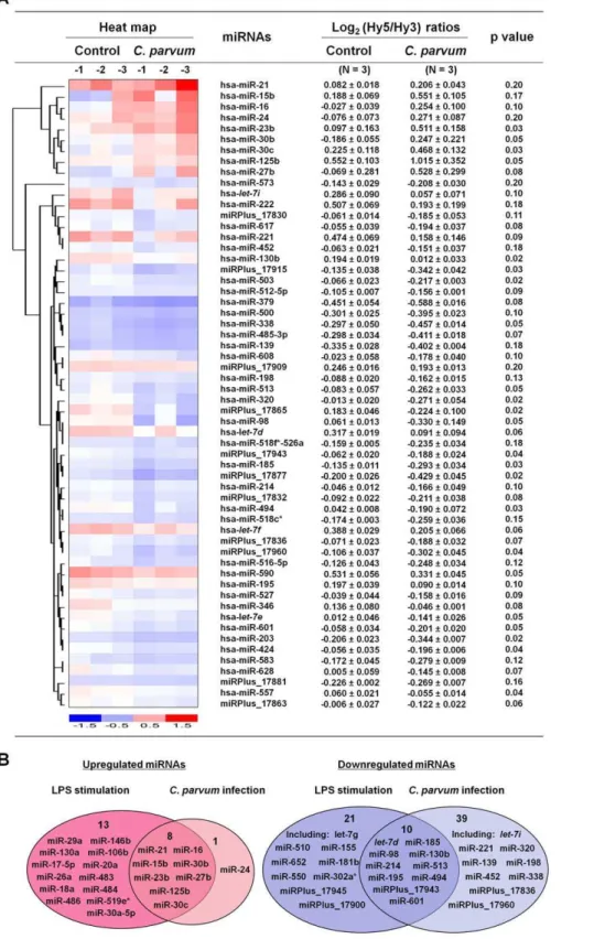
C. parvuminfection, we performed a microarray analysis of mature miRNA expression in H69 cells [26]. The miRCURYTM LNA human microRNAs assays (version 8.1; Exiqon; Vedbaek, Denmark) covers a total of up to 600 known human mature miRNAs and were used as previously described [27]. The quality of the RNA was verified using an Agilent 2100 Bioanalyzer (Figure S1). A total of 383 mature miRNAs were detected in the uninfected H69 cells. Of the miRNAs expressed, miR-23b, miR-30b, miR-30c, and miR-125b expression were significantly increased in H69 cells after exposure to liveC. parvuminfection for 12 h (p,= 0.05; Figure 1A and Table S1). Five additional miRNAs (miR-15b, miR-16, miR-27b, miR-24, and miR-21) showed a tendency to increase (0.05,p,= 0.20) (Figure 1A). A total of 19 miRNAs were significantly downregulated (p,= 0.05) and 30 additional miRNAs showed a tendency to decrease (0.05,p,= 0.20) followingC. parvuminfection (Figure 1A and Table S1). Sham-infected control cells (H69 cells exposed to heat-inactivated C. parvum oocysts after incubation at 65uC for 30 min) displayed a similar miRNA expression profile as non-infected control samples (Table S1). Microarray analysis of mature miRNAs was also performed on H69 cells treated with LPS (1mg/ ml for 8 h). Interestingly, most of the miRNAs upregulated byC. parvumalso displayed an increased expression in cells treated by LPS (Figure 1B and Table S1). Nevertheless, increased expression of additional 13 miRNAs was identified in LPS-treated cells but not in cells exposed toC. parvum. A total of 31 miRNAs showed a decreased expression in LPS-treated cells and 10 of them were also downregulated byC. parvum (Figure 1B and Table S1). No LPS contamination in theC. parvumpreparation was detected using the Limulus Amebocyte Lysate (LAL) test kit (Bio-Whittaker) (data not shown). All microarray data were described in accordance with MIAME guidelines and deposited at ArrayExpress (accession number: E-MEXP-2050 and E-MEXP-2052).
Real-time PCR analysis using primers and probes for mature miRNAs (Ambion) was performed to assess the kinetics of selected miRNAs in H69 cells following C. parvum infection. Increased expression of miR-125b, miR-21, miR-23b, miR-30b and miR-16 was detected in H69 cells followingC. parvuminfection for 12 h to 24 h, but not in the early time points (2 h to 8 h) (Figure 2A). Increased expression of miR-125b, miR-16, miR-23b, miR-21 and miR-30b, as well as decreased expression of miR-98, was further
Author Summary
Figure 1. Expression profiling of mature miRNAs in cholangiocytes followingC. parvuminfection and LPS stimulation.(A) miRNA expression profile in H69 cells followingC. parvuminfection. The left panel shows a heat-map of selected miRNAs that showed changes in expression in H69 cells followingC. parvuminfection. The horizontal axis indicates samples of non-infected cells (n = 3; Control-1, -2, and -3) and cells after exposure to liveC. parvumfor 12 h (n = 3,C. parvum-1, -2, and -3). The right panel shows expression of miRNAs in H69 cells followingC. parvum
infection. Cellular levels of miRNAs were presented as the log2(Hy5/Hy3) ratios which passed the filtering criteria variation across the samples.p values are from the t’ test. hsa =Homo sapiens. (B) Comparison of miRNA expression patterns in H69 cells followingC. parvuminfection for 12 h and LPS stimulation for 8 h. Graphics indicate those miRNAs showing an increased or decreased expression (including those significant change when p,= 0.05 and those with a tendency to change when 0.05,p,= 0.20) in cells after treatment with LPS (n = 3) or exposure toC. parvum(n = 3). A complete description of miRNA expression profiles in cells was listed in Table S1.
confirmed in cells followingC. parvuminfection for 12 h by Northern blot (Figure 2B). An increased expression of the precursors for miR-125b, miR-16, miR-21 and miR-23b was also detected in cells following C. parvum infection by Northern blot (Figure 2B). No positive signal for the above human miRNAs was detected inC. parvumRNA using the probes or primers for miRNA real-time PCR (data not shown) and Northern blot (Figure 2C), demonstrating the specificity of these probes for human miRNAs. Downregulation of selected miRNAs induced byC. parvum, including miR-98, miR-320 and miR-424, was further confirmed by bead-based multiplexed miRNA expression assay using the FlexmiRTM Select kit (Figure 2D). For those miRNAs that did not show significant alterations in cells followingC. parvuminfection as revealed by the microarray analysis, we selected miR-326 for bead-based multi-plexed analysis and no change was detected inC. parvuminfected cells (Figure 2D), further confirming the accuracy of the array data.
Database analysis of upregulated miRNAs in
cholangiocytes followingC. parvuminfection reveals potential NF-kB binding sites in their promoter elements
Differential alterations in the mature miRNA expression profile of C. parvum-infected H69 cells suggest that miRNA gene expression is finely controlled in epithelial cells in response toC. parvuminfection. One potential mechanism for selectively altering miRNA levels is through activation of distinct intracellular signaling pathways and nuclear transcription factors [15,16]. This mechanism is consistent with our previous data demonstrat-ing that C. parvum infection activates the NF-kB pathway in cholangiocytes through microbial recognition of TLR4 and TLR2 [28]. We hypothesized that activation of the NF-kB pathway is involved in the transcription of select miRNAs upregulated byC. parvum. Based on TFSEARCH (http://www.cbrc.jp/research/db/ TFSEARCH.html) and MOTIF (http://motif.genome.jp/) data-base searches [29,30], many of these miRNA genes have putative NF-kB binding sites in their potential promoter elements [31–34] (Table 1). Several miRNAs upregulated in H69 followingC. parvum
infection are cluster miRNAs; e.g., 23b, 27b and miR-24 are from themir-23b-27b-24-1gene cluster and miR-15b and miR-16 from themir-15b-16-2cluster [31,32]. The promoters of themir-125b-1andmir-30bgenes have not been characterized and it is unknown whether they have potential NF-kB binding sites. Transactivation of most NF-kB-dependent genes requires NF-kB p65 binding to the promoter [8] and nuclear translocation of p65 was demonstrated followingC. parvum infection of cholangiocytes [28]. Coupled with the results showing some similar changes in miRNA expression in H69 cells treated with LPS (which activates TLR4/NF-kB signaling in H69 cells), we then focused on determining whether p65 binds to the promoter and transactivates the miRNA genes upregulated byC. parvuminfection.
Differential expression of primary transcripts ofC. parvum-upregulated mature miRNAs in H69 cells
We then analyzed the kinetics of alterations of the primary transcripts (pri-miRNAs) for select mature miRNAs upregulated byC. parvum as listed in Table 1. H69 cells were exposed toC. parvum for various periods of time and pri-miRNAs of interests were quantified by real-time PCR (primers listed in Table S2). Expression of pri-miR-125b-1, pri-miR-21, 23b-27b-24-1, 30b, 30c-23b-27b-24-1, 15a-16-23b-27b-24-1, and pri-miR-15b-16-2 showed a time-dependent increase in cells followingC. parvuminfection, with a peak at 8 h or 12 h after exposure to the parasite (Figure 3). In contrast, no significant increase of pri-miR-125b-2 and pri-miR-30c-2 was detected in cells after exposure to
Figure 2. Altered expression of selected miRNAs confirmed by real-time PCR, Northern blot and Luminex bead analyses.(A) Alterations of selected miRNA expression in cells after exposure toC. parvumfor various periods of time as assessed by real-time PCR. The amount of mature miRNAs was obtained by normalizing to the level of snRNA RNU6B in the samples. Data are expressed as the amount of mature miRNAs in the infected samples relative to the control uninfected samples and representative of three independent experi-ments. (B) Alterations of selected miRNA expression in cells after exposure toC. parvumfor 12 h as determined by Northern blot. snRNA RNU6B was used as a control to ensure equal loading. Representative Northern blots (C. parvuminfected cells vs non-infected control) from three independent experiments are shown. (C) Total RNA isolated from
C. parvumoocysts was also blotted to demonstrate the specificity of the probes. (D) Alterations of selected miRNA expression in cells after exposure to C. parvumfor 12 h as assessed by bead-based miRNA Luminex analysis. The amount of mature miRNAs was obtained by normalizing the samples to the positive control beads provided by the company (Luminex). Data are representative of three independent experiments. *, p,0.05 vs. the non-infected control.
C. parvuminfection (Figure 3), suggesting a differential expression of the primary transcripts ofC. parvum-upregulated miRNAs.
Promoter binding of NF-kB p65 subunit is required for the transcription of select miRNA genes induced byC. parvumin H69 cells
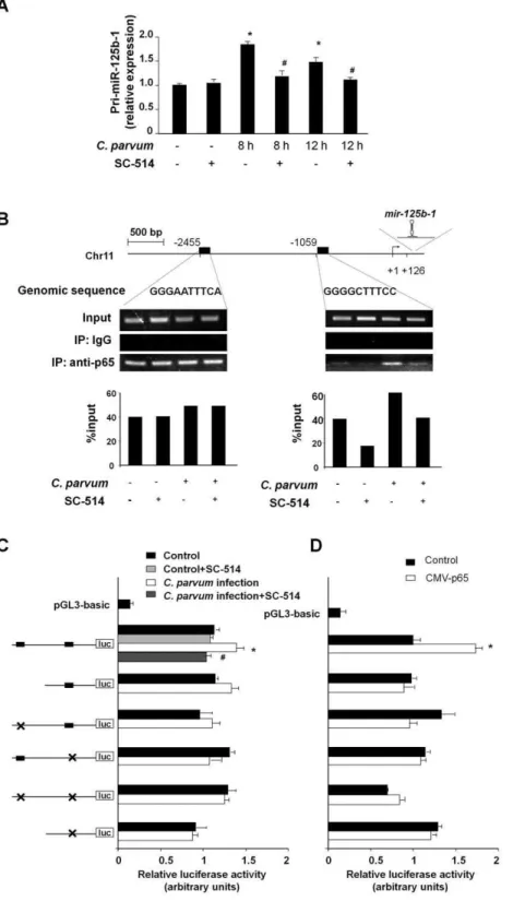
To test whether NF-kB p65 subunit is involved in C. parvum -induced transactivation of pri-miR-125b-1, we exposed H69 cells to C. parvum infection in the presence of SC-514, an IKK2
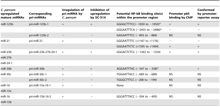
inhibitor that prevents p65-associated transcriptional activation of the NF-kB pathway [35]. SC-514 blocked theC. parvum-induced increase of pri-miR-125b-1 (Figure 4A). To further test the potential transactivation ofmir-125b-1gene by p65 subunit, rapid amplification of 59 complementary DNA ends (59-RACE) PCR was used to identify the 59end of pri-miR-125b-1. Primers were designed to amplify pri-miR-125b-1 based on the sequence obtained from the Sanger miRNA Registry (Table S2). Database analysis revealed two potential p65 binding sites in the upstream Table 1.Analysis ofC. parvum-upregulated miRNAs in cholangiocytes reveals potential transactivation of their genes by NF-kB.
Mature miRNAs miRNA genes (or cluster) Chromosome (strand) Host gene Predicted NF-kB binding sites (from miRNA TSS) Reference
miR-125b mir-125b-1 11 (2) None Promoter element unknown [31]
mir-125b-2 21 (+) C21orf34 GAGAATTTCC (2893 to2884)
miR-21 mir-21 17 (+) None GGGAATTTTC (+1167 to+1176) [33,34]
GGGAATTCTC (+1395 to+1404)
miR-23b mir-23b-27b-24-1 9 (+) C9orf3 GGGACTCTCC (21263 to21254) [31]
miR-27b
miR-24
miR-30b mir-30b 8 (2) None Promoter element unknown
miR-30c mir-30c-1 1 (+) NFYC TGGAATTACC (2689 to2680) [31,32]
mir-30c-2 6 (2) C6orf155 TGGGCTTTCC (2208 to2199)
miR-16 mir-15a-16-1 13 (2) DLEU2 None [32]
miR-15a
miR-16 mir-15b-16-2 3 (+) SMC4 GGGATTTACC (2504 to2495) [32]
miR-15b
MicroRNA genes related toC. parvum-upregulated mature miRNAs, their chromosomal location and co-expressed host genes were identified by the miRBase (http:// microrna.sanger.ac.uk/) database search and confirmed by previous studies as referred. Potential promoter element for each miRNA was based on the referred studies and potential NF-kB binding sites were identified by the TFSEARCH (http://www.cbrc.jp/research/db/TFSEARCH.html) and MOTIF (http://motif.genome.jp/) search. doi:10.1371/journal.ppat.1000681.t001
Figure 3. Differential expression of primary transcripts ofC. parvum-upregulated mature miRNAs in H69 cells.H69 cells were exposed toC. parvumfor 2 h to 24 h and primary transcripts (pri-miRNAs) of select miRNAs were quantified by real-time PCR. The amount of pri-miRNAs was obtained by normalizing to the level of GAPDH in the samples. Data are expressed as the amount of pri-miRNAs in the infected samples relative to the control uninfected samples and representative of three independent experiments. *, p,0.05 vs. the non-infected control.
Figure 4. Promoter binding of p65 transactivatesmiR-125b-1gene to increase miR-125b expression followingC. parvuminfection.(A) p65-dependent upregulation of pri-miR-125b-1 in cholangiocytes followingC. parvuminfection. Data are presented as the relative expression level of pri-miR-125b-1 in H69 cells followingC. parvuminfection in the presence or absence of SC-514 as assessed by real-time PCR. (B)C. parvumincreases promoter element binding of p65 tomir-125b-1gene. The schematic diagram shows two potential binding sites in the putative promoter element of
mir-125b-1gene. ChIP analysis revealed increased binding of p65 to the binding site at21059, but not at22455, ofmir-125b-1promoter element in cells following infection. Representative ChIP gels are shown in the upper panel and densitometry analysis of the gels in the lower panel. (C) H69 cells were transfected with various luciferase reporter constructs spanning the potential p65 binding sites of themir-125b-1promoter. The transfected cells were exposed toC. parvumin the presence or absence of SC-514. Luciferase activity was measured and presented as the ratio of the activity of the test construct with the control luciferase reporter construct. Six reporter constructs containing the mutants of the two potential NF-kB binding sites were also utilized for the analysis as indicated. (D) H69 cells were co-transfected with the pCMV-p65 to overexpress p65 and the luciferase reporter construct containing themir-125b-1promoter for 24 h followed by measurement of luciferase activity. *, p,0.05 vs. the non-infected control (in A and C) or empty pCMV vector control (in D);#
sequence ofmir-125b-1(Figure 4B). Increased binding of p65 to the binding site at 21059, but not the putative binding site at 22455, in the promoter element ofmir-125b-1 gene (Figure 4B) was demonstrated by chromatin immunoprecipitation (ChIP) analysis using specific primers for each putative binding site (Table S2). C. parvum-induced transactivation of the mir-125b-1
gene by p65 was further confirmed by using luciferase reporter gene constructs that spanned themir-125b-1promoter (Figure 4C).
C. parvuminfection increased luciferase activity in cells transfected with the luciferase constructs that encompassed the binding site for p65 at 21059 in the promoter region of mir-125b-1 gene. A mutant of the p65 binding site at 21059 blocked C. parvum -induced luciferase activity. In addition, SC-514 significantly inhibited the increase of luciferase activity induced by C. parvum
infection (Figure 4C). Moreover, p65-associated transactivation of themir-125b-1promoter was also confirmed by the upregulation of luciferase activity after p65 overexpression in the cells (Figure 4D). As an additional control, we analyzedIL-8transactivation, a p65-dependent process induced by C. parvum in epithelial cells [36]. NF-kB p65-dependent increase of IL-8 mRNA expression and binding of p65 to the promoter ofIL-8gene in cells exposed toC. parvum were confirmed (Figure S2). Together, these data demonstrate that p65 binding to the promoter element of the
mir-125b-1 gene mediatesmir-125b upregulation in H69 cells in response to C. parvum infection. The dynamics of p65 nuclear translocation were confirmed by Western blot analysis of p65 in the nuclear extracts from H69 cells followingC. parvum infection (Protocol S1 and Figure S3), correlated to the kinetics ofC. parvum
-induced expression of pri-miRNAs in cells (Figure 3). Consistent with previous results, maximal p65 translocation was observed at 8 h after exposure toC. parvum[5].
Using the same approaches, we analyzed p65 promoter element binding inC. parvum-induced transcription of 21, miR-23b-27b-24-1, miR-30b, miR-30c-1, miR-30c-2, pri-miR-15a-16-1, and pri-miR-15b-16-2. Our data are summarized in Table 2 and presented in detail in Figures S4, S5, S6 and S7. Specifically, p65 binding to the putative p65 binding site around +1395 of themir-21gene appears to be associated withC. parvum -induced transcription of pri-miR-21 (Figure S4). C. parvum
increases transcription of pri-miR-23b-27b-24-1 cluster, as well as the host gene transcript, C9orf3, via promoter binding of p65 to a binding site at21254 of the immediate upstream of the gene (Figure S5). Increased transcription of pri-miR-30b induced byC. parvumis p65-dependent (Figure S6). Nevertheless, it appears that
C. parvuminfection increases transcription of miR-30c-1, pri-miR-15a-16-1 and pri-miR-15b-16-2 in cholangiocytes through a p65-independent mechanism (Figure S7).
Functional inhibition of selected p65-dependent miRNAs in cholangiocytes increasesC. parvuminfection burden
To test whether miRNAs are involved in cholangiocyte defense responses againstC. parvuminfection, we assessed parasite burden over time in cultured cholangiocytes transfected with various anti-miRs thereby inhibiting function of specificC. parvum-upregulated miRNAs. Anti-miRs (anti-miRTM miRNA inhibitors) are com-mercially available, chemically modified single stranded nucleic
Table 2.Promoter binding of NF-kB p65 subunit inC. parvum-induced transactivation of miRNA genes in H69 cells.
C. parvum -upregulated mature miRNAs
Corresponding pri-miRNAs
Uregulation of pri-miRNA by C. parvum
Inhibition of upregulation by SC-514
Potential NF-kB binding site(s) within the promoter region
Promoter p65 binding by ChIP
Conformed by promoter reporter assay
miR-125b pri-miR-125b-1 + + GGGGCTTTCC(21059 to21050)* + +
GGGAATTTCA (22455 to22446)* 2 2
pri-miR-125b-2 2 2 GAGAATTTCC (2893 to2884) NS NS
miR-21 pri-miR-21 + + GGGAATTTTC (+1167 to+1176) 2 2
GGGAATTCTC (+1395 to+1404) + +
miR-23b pri-miR-23b-27b-24-1 + + GGGACTCTCC (21263 to21254) + +
miR-27b
miR-24-1
miR-30b pri-miR-30b + + AGGAATTTAC (2347 to2338)* + +
miR-30c pri-miR-30c-1 + 2 TGGAATTACC (2689 to2680) NS NS
pri-miR-30c-2 2 2 TGGGCTTTCC (2208 to2199) NS NS
miR-16 pri-miR-15a-16-1 + 2 None NS NS
miR-15a
miR-16 pri-miR-15b-16-2 + 2 GGGATTTACC (2504 to2495) NS NS
miR-15b
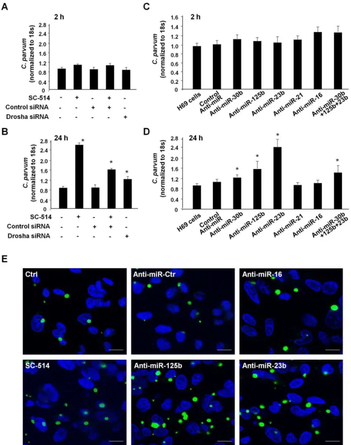
acids designed to specifically bind to and inhibit endogenous miRNAs [21]. Cells were transfected with specific anti-miRs (30 nM, Ambion) or a mixture of anti-miRs to 125b, miR-23b and miR-30b (a total of 30 nM with 10 nM for each), and then exposed toC. parvum. Following incubation with a constant number ofC. parvumsporozoites for 2 h to allow sufficient host-cell attachment and cellular invasion [3,24], cells were washed with culture medium to remove non-attached and non-internalized parasites. Cells were then cultured for an additional 2 h or 22 h. Parasite burden was assessed in the samples using a real-time PCR approach as we previously reported [24]. The parasite burden following exposure toC. parvumfor 2 h was similar in all cultures, including those transfected with the siRNA to Drosha or the specific anti-miRs (Figure 5A and 5C), suggesting that those miRNAs do not affect initial parasite host cell attachment and cellular invasion. Additionally, SC-514 treatment did not impact parasite burden at this time point (Figure 5A). Consistent with our previous studies [24], a significant increase in parasite burden was identified in SC-514-treated H69 cells 24 h after initial infection (Figure 5B). Cells transfected with the siRNA to Drosha displayed an increased parasite burden as compared to control cells (Figure 5B). Interestingly, we also detected a significantly higher parasite burden 24 h after initial infection in cells treated with the anti-miRs to miR-125b, miR-23b, and miR-30b, as well as a mixture of three anti-miRs, compared with that in control cells (Figure 5D); anti-miRs to miR-16 and miR-21 did not impact infection burden (Figure 5D). Increase of parasite burden 24 h after initial infection in H69 cells treated with SC-514 or select anti-miRs was further confirmed by immunofluorescent micros-copy (Figure 5E).
The targets of a majority of known miRNAs are still yet to be identified. C. parvum-responsive miRNAs may regulate the expression of proteins of various functions related to epithelial anti-C. parvumdefense. Using computational analyses as previously reported [19,37–39], we identified a variety of potential targets of
C. parvum-responsive miRNAs selected on the basis of their known involvement in immune related responses (Table S3).
Discussion
There is emerging evidence that miRNAs play a critical role in the regulation of both innate and adaptive immunity [17–19]. A better understanding miRNA expression changes in epithelial cells followingC. parvuminfection will provide new insights in miRNA-associated epithelial defense toC. parvum. Using anin vitromodel of human biliary cryptosporidiosis, we report significant alterations in miRNA expression profiles in epithelial cells following C. parvum
infection. Our analysis of miRNAs upregulated byC. parvumin H69 cells revealed that mir-125b-1, mir-23b-27b-24-1, mir-21, and mir-30bgenes are transactivated via potential promoter binding of the NF-kB p65 subunit. These data provide several insights relevant to miRNA expression regulation in cholangiocytes followingC. parvum
infection. First, similar to the regulation of miRNA genes in other cells [16,40,41], promoter binding of transcription factors regulates miRNA genes in epithelial cells in response toC. parvuminfection. Therefore, transcription factor-mediated miRNA expression and subsequent gene regulation at the posttranscriptional level through miRNA targeting may be an important element of host responses against C. parvum infection. Since similar alterations in miRNA expression profile were identified in LPS-treated cells, this observation may also be relevant to cellular gene regulation in general. Second, transactivation of miRNA genes that produce the same mature miRNA can be differentially controlled. Specifically, bothmir-125b-1andmir-125b-2genes can produce mature
miR-125b, but only transactivation ofmir-125b-1gene was detected in cells followingC. parvuminfection. Indeed, differential activation of genes for the same mature miRNA molecule has been previously reported [31]. Finally, transactivation of genes of cluster miRNAs or as introns in other gene alleles may be controlled by the same promoter element. Of note, miR-23b, miR-27b and miR-24 are cluster gene miRNAs and co-transcribed with a host gene,C9orf3
[31].C. parvuminfection upregulates expression of the mature forms of these three miRNAs, as well as pri-miR-23b-27b-24-1 and the host gene transcript, C9orf3. Our data are consistent with recent studies demonstrating transcriptional control of genes that code cluster miRNAs or that encode both miRNAs and other host transcripts [31,32].
The NF-kB family of transcription factors consists of five members, p50, p52, p65 (RelA), c-Rel, and RelB [8]. The transcription activation domain (TAD) necessary for the positive regulation of gene expression is present only in p65, c-Rel, and RelB [8]. Thus, promoter binding of p65, c-Rel and RelB is usually associated with gene transactivation [8,42–44]. Because they lack TADs, p50 and p52 may repress transcription unless they associate with a TAD-containing NF-kB family member or another protein capable of coactivator recruitment [8,45,46]. Increased nuclear translocation of p65 and p50 was previously reported in human cholangiocytes following C. parvuminfection [28]. In this study, we demonstrated that promoter binding of the NF-kB p65 subunit is required for transactivation of the mir-125b-1,mir-23b-27b-24-1,mir-21andmir-30bgenes in cells followingC. parvum infection. Although transactivation of mir-30c-1 and mir-15b-16-2 genes was observed in C. parvum-infected cells and potential NF-kB binding sites were identified in their promoter elements, inhibition of p65 activation failed to inhibit transactiva-tion of eithermir-30c-1ormir-15b-16-2 in H69 cells followingC. parvum-infection. In addition, miR-146b, miR-155, and miR-9 have been reported to be NF-kB-dependent miRNAs in monocytes or neutrophils [15,47,48]. Although miR-146b and miR-155 are expressed in cholangiocytes, no upregulation of either miR-146b or miR-155 was detected in H69 cells following
C. parvum infection. Given the complexity and variability in the gene structure for each miRNA, it is obvious that multiple mechanisms are involved in the transcriptional regulation of human miRNA genes [32,33,49]. Therefore, transcription of miRNA genes is expected to be a dynamic process in response to the constant alterations in intracellular signals. miRNA expression thus reflects the final integrated result of multiple interrelated signals on miRNA transcription. In this regard, other transcription factors, such as AP-1, c-myc, C/EBPa, may also be involved in the transcriptional regulation of miRNA genes in epithelial cells in response to C. parvum infection. Future studies will focus on whether nuclear translocation of p50 is involved in theC. parvum -induced down-regulation of miRNA expression.
Figure 5. Functional inhibition of selected p65-dependent miRNAs in cholangiocytes increasesC. parvuminfection burdenin vitro. (A) A similar number of parasites was detected in cells transfected with Drosha siRNA or treated with SC-514 after initial exposure toC. parvumfor 2 h as quantified by real-time PCR. (B) Transfection of cells with Drosha siRNA or treated with SC-514 increased C. parvum infection burden in cholangiocytesin vitro24 h after initial exposure to the parasite. (C) Effects of anti-miRs onC. parvumafter initial exposure toC. parvumfor 2 h. (D) Transfection of cells with anti-miRs onC. parvuminfection burden in cholangiocytes 24 h after initial exposure to the parasite. *,p,0.05 vs. non-treated cells or cells transfected with a control siRNA (in B) or non-specific control anti-miR (in D). (E) Effects of anti-miRs or SC-514 onC. parvum
burden in cholangiocytesin vitro24 h after initial exposure to the parasite as assessed by immunofluorescent microscopy.C. parvumparasites were stained in green and nuclei in blue. Bars = 5mm.
The TLR/NF-kB signaling is critical to innate epithelial immune defenses to microbial infection including parasites [4,52]. We previously demonstrated that TLR4 and TLR2 are involved in cholangiocyte immune response toC. parvuminfection via activation of NF-kB [5]. Here, we expanded our previous studies by demonstrating that miRNAs may also regulate TLR/ NF-kB-mediated epithelial anti-C. parvumdefense. We indentified a panel of miRNA genes that are transactivated via p65 promoter binding in cholangiocytes in response to C. parvum infection. Transfection of cells with anti-miRs to miR-125b, miR-23b or miR-30b, but not anti-miRs to miR-16 or miR-21, significantly increased parasite burden in cholangiocytes. The molecular mechanisms by which C. parvum-responsive miRNAs modulate epithelial anti-C. parvum defense are largely unclear. Previous studies demonstrated thatlet-7regulates TLR4 expression and is involved in epithelial defense against C. parvum [24]. Various immune related genes are identified as potential targets for these
C. parvum-responsive miRNAs using computational analyses. The concept that a pathogen encodes mRNAs targeted by host miRNAs has recently emerged as an important mechanism of host anti-viral defense [21]. Likewise, it is of interest to test the possibility that host cell miRNAs target the internalized parasite mRNAs and silence genes of the pathogen. The directC. parvum -host cell cytoplasmic tunnel-connection [53] could mediate exchange of molecules, including miRNAs, between the host cells and internalized parasite. Further investigation should test whether p65 promoter binding transactivates LPS-responsive miRNA genes. This also raises the possibility that transactivation of miRNA genes through promoter binding of NF-kB subunits may be involved in host anti-microbial responses in general.
In summary, this first miRNA profiling in cholangiocytes in response to C. parvum infection in vitro revealed significant alterations in cellular miRNA expression. The mechanism by which C. parvum induces upregulation of a panel of miRNAs in cholangiocytes involves transactivation of miRNA genes through promoter binding of the NF-kB p65 subunit. In addition, functional inhibition of the upregulated miRNAs increases C. parvum infection burden in cholangiocytes in vitro thereby implicating these miRNAs in host cell defense to the parasite. These data demonstrate a key role for miRNAs in epithelial immune responses against C. parvum infection and may provide new insights into general mechanisms of the regulation of epithelial anti-microbial immunity.
Materials and Methods
C. parvumand human cholangiocyte cell line
C. parvum oocysts of the Iowa strain were purchased from a commercial source (Bunch Grass Farm, Deary, ID). H69 cells (a gift of Dr. D. Jefferson, Tufts University) are SV40 transformed normal human cholangiocytes originally derived from liver harvested for transplant. These cholangiocytes continue to express biliary epithelial cell markers, including cytokeratin 19, gamma glutamyl transpeptidase and ion transporters consistent with biliary function and have been extensively characterized [26].
In vitroinfection model and infection assay
Anin vitromodel of human biliary cryptosporidiosis using H69 cells was employed in these studies. Before infecting cells,C. parvum
oocysts were treated with 1% sodium hypochlorite on ice for 20 min followed by extensive washing with DMEM-F12 medium. Oocysts were then added to the cell culture to release sporozoites to infect cells [54]. Infection was performed in culture medium (DMEM-F12 with 100 U/ml penicillin and 100mg/ml
strepto-mycin) containing viableC. parvumoocysts (oocysts with host cells in a 5:1 ratio). Inactivated organisms (treated at 65uC for 30 min) were used for sham infection controls. All experiments were performed in triplicate. For the inhibition experiments, SC-514 (Calbiochem) was added to the medium. Cells were pre-treated with SC-514 for 1 h prior toC. parvuminfection. SC-514 was used at a concentration of 100mM, which showed no cytotoxic effects on H69 cells or onC. parvumsporozoites, in these studies.
Real-time PCR and immunofluorescent microscopy were used to assayC. parvum infection as previously reported [24]. Briefly, primers specific for C. parvum 18s ribosomal RNA (forward: 59 -TAGAGATTGGAGGTTGTTCCT-39and reverse: 59 -CTCCA-CCAACTAAGAACGGCC-39) were used to amplify the cDNA specific to the parasite. Primers specific for human plusC. parvum
18s were used to determine total 18s cDNA [24]. Data were expressed as copies ofC. parvum 18s vs total 18s. For immuno-fluorescent microscopy, cells were fixed with 2% paraformalde-hyde and incubated with a polyclonal antibody againstC. parvum(a gift from Dr. Guan Zhu, Texas A&M University) followed by anti-rabbit FITC-conjugated secondary antibody (Molecular Probes) and co-staining with 49, 6-diamidino-2-phenylindole (DAPI, 5mM) to stain cell nuclei. Labeled cells were assessed by confocal laser scanning microscopy.
miRCURYTMLNA array analysis of miRNAs
The Exiqon (Vedbaek, Denmark) miRCURY LNA microRNA arrays and service to process the samples were used [27]. Briefly, H69 cells were grown to 80% confluence and exposed toC. parvum
oocysts for 12 h or LPS (1mg/ml) for 8 h. Total RNAs from H69 cells or C. parvum oocysts were prepared with the mirVanaTM miRNA Isolation Kit according to the manufacturer’s instruction (Ambion). The quality of isolated RNAs was verified by an Agilent 2100 Bioanalyzer profile (Figure S1). A mixture of equal amounts of total RNAs from the control andC. parvum-infected cells were used as the reference pool. A total of 2mg RNA from each sample was then labeled with the Hy5TM fluorescent label and the reference pool labeled with Hy3TM using the miRCURYTM LNA Array labeling kit (Exiqon). The labeled samples and reference pool were then mixed pair-wise and hybridized to the miRCURYTM LNA array containing capture probes targeting all human miRNAs listed in the miRBASE version 8.1 (Exiqon). After hybridization, the slides were scanned and quantified signals normalized by Exiqon using the global Lowess (Locally Weighted Scatterplot Smoothing) regression algorithm. Normalized Hy5/Hy3 ratios were used for further analysis as previously reported [55–57].
Bead-based multiplex sandwich immunoassays
A bead-based multiplex sandwich immunoassay, read with a Luminex 200 system (Luminex), was used to measure the concentrations of selected miRNAs as previously reported [57]. Briefly, total cellular RNAs are isolated using the mirVanaTM miRNA Isolation Kit (Ambion). An amount of 0.5mg of total RNAs was used for Biotin-labeling using the FlexmiR MicroRNA Labeling Kit for selected miRNAs (Luminex). Signals for miRNAs were recorded and standardized to the standard beads according to the manufacturer’s instructions (Luminex).
Real-time PCR
the Applied Biosystems 7500 FAST real-time PCR System. Mature miRNA-specific primers and probes were obtained from Applied Biosystems. Normalization was performed by using RNU6B primers and probes. Relative expression was calculated by using the comparative CT method [56].
For analysis of pri-miRNAs, total RNA was isolated from cells with Trizol reagent (Ambion). RNAs were treated with DNA-freeTM Kit (Ambion) to remove any remaining DNA. Compar-ative real-time PCR was performed by using the SYBR Green PCR Master Mix (Applied Biosystems). Specific primers for pri-miRNAs were listed in Table S2. All reactions were run in triplicate. The Ct values were analyzed using the comparative Ct (DDCt) method and the amount of target was obtained by normalizing to the endogenous reference (GAPDH) and relative to the control (non-treated cells) [58].
Northern blot
Total RNAs harvested as above were run on a 15% Tris/ Borate/EDTA (90 mM Tris/64.6 mM boric acid/2.5 mM EDTA, pH 8.3)–urea gel (Invitrogen) and transferred to a Nytran nylon transfer membrane (Ambion). LNA DIG-probes for selected miRNAs (Exiqon) were hybridized using UltraHyb reagents (Ambion) according to the manufacturer’s instructions with blotted snRNA RNU6B as a control.
59-RACE PCR
59-RACE PCR was utilized to identify 59end of miRNA primary transcripts to localize the start sites ofmir-125b-1,mir-30band mir-30d. Primer sequences are listed in Table S2. The SMARTTM RACE cDNA Amplification Kit (Clontech) was used for the analysis. Total RNA was isolated for H69 cells treated with a Drosha siRNA (Santa Cruz biotechnology) as previously reported [32].
ChIP
ChIP analysis was performed with a commercially available ChIP Assay Kit (Upstate Biotechnologies) in accordance with the manufacturer’s instructions. In brief, 16106 H69 cells were cultured in 15-cm culture dishes and exposed toC. parvumin the presence or absence of SC-514 for 8 h. The chromatin fraction was immunoprecipitated for overnight at 4uC using anti-NF-kB p65 (Upstate Biotechnologies). PCR amplification was performed in a total volume of 25ml with specific primers. The forward and reverse primers used for each gene were listed in Table S2.
Luciferase reporter constructs and luciferase assay Promoters of miRNAs were amplified by PCR from human genomic DNA. PCR primers were listed in Table S2. The PCR products were separated by agarose gel electrophoresis, and the DNA fragments then isolated and cloned in the restriction enzyme digested pGL3 Basic Vector (Promega) using T4 DNA ligase (Fisher scientific). All constructs were confirmed by sequencing. Mutations were introduced into the NF-kB binding sites using the QuikChange site-directed mutagenesis kit (Stratagene). H69 cells were transfected with each reporter construct for 24 h and then exposed toC. parvumoocysts for 8 h in the presence or absence of SC-514 followed by assessment of luciferase activity. Luciferase activities were then measured and normalized to the controlb-gal level. The luciferase activity of each construct was compared with that of the promoterless pGL3 basic vector.
Supporting Information
Table S1 miRNA expression profile in cholangiocytes following
C. parvum infection and LPS stimulation. Data represent the
mean6SE of the log2 (Hy5/Hy3) ratios from non-infected cell cultures (n = 3), C. parvum infected cultures (n = 3), LPS treated cultures (n = 3), and one cell culture exposed to heated-inactivated
C. parvum(Sham) by using the miRCURYTMLNA Array (Version 8.1). a, p,= 0.05; b, 0.05,p,= 0.20, compared with non-infected cells; NA = not detectable.
Found at: doi:10.1371/journal.ppat.1000681.s001 (0.09 MB PDF)
Table S2 Primers used for PCR and construct generating. Listed in this table are all the primers used in this study for the real-time PCR and RACE PCR, as well as those for ChIP analysis and construct generating.aRestriction enzyme sites were indicated by lowercase letters.
Found at: doi:10.1371/journal.ppat.1000681.s002 (0.02 MB PDF)
Table S3 Prediction of immune-related target genes of C. parvum-responsive miRNAs. Prediction of immune-related target genes forC. parvum-responsive miRNAs was performed with the computerlized predictive algorithms as previously reported [19,37–39]. Some of the predicted targets have been experimen-tally confirmed [24,25,27,59,60] and the corresponding miRNAs are in red font.
Found at: doi:10.1371/journal.ppat.1000681.s003 (0.02 MB PDF)
Figure S1 Quality control of RNA. Total RNAs from cells were prepared with the mirVanaTMmiRNA Isolation Kit according to the manufacturer’s instructions (Ambion). The quality of the isolated RNAs was verified by examining the Agilent 2100 Bioanalyzer profile of the sample. Representative RNA profiles from non-infected H69 cells (A), cells exposed to live (B) and heat-inactivatedC. parvumoocysts (C) are shown.
Found at: doi:10.1371/journal.ppat.1000681.s004 (1.62 MB TIF)
Figure S2 Promoter binding of p65 transactivates theIL-8gene in cholangiocytes in response to C. parvum infection. (A) p65-dependent upregulation of IL-8 mRNA in cholangiocytes following C. parvum infection. Bars represent the levels of IL-8 mRNA in cells followingC. parvum infection in the presence or absence of SC-514 as assessed by real-time PCR. (B) A schematic diagram shows the structure of IL-8 gene. ChIP analysis demonstrated increased binding of p65 to the binding site atIL-8
promoter in cells following infection. *, p,0.05 vs. non-infected cells;#, p,0.05 vs.C. parvuminfected cells.
Found at: doi:10.1371/journal.ppat.1000681.s005 (0.28 MB TIF)
Figure S3 Nuclear translocation of p65 in cholangiocytes cells induced byC. parvum. Cells were exposed toC. parvumfor various periods of time and nuclear extracts obtained as described in Protocol S1. The NF-kB p65 subunit was detected by Western blot. Actin was used as a loading control. Representative Western blots are shown.
Found at: doi:10.1371/journal.ppat.1000681.s006 (0.61 MB TIF)
Figure S4 Promoter binding of p65 transactivatesmir-21gene to increase miR-21 expression in biliary epithelial cells in response to
mutant at +1395 blocked C. parvum-induced luciferase reporter activity in transfected cells. (D) H69 cells were co-transfected with the pCMV-p65 to overexpress p65 and the luciferase reporter construct containing the mir-21 promoter. Different from the results inC. parvum-infected cells, a significant increase of luciferase reporter activity was detected in cells co-transfected with the pCMV-p65 and the mutant at +1167. *, p,0.05 vs. the non-infected control (in A and C) or empty pCMV vector control (in D);#, p,0.05 vs.C. parvuminfected cells (in A and C).
Found at: doi:10.1371/journal.ppat.1000681.s007 (0.63 MB TIF)
Figure S5 Promoter binding of p65 transactivates the mir-23b-27b-24-1 cluster gene in cholangiocytes in response toC. parvum
infection. (A) p65-dependent upregulation of pri-miR-23b-27b-24-1 in cholangiocytes following C. parvum infection. A schematic diagram shows the structure of themir-23b-27b-24-1cluster gene. Real-time PCR was used to assess the expression levels of pri-miRNA-23b-27b-24-1 and C9orf3 followingC. parvuminfection in the presence or absence of SC-514. (B) C. parvum increases promoter binding of p65 to themir-23b-27b-24-1cluster gene. The schematic diagram shows one potential NF-kB binding site in the promoter element of mir-23b-27b-24-1. ChIP analysis revealed increased binding of p65 to the binding site at 21254 of the promoter in cells following infection. (C) H69 cells were transfected with luciferase gene reporter constructs with or without mutations in the p65 binding site of the promoter and then exposed to C. parvumin the presence or absence of SC-514. (D) H69 cells were co-transfected with the pCMV-p65 to overexpress p65 and the luciferase reporter gene construct containing the promoter. Cells were then cultured for 24 h followed by measurement of luciferase activity. *, p,0.05 vs. the non-infected control (in A and C) or empty pCMV vector control (in D);#, p,0.05 vs.C. parvuminfected cells (in A and C).
Found at: doi:10.1371/journal.ppat.1000681.s008 (0.76 MB TIF)
Figure S6 Promoter binding of p65 transactivates the mir-30b
gene to increase miR-30b expression in biliary epithelial cells following C. parvuminfection. (A) p65-dependent upregulation of pri-miR-30b, but not pri-miR-30d, in cholangiocytes followingC. parvum infection. The expression levels of pri-miRNAs in cells followingC. parvuminfection were assessed by real time PCR in the presence or absence of SC-514. Treatment of cells with SC-514 blocked C. parvum-induced increase of pri-miR-30b, suggesting p65-dependent miR-30b expression. (B) We performed 59-RACE PCR to identify the 59end of pri-miR-30d and identified a potential p65 binding site at2472 of its upstream sequence. H69 cells were transfected with the luciferase gene reporter construct covering the potential p65 binding site within the putative promoter of mir-30d and then exposed toC. parvum. These results support that pri-miR-30b and pri-miR-30d are not transcribed
from the same gene in human cholangiocytes, inconsistent with previous results suggesting that pri-miR-30b and pri-miR-30d may be transcribed from the same gene on chr8 [31,40]. (C) To clarify how p65 is involved in the transactivation of miR-30b gene transactivation, we performed 59-RACE PCR but failed to amplify the corresponding sequence (data not shown). Nevertheless, database analysis revealed one potential binding site for NF-kB in the upstream sequence of miR-30b precursor. ChIP analysis detected an increased binding of p65 to this region in cells followingC. parvuminfection. (D) Luciferase reporter gene analysis demonstrated a significant increase in luciferase reporter activity in cells following C. parvum infection or overexpressed with p65. *, p,0.05 vs. the non-infected control (in A and D) or empty pCMV vector control (in E);#, p,0.05 vs.C. parvuminfected cells (in A and D).
Found at: doi:10.1371/journal.ppat.1000681.s009 (0.73 MB TIF)
Figure S7 p65-independent expression of miR-30c and miR-16 in cholangiocytes in response to C. parvuminfection. (A and B) miR-30c is transcribed from two genes,mir-30c-1and mir-30c-2, localized on chr1 and chr6, respectively [31]. Real-time PCR analysis revealed an increase of miR-30c-1(A), but not pri-miR-30c-2 (B), in H69 cells following C. parvum infection. Treatment of cells with SC-514 failed to blockC. parvum-induced expression of pri-miR-30c-1 (A). (C and D) miR-16 is transcribed from two genes,mir-15a-16-1andmir-15b-16-2localized on chr13 and chr3 and clustered with miR-15a and miR-15b, respectively [31]. Increased expression of pri-miR-15a-16-1 (at 12 h; C) and pri-miR-15b-16-2 (at 8 h and 12 h; D) was detected in H69 cells afterC. parvuminfection. Treatment of cells with SC-514 failed to block either pri-miR-15a-16-1 (C) or pri-miR-15b-16-2 (D). *, p,0.05 vs. the non-infected control.
Found at: doi:10.1371/journal.ppat.1000681.s010 (0.58 MB TIF)
Protocol S1 Nuclear translocation of p65.
Found at: doi:10.1371/journal.ppat.1000681.s011 (0.01 MB PDF)
Acknowledgments
We thank O’Hara SP (Mayo), Splinter PS (Mayo), Li X (Union Hospital, Wuhan) and Zhu G (Texas A&M) for helpful and stimulating discussions, Fang X (Creighton) for performing the statistic analysis, and Badley AD (Mayo) for providing the p65 plasmid and LaRusso NF (Mayo) for the IL-8 luciferase reporter construct.
Author Contributions
Conceived and designed the experiments: RZ GH XMC. Performed the experiments: RZ GH AYG XMC. Analyzed the data: RZ GH JL AYG KMD XMC. Contributed reagents/materials/analysis tools: GH KMD. Wrote the paper: RZ KMD XMC.
References
1. Chen XM, Keithly JS, Paya CV, LaRusso NF (2002) Cryptosporidiosis. N Engl J Med 346: 1723–1731.
2. Wanyiri J, Ward H (2006) Molecular basis of Cryptosporidium-host cell interactions: recent advances and future prospects. Future Microbiol 1: 201–208. 3. Chen XM, Levine SA, Tietz P, Krueger E, LaRusso NF (1998)Cryptosporidium parvumis cytopathic for cultured human biliary epithelia via an apoptotic mechanism. Hepatology 28: 906–913.
4. Rogers KA, Rogers AB, Leav BA, Sanchez A, Vannier E, et al. (2006) MyD88-dependent pathways mediate resistance toCryptosporidium parvuminfection in mice. Infect Immun 74: 549–556.
5. Chen XM, Nelson JB, O’Hara SP, Splinter PL, Small AJ, et al. (2005) Multiple Toll-like Receptors are expressed in human cholangiocytes and mediate host epithelial responses to Cryptoaporidium parvum via activation of NF-kappaB. J Immunol 175: 7447–7456.
6. Akira S, Takeda K (2004) Toll-like receptor signalling. Nat Rev Immunol 4: 499–511.
7. Iwasaki A, Medzhitov R (2004) Toll-like receptor control of the adaptive immune responses. Nat Immunol 5: 987–995.
8. Hayden MS, Ghosh S (2008) Shared principles in NF-kB signaling. Cell 132: 344–362.
9. Bartel DP (2004) MicroRNAs: genomics, biogenesis, mechanism, and function. Cell 116: 281–297.
10. Ambros V (2004) The functions of animal microRNAs. Nature 431: 350–355. 11. Lee Y, Kim M, Han J, Yeom KH, Lee S, et al. (2004) MicroRNA genes are
transcribed by RNA polymerase II. EMBO J 23: 4051–4060.
12. Ozsolak F, Poling LL, Wang Z, Liu H, Liu XS, et al. (2008) Chromatin structure analyses identify miRNA promoters. Genes Dev 22: 3172–83.
13. Kim YK, Kim VN (2007) Processing of intronic microRNAs. EMBO J 26: 775–783.
15. Taganov KD, Boldin MP, Chang KJ, Baltimore D (2006) NF-kappaB-dependent induction of microRNA miR-146, an inhibitor targeted to signaling proteins of innate immune responses. Proc Natl Acad Sci U S A 103: 12481–12486.
16. Fazi F, Rosa A, Fatica A, Gelmetti V, De Marchis ML, et al. (2005) A minicircuitry comprised of microRNA-223 and transcription factors NFI-A and C/EBPalpha regulates human granulopoiesis. Cell 123: 819–831.
17. Baltimore D, Boldin MP, O’Connell RM, Rao DS, Taganov KD (2008) MicroRNAs: new regulators of immune cell development and function. Nat Immunol 9: 839–845.
18. Liu J, Drescher KM, Chen XM (2009) MicroRNAs and Epithelial Immunity. Int Rev Immunol 28: 139–154.
19. Asirvatham AJ, Gregorie CJ, Hu Z, Magner WJ, Tomasi TB (2008) MicroRNA targets in immune genes and the Dicer/Argonaute and ARE machinery components. Mol Immunol 45: 1995–2006.
20. Muljo SA, Ansel KM, Kanellopoulou C, Livingston DM, Rao A, et al. (2005) Aberrant T cell differentiation in the absence of Dicer. J Exp Med 202: 261–269. 21. Pedersen IM, Cheng G, Wieland S, Volinia S, Croce CM, et al. (2007) Interferon modulation of cellular microRNAs as an antiviral mechanism. Nature 449: 919–922.
22. O’Connell RM, Taganov KD, Boldin MP, Cheng G, Baltimore D (2007) MicroRNA-155 is induced during the macrophage inflammatory response. Proc Natl Acad Sci U S A 104: 1604–1609.
23. Friedman RC, Farh KK, Burge CB, Bartel DP (2009) Most mammalian mRNAs are conserved targets of microRNAs. Genome Research 19: 92–105. 24. Chen XM, Splinter PL, O’Hara SP, LaRusso NF (2007) A cellular miRNA,
let-7i, regulates toll-like receptor 4 expression and contributes to cholangiocyte immune responses againstCryptosporidium parvuminfection. J Biol Chem 282: 28929–28938.
25. Hu G, Zhou R, Liu J, Gong A-Y, Eischeid A, et al. (2009) MicroRNA-98 and let-7 confer cholangiocyte expression of cytokine-inducible Src homology 2-containing protein in response to microbial challenge. J Immunol 183: 1617–1624.
26. Grubman SA, Perrone RD, Lee DW, Murray SL, Rogers LC, et al. (1994) Regulation of intracellular pH by immortalized human intrahepatic biliary epithelial cell lines. Am J Physiol 266 (6 Pt 1): G1060–1070.
27. Gong A-Y, Zhou R, Hu G, Li X, Splinter PL, et al. (2009) MicroRNA-513 regulates B7-H1 translation and is involved in interferon-gamma-induced B7-H1 expression in cholangiocytes. J Immunol 182: 1325–1333.
28. Chen XM, Levine SA, Splinter PL, Tietz PS, Ganong AL, et al. (2001) Cryptosporidium parvum activates nuclear factor kappaB in biliary epithelia preventing epithelial cell apoptosis. Gastroenterology 120: 1774–1783. 29. Kast C, Wang M, Whiteway M (2003) The ERK/MAPK pathway regulates the
activity of the human tissue factor pathway inhibitor-2 promoter. J Biol Chem 278: 6787–6794.
30. Musikacharoen T, Matsuguchi T, Kikuchi T, Yoshikai Y (2001) NF-kappa B and STAT5 play important roles in the regulation of mouse Toll-like receptor 2 gene expression. J Immunol 166: 4516–4524.
31. Rodriguez A, Griffiths-Jones S, Ashurst JL, Bradley A (2004) Identification of mammalian microRNA host genes and transcription units. Genome Res 14: 1902–1910.
32. Chang TC, Yu D, Lee YS, Wentzel EA, Arking DE, et al. (2008) Widespread microRNA repression by Myc contributes to tumorigenesis. Nat Genet 40: 43–50.
33. Lo¨ffler D, Brocke-Heidrich K, Pfeifer G, Stocsits C, Hackermu¨ller J, et al. (2007) Interleukin-6 dependent survival of multiple myeloma cells involves the Stat3-mediated induction of microRNA-21 through a highly conserved enhancer. Blood 110: 1330–1333.
34. Cai X, Hagedorn CH, Cullen BR (2004) Human microRNAs are processed from capped, polyadenylated transcripts that can also function as mRNAs. RNA 10: 1957–1966.
35. Kishore N, Sommers C, Mathialagan S, Guzova J, Yao M, et al. (2003) A selective IKK-2 inhibitor blocks NF-kappa B-dependent gene expression in interleukin-1 beta-stimulated synovial fibroblasts. J Biol Chem 278: 32861–32871.
36. Laurent F, Eckmann L, Savidge TC, Morgan G, Theodos C, et al. (1997) Cryptosporidium parvuminfection of human intestinal epithelial cells induces the polarized secretion of C-X-C chemokines. Infect Immun 65: 5067–5073.
37. Schmidt WM, Spiel AO, Jilma B, Wu¨ller M, et al. (2009) In vivo profile of the human leukocyte microRNA response to endotoxemia. Biochem Biophys Res Commun 380: 437–441.
38. Grimson A, Farh KK, Johnston WK, Garrett-Engele P, Lim LP, et al. (2007) MicroRNA targeting specificity in mammals: determinants beyond seed pairing. Mol Cell 27: 91–105.
39. Betel D, Wilson M, Gabow A, Marks DS, Sander C (2007) The microRNA.org resource: targets and expression. Nucleic Acid Res 36: D149–153.
40. Marson A, Levine SS, Cole MF, Frampton GM, Brambrink T (2008) Connecting microRNA genes to the core transcriptional regulatory circuitry of embryonic stem cells. Cell 134: 521–533.
41. Song G, Wang L (2008) Transcriptional mechanism for the paired miR-433 and miR-127 genes by nuclear receptors SHP and ERRc. Nucleic Acid Res 36: 5727–5735.
42. Abreu MT, Fukata M, Arditi M (2005) TLR signaling in the gut in health and disease. J Immunol 174: 4453–4460.
43. Han J, Ulevitch RJ (2005) Limiting inflammatory responses during activation of innate immunity. Nat Immunol 6: 1198–1205.
44. Harada K, Ohira S, Isse K, Ozaki S, Zen Y, et al. (2003) Lipopolysaccharide activates nuclear factor-kappaB through toll-like receptors and related molecules in cultured biliary epithelial cells. Lab Invest 83: 1657–1667.
45. Poppelmann B, Klimmek K, Strozyk E, Voss R, Schwarz T, et al. (2005) NF{kappa}B-dependent down-regulation of tumor necrosis factor receptor-associated proteins contributes to interleukin-1-mediated enhancement of ultraviolet B-induced apoptosis. J Biol Chem 280: 15635–15643.
46. Kim S, Domon-Dell C, Kang J, Chung DH, Freund JN, et al. (2004) Down-regulation of the tumor suppressor PTEN by the tumor necrosis factor-alpha/ nuclear factor-kappaB (NF-kappaB)-inducing kinase/NF-kappaB pathway is linked to a default IkappaB-alpha autoregulatory loop. J Biol Chem 279: 4285–4291.
47. Bazzoni F, Rossato M, Fabbri M, Gaudiosi D, Mirolo M, et al. (2009) Induction and regulatory function of miR-9 in human monocytes and neutrophils exposed to proinflammatory signals. Proc Natl Acad Sci U S A 106: 5282–5287. 48. Tili E, Michaille JJ, Cimino A, Costinean S, Dumitru CD, et al. (2007)
Modulation of miR-155 and miR-125b levels following lipopolysaccharide/ TNF-alpha stimulation and their possible roles in regulating the response to endotoxin shock. J Immunol 179: 5082–5089.
49. Saini HK, Griffiths-Jones S, Enright AJ (2007) Genomic analysis of human microRNA transcripts. Proc Natl Acad Sci U S A 104: 17719–177124. 50. Xue X, Sun J, Zhang Q, Wang Z, Huang Y, et al. (2008) Identification and
characterization of novel microRNAs from Schistosoma japonicum. PLoS ONE 3: e4034. doi:10.1371/journal.pone.0004034.
51. Cerutti H, Casas-Mollano JA (2006) On the origin and functions of RNA-mediated silencing: from protists to man. Curr Genet 50: 81–99.
52. Zaph C, Troy AE, Taylor BC, Berman-Booty LD, Guild KJ, et al. (2007) Epithelial-cell-intrinsic IKK-beta expression regulates intestinal immune ho-meostasis. Nature 446: 552–556.
53. Huang BQ, Chen XM, LaRusso NF (2004)Cryptosporidium parvumattachment to and internalization by human biliary epitheliain vitro: a morphologic study. J Parasitol 90: 212–221.
54. Verdon R, Keusch GT, Tzipor S, Grubman SA, Jefferson DM, et al. (1997) An in vitromodel of infection of human biliary epithelial cells byCryptosporidium parvum. J Infect Di 175: 1268–1272.
55. Castoldi M, Schmidt S, Benes V, Noerholm M, Kulozik AE, et al. (2006) A sensitive array for microRNA expression profiling (miChip) based on locked nucleic acids (LNA). RNA 12: 913–920.
56. Loscher CJ, Hokamp K, Kenna PF, Ivens AC, Humphries P, et al. (2007) Altered retinal microRNA expression profile in a mouse model of retinitis pigmentosa. Genome Biol 8: R248.
57. Lu J, Getz G, Miska EA, Alvarez-Saavedra E, Lamb J, et al. (2005) MicroRNA expression profiles classify human cancers. Nature 435: 834–838.
58. Davis BN, Hilyard AC, Lagna G, Hata A (2008) SMAD proteins control DROSHA-mediated microRNA maturation. Nature 454: 56–61.
59. Cimmino A, Calin GA, Fabbri M, Iorio MV, Ferracin M, et al. (2005) miR-15 and miR-16 induce apoptosis by targeting BCL2. Proc Natl Acad Sci U S A 102: 13944–12949.