The presence of asymptomatic third molars can represent a potential problem in the mandible when these teeth are retained and the patient has lost all normally erupted teeth. Once the mandibular first and second molars are removed, the mandibular body becomes weaker with time, increasing the complexity, morbidity and incidence of complication in the surgical procedure to remove the retained third molar. This paper reports a case where the mandibular third molars retained in a severely resorbed mandible were removed in a 54-year-old female patient. The treatment plan was based on the safe surgical removal of the teeth and prosthetic rehabilitation with an implant-supported milled bar overdenture and a bone-mucous-supported complete denture in the mandibular and maxillary arch, respectively. If the removal of a retained third molar is indicated in a severely resorbed edentulous mandible, the treatment plan must involve not only preventive measures in order to avoid mandible fracture during or after tooth removal, but also alternatives that allow an adequate mandibular rehabilitation.
Retained Third Molars Removal in
a Severely Resorbed Edentulous
M a n d i b l e . A C a s e R e p o r t
Cassio Edvard Sverzut, Alexandre Elias Trivellato, Alexander Tadeu Sverzut, Marcelo Rodrigues Azenha, Marco Aurélio Kenichi Yamaji, Andre Oliveira Pepato
Department of Oral and Maxillofacial Surgery and Periodontology, Dental School of Ribeirão Preto, University of São Paulo, Ribeirão Preto, SP, Brazil
Correspondence: Prof. Dr. Cassio Edvard Sverzut, Avenida do Café S/N, Monte Alegre, 14040-904 Ribeirão Preto, SP, Brasil. Tel: + 55-16-3602-3980. e-mail: [email protected]
Key Words: third molar, atrophic mandible, edentulous.
Introduction
Approximately 65% of the human population has at least one impacted third molar at 20 years of age (1). Reports indicate that 18-40% of all extracted third molars are asymptomatic (2), and there is considerable controversy regarding the best option for managing such cases. Adverse impacts of oral health on quality of life can be expected for one in ten patients with asymptomatic third molars. Nonetheless, for patients who develop pain and swelling related to these teeth, the odds of experiencing adverse impacts increase threefold (3).
The presence of an asymptomatic third molar may present a potential problem in the mandible when these teeth are retained and the patient has lost all normally erupted teeth. According to the Wolff´s law, after tooth extraction, the alveolar bone loses its function and consequently bone mass with time, eventually resulting in a severely resorbed mandible (4). Furthermore, the life expectancy at birth for both genders in the year of 2006 in Japan was 83 years, while in the USA and Brazil it was 78 and 72 years, respectively (5). Therefore it may be supposed that there is a tendency to treat more often adult and elderly patients presenting a severely resorbed edentulous mandible with third molars retained or partially erupted. The purpose of this paper is to present a clinical case in which the removal of the mandibular third molars was indicated in a patient with a severely resorbed edentulous mandible.
Case Report
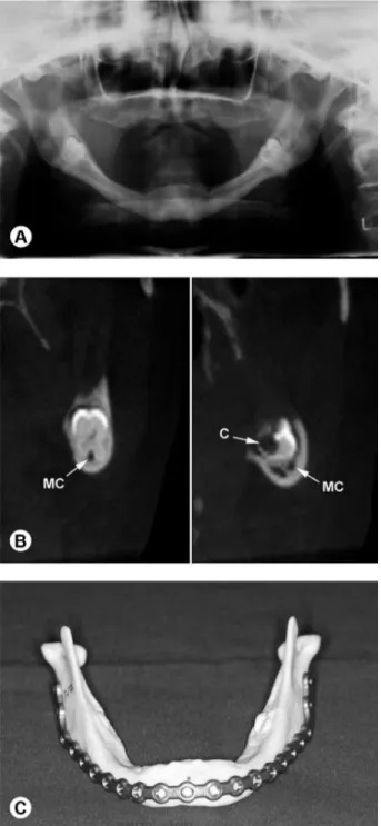
A 54-year-old Caucasian female patient came to our Dental School’s Clinic of Oral and Maxillofacial Surgery with impairment of her masticatory function associated with instability of the mandibular complete denture and recurrent episodes of acute pain in her left mandibular angle, which had began about two months before. The patient reported no contributory medical history. The clinical examination revealed complete edentulism in both arches, except for the mandibular left third molar that was partially erupted and decayed. During physical examination, a small discharging fistula with little amount of purulent secretion was noted around the left third molar and two remarkable prominences were observed on the mandibular base at both third molars location, being more palpable on the left side. An orthopantomograph previously requested by the general dentist confirmed the clinical exam about the mandibular left third molar, but it also revealed retention of the mandibular right third molar (Fig. 1). Furthermore, it revealed severe reduction of the residual ridges and Class IV mandible atrophy (4). Cone beam computed tomography (CBCT) scanning revealed close relationship between the teeth and the mandibular canals, reduced bone volume around the teeth on both sides and confirmed the carious lesion in the left molar (Fig. 2A and B). The preeminence of the tooth roots on the mandibular base was evident, mainly the mesial root of the left molar.
Resorbed mandible
the prophylactic removal of the right molar under general anesthesia were the combined procedures of choice. Two reasons were considered for establishing this treatment plan: first, the reduced amount of bone around the teeth increased the risk of mandibular fracture during or after
the surgery, and second, the possibility of a similar future occurrence related to the right molar. The DICOM files acquired from the CBCT scanning were applied to obtain a customized rapid prototype model of the mandible, which that was used to adapt a 2.4-mm locking reconstruction plate (Neo-ortho, Curitiba, PR, Brazil) to the mandible and also to select the screw length previous to the surgical procedure (Fig. 3). The advantages and disadvantages of the extra- and trans-oral surgical approaches were discussed with the patient and she decided for the trans-oral approach. Under general anesthesia by nasotracheal intubation, the surgical approach consisted in a linear incision on the crest of the alveolar residual ridge from one side to another. The posterior limits were the anterior border of the coronoid process. The anatomical structures related to the mentum foramens were identified bilaterally, dissected and transected in order to ensure an adequate surgical field to put the plate on the mandible.
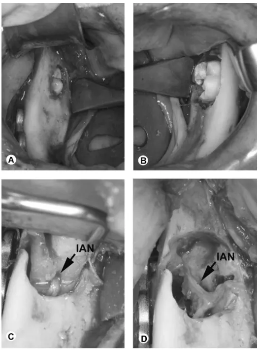
The plate was passively adapted on the bone surface and fixated first at the symphysal area by an intra-oral approach and next at the posterior area through trans-oral approach applying a trocater. The placement of screws on the symphysal area was planned in order to allow an adequate dental implant positioning in the future. After plate fixation, the teeth were removed implying odontectomy and minimum of ostectomy (Fig. 4A and B). No signal of mandibular fracture was observed. As revealed by the CBCT scanning, the inferior alveolar nerve was visualized passing through the roots of the right molar and its continuity was preserved (Figs. 5 A and B). A neurorrhaphy was carried out bilaterally in order to reconstruct the previously transected mentum nerves. The surgical field was copiously irrigated with sterile saline solution and the intra-oral suture was performed using a 4-0 Vicryl™ absorbable thread (Ethicon Ltd., Johnson & Johnson, São Paulo, SP, Brazil) and the extra-oral suture was placed using 5-0 Mononylon™ (Ethicon Ltd.).
The immediate postoperative orthopantomograph showed regular mandibular anatomy and adequate plate and screw positioning. The postoperative period occurred uneventful and as expected the patient complained about the sensory deficit in the lower lip and chin area related to the mental nerves handling. After 16 months of postoperative period the patient reported a noteworthy improvement in the sensory deficit and a new CBCT scanning was requested. The new CBCT scanning showed an adequate bone repair at the dental alveolus and the placement of dental implants was planned.
Four dental implants (3.75 x 11 mm) were placed concomitant to the vestibuloplasty procedure by modified Kazanjian technique. The repaired structures related to the mental foramens were visualized and showed regular Figure 1. A: Previous orthopantomograph revealing severe reduction of the
534
C.E. Sverzut et al.
clinical features. Four months later, the implants were exposed and the healing abutments were placed and maintained for 3 weeks. In sequence, the patient was referred to the Prosthetic Oral Rehabilitation Clinic and received an implant-supported milled bar overdenture in the mandibular arch and a bone-mucous-supported complete denture in the maxillary arch (Fig. 6A and B).
Discussion
The prophylactic removal of impacted third molars is basically indicated to avoid the morbidity associated with
tooth removal in elderly patients, while in the conservative approach removal is performed only if some pathological lesion is detected at regular follow-up visits. Nonetheless, maintaining a retained third molar in a patient that is gradually losing the other normally erupted teeth can become a serious problem in the future.
Despite advances in preventive dentistry, the location of third molars in the dental arches makes them difficult to care for, and their frequent impaction exposes patients to related degenerative conditions (1). Blakey et al. (6) evaluated 329 patients with asymptomatic retained third
Resorbed mandible
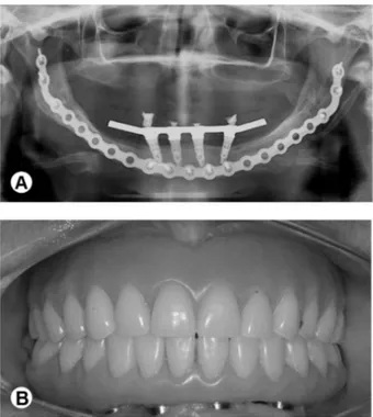
Figure 3. A: Orthopantomograph taken after prosthetic oral rehabilitation showing an adequate dental implants positioning. B: Prosthetic oral rehabilitation with an implant-supported milled bar overdenture in the mandibular arch and a bone-mucous-supported complete denture in the maxillary arch.
molars and found that 82 patients (25%) had at least one periodontal pocket equal to or greater than 5 mm on the distal surface of the second molar or around the third molar. The presence of visible third molars was associated with twice the odds of having periodontal pockets with at least 5 mm probing depth on the adjacent second molar (7) and the third molars may remain a negative impact on periodontal health long into later life (8).
The prevalence of caries in third molars has been shown to be high in patients aged 25 years and older, but not restricted to third molars (9). In another study, the presence of caries in first/second molars at baseline was highly predictive of the development of third molar caries during the ensuing 3 years (10). Therefore, if the mandibular first and/or second molars are carious, the patient must be aware of the necessity to take care of these teeth and the indication of removing the retained mandibular third molars should be discussed. Furthermore, if the carious lesions resulted in the indication of removing the first and/ or second molars, the removal of the third molar should be performed. With the loss of the first and second molars, the mandibular body becomes weaker with time as a result of bone atrophy (4), increasing the complexity, morbidity and incidence of complication in the surgical procedure to remove the retained third molar.
A routine follow-up may be proposed for an
asymptomatic third molar retained in severely resorbed mandible if any pathological condition is associated with the tooth (11). Nonetheless, the patient must be aware that this treatment involves periodic appointments and imaging exams and does not eliminate the possibility of a surgical procedure. The patients should be aware of the risks and benefits of conservative and interventionist management approaches, and patients’ perceptions should also be included in the decision-making process to provide a more comprehensive assessment of the effectiveness and value of third molar surgery (2,13).
Although a significant deterioration was observed in oral health-related quality of life in the immediate postoperative period following third molar surgery (14), an improvement of these parameters was observed in the later postoperative periods (13). This has implications for patients deciding on third molar surgery and giving informed consent, and for understanding the value of surgery from patients’ perspective and assessing health gain. Once the prophylactic removal of the third molars is indicated it should be performed at a proper surgical moment. The ideal time for removal of retained third molars is after the roots are one third formed and before they are two thirds formed (between ages 16 and 18) (15). Mesotten et al. (16) observed that the maxillary third molar formation was slightly advanced over the mandibular third molar and completion of third molar formation occurred earlier in males than females; the authors highlighted that even before the age of 18 some or all third molars may reach complete root development. Furthermore, increasing age is associated with a delayed recovery for clinical outcomes. The odds for delayed recovery of a patient older than 24 years is approximately four times the odds of a patient 18 years old or younger (17).
The surgeon’s skills and knowledge are also important because they are related to the surgery time. A 30-min or longer surgical time increases the odds of a prolonged recovery for early symptoms and lifestyle (17). Although factors are somewhat variable depending on the removed type of tooth, mandibular third molars can be more difficult to remove than their maxillary counterparts (18).
The CBCT scanning is a valuable image examination essential to establish the treatment plan and the DICOM files can be additionally applied to obtain a rapid mandible prototype model. The rapid prototype model materializes the real dimensions of the problem and early plate bending and screw length selection can be done before the surgery, decreasing the surgical time in the operating room.
536
C.E. Sverzut et al.
uncontrolled damage to the mental nerves. Actually, the intentional transaction of the mental nerves and its posterior neurorrhaphy resulted in a better surgical field and prevented a permanent damage to the mental nerve during the plate insertion. The treatment plan was based on the possibility of a bilateral mandibular fracture, not only in the intraoperative and immediate postoperative periods, but also in the near future. The mandibular bone mass decreases over time as a result of the loss of bone function, increasing the rate of mandibular fracture that is more critical in the mandibular body area. Furthermore, the screws must be placed in areas of adequate bone quality, which, in a severely resorbed mandible, are the symphysis and angle (19,20). If the single plate is sectioned at the symphysis area or two plates are applied, it will be impossible to place an adequate number of screws on the anterior portion of each plate.
Finally, if the removal of a retained third molar is indicated in a severely resorbed edentulous mandible, the treatment plan must involve not only preventive measures in order to avoid mandible fracture during or after the teeth removal, but also alternatives that allow an adequate mandibular rehabilitation. Fixed prostheses supported exclusively by osseointegrated implants are indicated for such cases because they offer better stability avoiding chronic trauma to the oral mucosa, mainly the mucosa covering the reconstruction plate.
Resumo
A presença de terceiros molares inclusos assintomáticos pode representar um grande problema quando estes dentes encontram-se inclusos em um paciente desdentado total. Uma vez que os primeiros e segundos molares foram extraídos, o corpo mandibular torna-se mais frágil com o passar do tempo, o que aumenta a complexidade, a morbidade e a incidência de complicações nas cirurgias de remoção de terceiros molares inclusos. Neste artigo é apresentado um caso de uma paciente de 54 anos de idade com severa reabsorção do osso mandibular onde os terceiros molares mandibulares encontravam-se inclusos e com necessidade de extração. O plano de tratamento objetivou a extração segura dos dentes e a reabilitação mandibular com o uso de uma prótese implanto-suportada. Se a extração dos terceiros molares inclusos é indicada em pacientes que apresentam mandíbula atrófica, o plano de tratamento deve incluir não apenas as medidas preventivas com o intuito de prevenir a fratura da mandíbula durante ou após a remoção dos dentes, mas também alternativas de tratamento que possibilitem uma adequada reabilitação.
Acknowledgements
Center of Technology of the Information “Renato Archer” linked to the Ministry of Science and Technology for supplying the mandible rapid prototype model, Portuguese Beneficent Society Charity Hospital (Ribeirão Preto, SP, Brazil) to support the surgical procedure and Professor Wilson Matsumoto for the oral prosthetic assistance.
References
1. Silvestri AR, Singht I. The unresolved problem of the third molar: would people be better off without it? J Am Dent Assoc 2003;143:450-455. 2. Liedholm R, Knutsson K, Lysell L, Rohlin M. Mandibular third molars:
oral surgeons’ assessment of the indication for removal. Br J Oral
Maxillofac Surg 1999;37:440-443.
3. Slade GD, Foy SP, Shugars DA, Phillips C, White Jr RP. The impact of third molars symptoms, pain, and swelling on oral health-related quality of life. J Oral Maxillofac Surg 2004;62:1118-1124.
4. Cawood JI, Howell RA. A classification of the edentulous jaws. Int J Oral Maxillofac Surg 1988;17:232-236.
5. http://data.un.org/Data.aspx?q=expectancy+of+life&d=WHO&f=MEA SURE_CODE:WHOSIS_000001.
6. Blakey GH, Marciani RD, Haug RH, Phillips C, Offenbacher S, Pabla T, et al.. Periodontal pathology associated with asymptomatic third molars. J Oral Maxillofac Surg 2002;60:1227-1233.
7. Elter JR, Cuomo CJ, Offenbacher S, White Jr RP. Third molars associated with periodontal pathology in the Third National Health and Nutrition Examination Survey. J Oral Maxillofac Surg 2004;62:440-445. 8. Elter JR, Offenbacher S, White Jr RP, Beck JD. Third molars associated
with periodontal pathology in older Americans. J Oral Maxillofac Surg 2005;63:179-184.
9. Shugars DA, Elter JR, Thomas Jacks M, White, Jr RP, Phillips C, Haug RH, et al.. Incidence of occlusal dental caries in asymptomatic third molars. J Oral Maxillofac Surg 2005;63:341-346.
10. Jacks MT, White RP Jr, Phillips C, Haug RH, Blakey GH. Occlusal caries in patients with asymptomatic third molars. J Oral Maxillofac Surg 2004;62:973-979.
11. Haug RH, Perrot DH, Gonzalez ML, Talwar RM. The American Association of Oral and Maxillofacial Surgeons age-related third molar study. J Oral Maxillofac Surg 2005;63:1106-1114.
12. Almendros-Marqués N, Alaejos-Algarra E, Quinteros-Borgarello M, Berini-Aytés L, Gay-Escoda C. Factors influencing the prophylactic removal of asymptomatic impacted lower third molars. Int J Oral Maxillofac Surg 2008;37:29-35.
13. McGrath C, Comfort MB, Lo ECM, Luo Y. Can third molar surgery improve quality of life? A 6 months cohort study. J Oral Maxillofac Surg 2003;61:759-763.
14. McGrath C, Comfort MB, Lo ECM, Luo Y. Changes in life quality following third molar surgery - the immediate postoperative period. Br Dent J 2003;194:265–268.
15. Peterson L. Principles of management of impacted teeth. In: Contemporary oral and maxillofacial surgery. St Louis: Mosby;1993:p 225-260.
16. Mesotten K, Gunst K, Carbonez A, Willems G. Dental age estimation and third molars: a preliminary study. Forensic Sci Int 2002;129:110–115. 17. Phillips C, White Jr RP, Shugars DA, Zhou X. Risk factors associated with
prolonged recovery and delayed healing after third molar surgery. J Oral Maxillofac Surg 2003;61:1436-1448.
18. Susala SM, Dodson TB. Risk factors for third molars extraction difficulty. J Oral Maxillofac Surg 2004;62:1363-1371.
19. Tiwana PS, Abraham MS, Kushner GM, Alpert B. Management of atrophic edentulous mandibular fractures: the case for primary reconstruction with immediate bone grafting. J Oral Maxillofac Surg 2009;67:882-887.
20. Kilic C, Kamburoglu K, Ozen T, Balcioglu HA, Kurt B, Kutoglu T, et al.. The position of the mandibular canal and histologic feature of the inferior alveolar nerve. Clin Anat 2010;23:34-42.