INTRODUCTION
Despite technological advances in dentistry in recent years, there still exist major challenges for restoring endodontically treated teeth in special cases where the root is weakened (1-7). The placement of posts creates an unnatural restored structure since it fills the root canal space with a material that, unlike the pulp, has a defined stiffness. Thus, the characteristics of the interface between restorative materials and dental structure (8) and the rigidity of the restorative materials are parameters that strongly influence the mechanical behavior of endodontically treated teeth (1,8-12).
For many years, cast posts and core were regarded
Effect of Post Type and Restorative Techniques
on the Strain and Fracture Resistance
of Flared Incisor Roots
Gisele Rodrigues da SILVA1 Paulo César de Freitas SANTOS-FILHO1
Paulo Cézar SIMAMOTO-JÚNIOR1 Luis Roberto Marcondes MARTINS2
Adérito Soares da MOTA1 Carlos José SOARES1
1Biomechanical Research Group, Department of Operative Dentistry and Dental Materials, Dental School, UFU - Federal University of Uberlândia, MG, Brazil
2Department of Dentistry, Piracicaba Dental School, UNICAMP - University of Campinas, Piracicaba, SP, Brazil
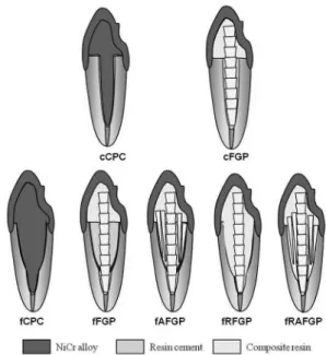
Restoring flared endodontically treated teeth continues to be a challenge for clinicians. This study evaluated the effect of post types and restorative techniques on the strain, fracture resistance, and fracture mode of incisors with weakened roots. One hundred five endodontically treated bovine incisors roots (15 mm) were divided into 7 groups (n=15). The two control groups were (C) intact roots restored with Cpc (cast posts and core) or Gfp (glass fiber posts). The five experimental groups were (F) flared roots restored with GfpAp (Gfp associated with accessory glass fiber posts), GfpRc (anatomic Gfp, relined with composite resin), and GfpRcAp (anatomized Gfp with resin and accessory glass fiber posts). All teeth were restored with metal crowns. Mechanical fatigue was performed with 3x10⁵/50 N. Specimens were loaded at 45o, and the strain values (μS) were obtained on root buccal and proximal surfaces. Following
that, the fracture resistance (N) was measured. One-way ANOVA and Tukey’s HSD tests (α=0.05) were applied, and failure mode
was checked. No significant difference in strain values among the groups was found. Cpc presented lower fracture resistance and more catastrophic failures in flared roots. Gfp associated with composite resin or accessory glass fiber posts seems to be an effective method to improve the biomechanical behavior of flared roots.
Key Words: flared endodontically treated teeth, strain, fracture resistance, cast posts and core, glass fiber post.
to be the treatment of choice for endodontically treated teeth without considering the quantity and quality of the remaining tooth tissue. These posts offer a good fit to the root canal because they are obtained from an impression taken directly from the root canal. However, these posts present only frictional retention in the canal, and they are dangerous for the root, potentially leading to its fracture because of great stiffness in homogeneity between metal and dentin (1,8-11). For these reasons, in the last decade, prefabricated glass fiber posts have gained popularity and have been used as a substitute for custom metallic posts (2-7,9,11). Glass fiber posts are easily bonded to the dental structure with the use of adhesive systems and resin cements (2,5,13,14),
and they have a modulus of elasticity closer to that of dentin (1,15). When bonded with dentin, glass fiber posts may provide adequate stress distribution on the tooth and may decrease the incidence of catastrophic root fractures (1,8,9,11).
The quantity of coronal and root dentin that remains after root canal treatment and post space preparation plays an important role in the longevity of the tooth and restoration (2-7,9,11). Roots can become weakened if flared as a result of recurrent caries into the root dentin around the post, over-preparation and instrumentation of the root canal, or the fact that the pulp has become necrotic prior to completion of root formation in a young patient (1,2-7). The resulting flared root canals have thin dentin walls, leaving them too weak to withstand normal masticatory forces and hence susceptible to fractures, which makes the restorative procedure more difficult (2-7). The morphology of flared canals also results in very wide, tapered and non-retentive posts. In these situations, if a prefabricated post is used, the excess space within the root canal would be taken up with a bulk of luting cement. This results in a potentially weak area in the restoration, which may serve to compromise the long-term prognosis (2-7). In order to avoid the extraction of flared roots, filling of the radicular space with restorative materials, such as a glass-ionomer cement (16), composite resins (1-6,16), and accessory glass fiber posts (6,7) has been suggested.
The aim of this laboratory study was to compare the root strain and fracture resistance of flared roots restored with different post systems and restorative techniques. Two research hypotheses were tested. The first is that the post system influences the strain, fracture resistance, and failure mode of flared roots. The second is that reliningfiber posts with composite resin could increase the fracture resistance and decrease the root strain.
MATERIAL AND METHODS
One hundred five bovine roots of similar size and shape were selected by measuring the buccolingual and mesiodistal widths in millimeters, allowing a maximum deviation of 10% from the determined mean (11). The teeth were stored in 0.2% thymol solution (Pharmacia Biopharma Ltda., Uberlândia, MG, Brazil). The soft-tissue deposits were removed with a hand scaler (SS White Duflex, Rio de Janeiro, RJ, Brazil), and the teeth were cleaned using a rubber cup and fine pumice water
slurry. The coronal portion of each tooth was sectioned 15 mm coronally from the root apex, using a diamond double-faced disk (KG Sorensen, Barueri, SP, Brazil) in a slow-speed handpiece, cooled with air/water spray. Root canals were prepared with the use of Gates-Glidden burs (Maillefer, Ballaigues, Switzerland): burs no. 2 (Ø 0.54 mm) and 3 (Ø 0.83 mm) were used in the entire root canal length, and bur no. 4 (Ø 1.10 mm) was used only in the cervical third of the root canal. The canals were irrigated with 1% sodium hypochlorite solution (Asfer, Industrial Química, São Paulo, SP, Brazil); final irrigation was done with 0.9% sodium chloride solution; and canals were dried with absorbent paper points (Dentsply Ind. e Com. Ltda., Petrópolis, RJ, Brazil). Each canal was obturated by lateral condensation using gutta-percha points (Maillefer) and endodontic cement (Sealer 26, Dentsply). The gutta-percha was removed with hot pluggers (SS White Duflex) to remove 10 mm of the filling material, and then measured using a plastic stop. Post preparations were completed with a no. 5 reamer (Largo, Ø 1.5mm, Dentsply Ind. e Com. Ltda.). The external root surface was covered with a 0.2-0.3 mm layer of a polyether impression material (Impregum F, 3M ESPE, St. Paul, MN, USA) to simulate the periodontal ligament. The roots were embedded in a polystyrene resin (Cristal, Piracicaba, SP, Brazil) up to 4.0 mm below the cervical limit to simulate the alveolar bone (11,17).
composite resin); Group 7 (flared): FGfpRcAp, anatomic glass fiber post (the relining associate composite resin with accessory glass fiber posts).
The cast posts and cores (groups 1 and 3) were made with a direct technique using patterns in acrylic resin (Duralay, Reliance Dental MFG Company Worth, IL, USA). A 6.0 mm high pre-manufactured polycarbonate pattern (Nucleojet, Ângelus, Londrina, PR, Brazil) was used to standardize the coronal portion of the cast metal core. A Ni-Cr alloy (Kromalit, Knebel, Porto Alegre, RS, Brazil) was used to cast the post and core patterns.
The prefabricated glass fiber post (Reforpost RX no. 3, Ângelus) used in groups 2, 4, 5, 6, and 7, was 1.5 mm in diameter on the coronal and 1.1 mm on the apical portion. The glass fiber post was composed of 85% glass fiber and 15% epoxy resin. Accessory glass fiber posts (used in groups 5 and 7) were composed of 70% glass fiber and 30% epoxi resin (Reforpin, Ângelus), and its dimensions were as follows: no. 3 (coronary Ø 1.45 mm and apical Ø 0.55 mm) and no. 1 (coronary Ø 1.1 mm and apical Ø 0.5 mm).
In order to cement the fiber and metallic posts, the root canals were etched with 37% phosphoric acid (Dentsply) for 15 s, and then washed and dried with absorbent paper tips (Dentsply). Two consecutive layers of the primer (Adper Scotchbond Multi Purpose, 3M ESPE) were applied using a microbrush. They were then gently air dried for 20 s, and this was followed by the application of the adhesive (Adper Scotchbond Multi Purpose, 3M ESPE). Excess adhesive was removed from the canal using a clean microbrush. Light activation was performed for 20 s on the cervical root face, parallel to the long axis of the root, using a halogen light unit with an intensity of 800 mW/cm2 (XL 3000, 3M ESPE). The
post was cleaned with 70% alcohol in a single application
using a microbrush; after drying, a silane agent was applied (Silano, Ângelus). The self-curing resin cement (Cement Post, Ângelus) was mixed in accordance with the manufacturer’s instructions introduced into the canal with a lentulo spiral drill (Dentsply Maillefer) in a low-speed handpiece. Cement was placed on the post and the post was seated under a constant load of 500 g during 5 min. Excess cement was removed after 1 min. For the group5, before resin cement polymerization, the principal post (Reforpost RX no. 3) was positioned on the center of canal and four accessory posts no. 3 and three no. 1 posts were introduced around the root canal space. For groups 6 and 7,before the adhesive protocol, an impression of the flared canal was made with a polyether impression material (Impregum F, 3M ESPE), and after that, a silicone material (CIS - 745, Technology Inc., Taiwan.) was used to fabricate the mold of the root canal. In group 6, the main post was positioned in the center of the replica of the root canal, and a composite resin (Filtek Z250, 3M ESPE) was inserted and photoactivated with a halogen light-curing unit (XL 3000, 3M ESPE) for 40 s. The relined post was removed from the replica and over-polymerized for another 1 min. In group 7, the same protocol used on group 6 was repeated; however, before the polymerization, four accessory posts no. 3 and three accessory posts no. 1 were introduced in the canal replica for relining with composite resin. The relined posts of groups 6 and 7 were removed from molds and luted using the same
Figure 1. Schematic illustration of the specimen’s dimensions. A.
protocol described for the other groups.
The composite resin core was standardized using an acetate matrix constructed in a vacuum plasticizer using a polycarbonate pattern (Nucleojet, Ângelus). An incremental technique was used to place composite resin (Filtek Z250, 3M ESPE); each increment was polymerized for 40 s. In all groups that used glass fiber posts, the core build-up followed this method.
All specimens were prepared with a diamond round bur (#3215; KG Sorensen) in a high-speed handpiece with water spray cooling (Super Torque 625 Autofix; KaVo do Brazil Ind. e Com. Ltd, Joinville, SC, Brazil). Specimens were prepared to receive complete crowns with a 1.5 mm reduction and 2.0 mm ferrule. An impression of the specimens was made using a polyether impression material (Impregum F, 3M ESPE), and the impressions were poured with Type IV stone (Durone IV, Dentsply Ind. e Com. Ltda.). Wax patterns were formed using a silicone impression material (Aerojet, São Paulo, Brazil) mold made in the shape of a composite resin central incisor crown. This mold was used to fabricate all wax crown patterns. A standardized notch was placed across the palatal surface of each crown 3 mm from the incisal edge for load application in the mechanical tests. Wax (Degussa, Hanau, Germany) was then poured into the impression, and the tooth was inserted. After the wax cooled, the impression was removed, and the margins were perfected. The wax patterns were cast in a Ni-Cr alloy (Kromalit, Knebel) (9,11). Crowns were luted to the teeth following the same protocol as for post fixation. A mechanical fatigue (18) was performed with 3x10⁵ cycles of 50 N, at 37 ºC and 100% humidity (Erios,
São Paulo, SP, Brazil). The force was applied 3 mm below the incisal edge on the palatal surface of the crown at an angle of 45o (Fig. 3) (9,11). Root dentin strain was
measured with strain gauges PA-06-060BG-350LEN (Excel Sensores, SP, Brazil), which had an internal
electrical resistance of 350 Ω, a gauge factor of 2.12, and
a grid size of 4.2 mm2. The gauge factor is a proportional
constant between electrical resistance variation and strain. A 37% phosphoric acid solution (3M ESPE) was applied for 30 s to etch the gauge sites, which were then washed with water for 15 s and dried with air jets. The strain gauges were bonded to the tooth structure (n=5) with cyanoacrylate adhesive (SuperBonder; Loctite, Sao Paulo, Brazil), and the wires were connected to the data acquisition device (ADS0500IP; Lynx Tecnologia Eletrônica, São Paulo, SP, Brazil). In addition, two strain gauges were fixed to another specimen to which no load was applied to compensate for dimensional alterations due to temperature. This specimen is important for the measurement of strain, without the influence of environmental temperature. Each specimen was placed in a custom apparatus that allowed the specimen to be positioned at 45o to the long axis (9,11). The specimens
were submitted to 45o loading, using a universal testing
machine (EMIC DL 2000, São José dos Pinhais, PR, Brazil) until a force of 100 N was reached. Data were transferred to a computer that used specific acquisition, signal transformation, and data analysis software (AqDados 7.02 and AqAnalisys, Lynx). The strain values were evaluated statistically by one-way ANOVA.
A water circulation device (Federal University of Uberlândia, Uberlândia, MG, Brazil) was constructed to standardize temperature and moisture during the fracture resistance test. This device consists of an acrylic cylinder 150 mm in diameter and 200 mm high, fixed on a steel base with two water circulation directions. This device was linked to a water receptacle with a continuous water bombardment system and a digital heater (Quimis, São Paulo, SP, Brazil). Next, the temperature was standardized at 37oC and 100% of humidity (9,11).
The teeth were subjected to a 45o tangential
compressive loading with a metal knife blade tip at a
crosshead speed of 0.5 mm/min in a universal testing machine (EMIC DL 2000). The force required (N) to cause fracture was recorded by a 5-kN load cell hardwired to software (TESC; EMIC), which was able to detect any sudden load drop during compression (9,11). The fracture resistance data were analyzed by two-way ANOVA, comparing two control groups with two groups that used only posts without reinforcement (Cpc and Gfp), followed by Tukey’s Honestly Significant Difference (HSD) test. After that, the one-way ANOVA and Tukey’s HSD test were employed to compare all groups. All tests were considered significantly
statistically different at α=0.05 (SPSS software; SPSS
Inc., Chicago, IL, USA).
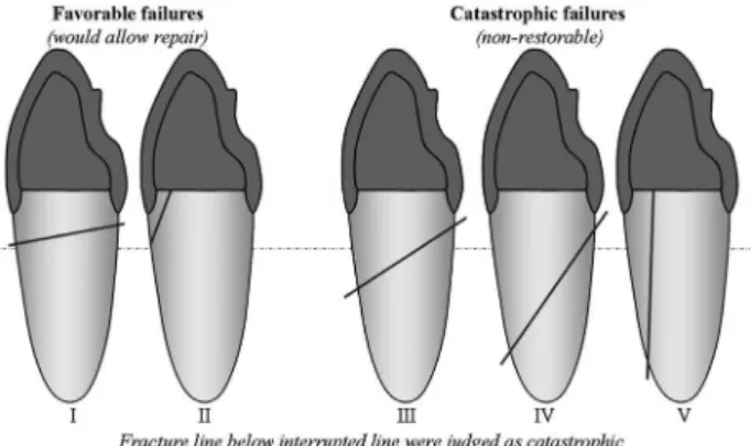
The mode of failure (Fig. 4) was recorded and classified in accordance with the degree of dental structure destruction (11) as either favorable (would allow repair) or catastrophic (non-restorable). The favorable fractures, the ones that would allow repair, were located on the cervical third of the root, obliquely extending from lingual to buccal surface (Type I) or vertically on the buccal surface (Type II). On the other hand, catastrophic fractures were observed obliquely from lingual to buccal surface involving the middle (Type III) or apical third (Type IV) of the roots and vertically on buccal surface (Type V).
RESULTS
The mean and standard deviation values for fracture resistance (N) are shown in Tables 1 and 2. The distribution of failure modes for all groups is represented in Table 3 and Figure 4.
Two-way ANOVA showed that there were significant differences in fracture resistance values for the interaction between post systems and characteristics of root canal (p=0.00). The Tukey HSD test indicated that the cast post and core groups presented lower fracture resistance values (Table 1) and prevalence of catastrophic failures in flared roots (Table 3). One-way ANOVA showed that groups restored with composite resin reeling or the groups restored with accessory glass fiber posts presented fracture resistance similar to the control groups (Table 2) and demonstrated more repairable fractures (Table 3).
Catastrophic fractures (Table 3) were present in the groups in the following order: FCpc, 66.7%; CCpc, 40%; FGfp and FGfpRcAp, 26.7%; FGfpAp and FGfpRc, 13.3%; and CGfp, 6.7%.
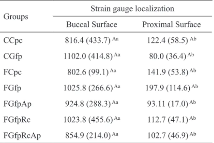
The mean strain and standard deviation values are shown in Table 4. There was no significant difference for the strain values among groups for proximal strain (p=0.140) or buccal strain (p=0.744). However, the proximal strain was lower than the buccal strain values for all groups.
DISCUSSION
The hypotheses were partially accepted, as post systems and restorative techniques do influence the fracture resistance and fracture mode of flared roots. On the other hand, they did not influence the strain measured on buccal or proximal surfaces. Cast posts and core posts, when used in flared teeth, resulted in lower fracture resistance than that of the control groups (Table 1) and prevalent catastrophic failures (Table 3). Restoring
Table 1. Mean and standard deviation values of fracture strength (N), and statistical categories for the comparison between two control groups and two flared groups restored only with cast post and core or glass fiber post (n=15).
Type of post
Root canal characteristics Non-weakened
root (Control)
Weakened root (Flared) Cast post and core 859.9 (199.3) Aa 625.3 (164.3) Ba Glass fiber post 627.1 (119.9) Ab 620.2 (164.2) Aa Different letters indicate statistically significant differences verified by Tukey’s HSD tests (p<0.05). Uppercase letters were used to compare groups in the rows (root condition); lowercase letters were used to compare groups in the columns (post systems).
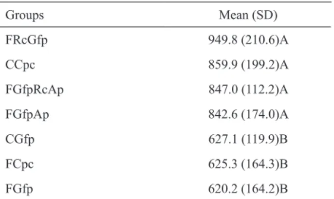
Table 2. Fracture resistance means (N) and standard deviation (SD) for all groups.
flared teeth with composite resin and accessory glass fiber posts presented higher fracture resistance than did flared teeth restored only with glass fiber posts (Table 2).
In the biomechanical analysis of tooth structures and restorative materials, destructive mechanical tests used to determine fracture resistance are important means of analyzing tooth behavior in situations of concentrated and high intensity load application (3,4,6,7,9,11,16,19,20). However, fracture tests have limitations with regard to obtaining information on the internal behavior of the tooth-restoration complex before the failure. Therefore, it is important to combine destructive tests with non-destructive methodologies (1,8-12), such as strain gauge measurement, for root strain analysis and its relation to fracture resistance and failure mode (9,11,19,20). This occurs because transmission of strain energy to the crack supplies energy for crack propagation, and the speed at which the crack is fed with energy will depend upon the rate of change in shape of the material adjacent to the crack. Consequently, the fracture resistance will be increased by any mechanism that increases the amount of energy required to propagate the primary crack (20). The ferule effect and the position of strain-gauge was not sufficient to permit measurement of differences on strain values among the groups, possibly because the strain gauges were placed 1 mm away from the cervical margin of the metal crown, and the majority of fractures occur apically in this region.
It is important to note that the presence of the post significantly altered the stress distribution on the tooth (1,8-12). When a system with different components of different rigidity is loaded, the more rigid components,
such as cast posts and core (NiCr- 200 GPa), are capable of concentrating the stress, thus resisting greater force without distortion, especially if it is not bonded to tooth structures (1,8-12). If stresses reach a critical value, a slowly growing crack causes a successive adhesive failure of the post-cement-root dentin interface. After loss of post adhesion, the post is more or less mobile within the root and is consequently allowed to act like a wedge (1,8). The energy accumulated in the inner post is transferred by the dentin. The fracture in dentine structure occurs from the inner region (adjacent to the root canal) to the outer surface of the tooth (20). If the dentin canal walls are thin or the resin cement is thick, less load is necessary to fracture the tooth, as occurred in groups 3 and 4 (Table 2). On the other hand, when a root canal was not flared and was restored with cast posts and core (group 1), the tooth obtained a higher fracture resistance mean (Table 2). This can be explained by the greater amount of dentin. The maximum load capability is affected by the strength of the surrounding hard tissue, which is directly correlated to the volume of dentin (20). During normal occlusal function, dentin exhibits considerable plastic deformation, resisting varying degrees and angles of load. However, loads exceeding the tensile strength or proportional limit of dentin can decrease the ability of dentin for plastic deformation, which leads to fractures (8,20). Since cast posts and core have more stiffness than glass fiber posts, they can withstand higher loads without fracturing (9,11), but
Table 4. Mean and standard deviation values of the microstrains (µS) of the buccal and proximal surfaces (n= 5) and statistical categories of all tested groups.
Groups Strain gauge localization Buccal Surface Proximal Surface CCpc 816.4 (433.7) Aa 122.4 (58.5) Ab
CGfp 1102.0 (414.8) Aa 80.0 (36.4) Ab
FCpc 802.6 (99.1) Aa 141.9 (53.8) Ab
FGfp 1025.8 (266.6) Aa 197.9 (114.6) Ab
FGfpAp 924.8 (288.3) Aa 93.11 (17.0) Ab
FGfpRc 1023.8 (455.6) Aa 112.7 (47.1) Ab
FGfpRcAp 854.9 (214.0) Aa 102.7 (46.9) Ab
Different letters indicate statistically significant differences verified by Tukey’s HSD tests (p<0.05). Uppercase letters were used to compare groups in the rows (root localization); lowercase letters were used to compare groups in the columns (tested groups). Table 3. Failure mode distribution in the groups (n=15).
Groups
Failure mode distribution Catastrophic failures (%) I II III IV V
the risk of irreparable root fractures would be increased (Table 3).
Using only glass fiber posts and resin cement in flared roots (group 4), fracture resistance was similar to that of control group 2. However, significant lower fracture resistance was verified when we compared this group with all groups that used composite resin or accessory glass fiber posts as a reinforcement of the flared dentin. The resin cement has monomers with functional groups that induce adhesion to the dentine; moreover, it is composed of filler particles (glass of barium and silica), which can improve its mechanical proprieties. Since luting cement is the weakest link in the tooth/post/core complex, the large amount of cement needed to cement the post may serve to compromise the long-term prognosis (2-7).
In the oral environment, the adhesive failure normally occurs as a result of cyclic loads associated with time and environmental factors such as temperature, pH, and microorganisms (18). To determine the longevity of restorations, a controlled clinical trial would be preferable. However, the number of parameters influencing the behavior can be extremely large, and they can take an extremely long time and a large patient group to collect sufficient data. A partial solution to this problem can be found by in vitro investigations using cyclic fatigue loading, which simulates accelerated mechanical deterioration (18). In this research, the teeth were loaded lingually at a 45º angle to the long axis. This angle reflects the positions, contacts, and loading characteristics of maxillary central incisors in class I occlusion (9,11). The fatigue loading was simulated using an intermittent force of 50 N during 3x10⁵ cycles,
resulting in approximately 67 h under load. This number of cycles was selected because Huysmans et al. (18) concluded that at least 105 load cycles are necessary
for fatigue and post failures to occur after 261,000 cycles. In the present study, after load cycles, no tooth cracking or displacement of the crown or the post was observed. New studies must be carried out to draw out the number of cycles necessary to determine with higher predictability the longevity of the restorative techniques in flared roots. Moreover, thermal cycles of fatigue could be carried out to create conditions more closely resembling the clinical situation.
With regard to the restoration of flared roots, composite resin is widely used as filling material for the radicular defects (1-6,16). However, the majority of the experiments or clinical cases are associated with
the use of light-transmitting plastic posts to carry out the radicular reinforcement (2-4). Composite resin, in association with or not with accessory glass fiber posts placed into the canal after reeling in indirect form (group 6 and 7), were tested in this study without the use of light-transmitting plastic post systems. Both restorative techniques revealed more efficient results in high values of fracture resistance than did the control Gfp group (Table 2). This technique could prevent the shrinkage stress in adhesive interface when compared with relining techniques used directly on the root canal. This good performance may have been a result of the intrinsic characteristics of composite resin. The resin used is a compact material, composed of 82% load particles (zirconium glass and silica) with high microhardness and diametral tensile strength.
treatment by reconstructing the tooth with adhesive materials in order to maintain the functioning tooth in the mouth. According to the results of this study, it may be concluded that the use of glass fiber posts associated with composite resin or accessory fiber glass posts seems to be an effective method to improve fracture resistance and increase repairable failures on flared root canals. However, additional clinical studies are necessary to clarify which are the best techniques and materials to closely mimic tooth structures, as well as to recover the mechanical properties and resistance to fracture of structurally compromised teeth by flared canals.
RESUMO
Restaurar dentes tratados endodonticamente continua sendo desafio para clínicos. Este estudo avaliou o efeito de pinos e técnicas na deformação, resistência à fratura e padrão de fratura de incisivos com canal radicular alargado. Cento e cinco raízes bovinas, tratadas endodonticamente (15 mm) foram divididas em 7 grupos (n=15). Os grupos controle (C), constituídos de raízes não alargadas, foram restauradas com Cpc (núcleo metálico fundido) ou Gfp (pino de fibra de vidro). Nos grupos experimentais os canais foram alargados (F) e restaurados com: GfpAp (Gfp associado com pinos de fibra de vidro acessórios); GfpRc (pino anatômico, reembasado com resina composta) e GfpRcAp (pino anatomizado com resina composta e pinos acessórios). Os dentes foram restaurados com coroas metálicas. Fadiga mecânica foi simulada com 3x10⁵/50 N ciclos. O teste foi realizado a 45o e a
deformação (μS) obtida nas superfícies vestibular e proximal. Em
seguida, a resistência à fratura (N) e o padrão de fratura foram
verificados. Aplicou-se ANOVA e Teste de Tukey (α=0,05). Não
houve diferença na deformação. Cpc resultou em menor resistência à fratura e com mais fraturas catastróficas em raízes fragilizadas. As técnicas de reembasamento do pino com resina composta ou o uso de pinos acessórios parecem ser efetivos para melhorar o comportamento biomecânico de raízes fragilizadas.
ACKNOWLEDGEMENTS
The authors are indebted to the financial support granted by FAPEMIG and CNPq. We also wish to thank 3M ESPE, Dentsply, and Ângelus for donating the products used in the research.
REFERENCES
1. Coelho CS, Biffi JC, Silva GR, Abrahão A, Campos RE, Soares CJ. Finite element analysis of weakened roots restored with composite resin and posts. Dent Mater J 2009;28:671-678.
2. Teixeira CS, Silva-Sousa YT, Sousa-Neto MD. Bond strength of fiber posts to weakened roots after resin restoration with different light-curing times. J Endod 2009;35:1034-1039.
3. Zogheib LV, Pereira JR, do Valle AL, de Oliveira JA, Pegoraro LF. Fracture resistance of weakened roots restored with composite resin and glass fiber post. Braz Dent J 2008;19:329-333.
4. Marchi GM, Mitsui FH, Cavalcanti AN. Effect of remaining dentine structure and thermal-mechanical aging on the fracture resistance of bovine roots with different post and core systems. Int Endod J 2008;41:969-976.
5. da Silveira Teixeira C, Santos Felippe MC, Silva-Sousa YT, de Sousa-Neto MD. Interfacial evaluation of experimentally weakened roots restored with adhesive materials and fibre posts: an SEM analysis. J Dent 2008;36:672-682.
6. Moosavi H, Maleknejad F, Kimyai S. Fracture resistance of endodontically-treated teeth restored using three root-reinforcement methods. J Contemp Dent Pract 2008;9:30-37. 7. Bonfante G, Kaizer OB, Pegoraro LF, do Valle AL. Fracture
strength of teeth with flared root canals restored with glass fibre posts. Int Dent J 2007;57:153-160.
8. Santos AF, Meira JB, Tanaka CB, Xavier TA, Ballester RY, Lima RG, et al.. Can fiber posts increase root stresses and reduce fracture? J Dent Res 2010;89:587-591.
9. da Silva NR, Raposo LH, Versluis A, Fernandes-Neto AJ, Soares CJ. The effect of post, core, crown type, and ferrule presence on the biomechanical behavior of endodontically treated bovine anterior teeth. J Prosthet Dent 2010;104:306-317.
10. Silva NR, Castro CG, Santos-Filho PC, Silva GR, Campos RE, Soares PV, et al.. Influence of different post design and composition on stress distribution in maxillary central incisor: Finite element analysis. Indian J Dent Res 2009;20:153-158. 11. Santos-Filho PC, Castro CG, Silva GR, Campos RE, Soares
CJ. Effects of post system and length on the strain and fracture resistance of root filled bovine teeth. Int Endod J 2008;41:493-501. 12. Soares CJ, Castro CG, Santos Filho PC, Soares PV, Magalhaes D, Martins LR. Two-dimensional FEA of dowels of different compositions and external surface configurations. J Prosthodont 2009;18:36-42.
13. Macedo VC, Faria e Silva AL, Martins LR. Effect of cement type, relining procedure, and length of cementation on pull-out bond strength of fiber posts. J Endod 2010;36:1543-1546.
14. Leitune VC, Collares FM, Werner Samuel SM. Influence of chlorhexidine application at longitudinal push-out bond strength of fiber posts. Oral Surg Oral Med Oral Pathol Oral Radiol Endod 2010;110:77-81.
15. Novais VR, Quagliatto PS, Bona AD, Correr-Sobrinho L, Soares CJ. Flexural modulus, flexural strength, and stiffness of fiber-reinforced posts. Indian J Dent Res 2009;20:277-281.
16. Marchi GM, Paulillo LA, Pimenta LA, De Lima FA. Effect of different filling materials in combination with intraradicular posts on the resistance to fracture of weakened roots. J Oral Rehabil 2003;30:623-629.
17. Soares CJ, Pizi EC, Fonseca RB, Martins LR. Influence of root embedment material and periodontal ligament simulation on fracture resistance tests. Braz Oral Res 2005;19:11-16.
18. Huysmans MC, Peters MC, Van der Varst PG, Plasschaert AJ. Failure behaviour of fatigue-tested post and cores. Int Endod J 1993;26:294-300.
19. Soares PV, Santos-Filho PC, Gomide HA, Araujo CA, Martins LR, Soares CJ. Influence of restorative technique on the biomechanical behavior of endodontically treated maxillary premolars. Part II: Strain measurement and stress distribution. J Prosthet Dent 2008;99:114-122.
20. Kishen A, Kumar GV, Chen NN. Stress-strain response in human dentine: rethinking fracture predilection in post-core restored teeth. Dent Traumatol 2004;20:90-100.