Despite several reports regarding tissue regeneration, including pulp repair induced by different light sources, only limited data have been reported concerning the effects of light-emitting diodes (LED) on stem cells from human exfoliated deciduous teeth (SHEDs). The aim of this study was to evaluate the effects of different energy densities of infrared LED on the cell viability, number of cells and mineralized tissue production by SHEDs. SHEDs were obtained from near-exfoliation primary teeth (n=3), seeded in plain DMEM (104
cells/cm2), and irradiated by a LED prototype (LEDTable 850 nm, 40 mW/cm2) delivering 0
(control), 2, 4, 8, 15 or 30 J/cm2 (n=9). Cell viability (MTT assay), cell proliferation (trypan
blue assay), and mineralized nodule (MN) formation (alizarin red stain) were assessed 12 and 72 h post-irradiation. Data were subjected to Kruskal-Wallis and Mann-Whitney tests (α=0.05). Cells irradiated with 2 or 4 J/cm2 exhibited higher metabolism at 72 h,
and all energy densities provided increase in cell proliferation after 12 h. Regarding MN formation, the best results were observed at 72 h after SHED irradiation with 8 and 15 J/cm2. It was concluded that the cell viability, cell number and MN formation by pulp
cells are enhanced after exposure to infrared LED irradiation. Overall, the greatest SHED biostimulation was obtained with 4 and 8 J/cm2.
D o s e - r e s p o n s e s o f S t e m C e l l s
f r o m H u m a n E x f o l i a t e d Te e t h
t o I n f r a r e d L E D I r r a d i a t i o n
Ana Paula Silveira Turrioni1, Liege Aldrovandi Montoro1, Fernanda Gonçalves Basso2, Leopoldina de Fátima Dantas de Almeida3, Carlos Alberto de Souza Costa2, Josimeri Hebling1
1Department of Pediatric Dentistry
and Orthodontics, Araraquara School of Dentistry, UNESP – Univ Estadual Paulista, Araraquara, SP, Brazil
2Department of Physiology and
Pathology, Araraquara School of Dentistry, UNESP - Univ Estadual Paulista, Araraquara, SP, Brazil
3Department of Restorative
Dentistry, Araraquara School of Dentistry, UNESP - Univ Estadual Paulista, Araraquara, SP, Brazil
Correspondence: Profa. Dra. Josimeri Hebling, Rua Humaitá, 1680. Centro, Caixa Postal 331, 14.801-903 Araraquara, SP, Brasil. Tel: + 55-16-3301-6334. e-mail: [email protected]
Key Words: infrared, phototherapy, pulp cells, metabolism.
Introduction
In addition to the lipopolysaccharides (LPS) released from bacteria during caries progression, the clinical procedure of caries removal may cause damage to the pulp-dentin complex. Also, some dental materials currently recommended as capping agents or liners might be cytotoxic and present poor physical or chemical properties, resulting in additional injury to the dental pulp (1,2).
To reduce pulp damage or improve healing of already damaged pulp tissue, different techniques, such as phototherapy, have been assessed and recommended (3,4). Low-level light therapy (LLLT), with lasers or light-emitting diodes (LED), has been widely used in dentistry for tissue and cell stimulation (5-11). Concerning the pulp tissue or pulp cells subjected to phototherapy, several authors have reported encouraging scientific data, such as increased deposition of tertiary dentin (5), enhanced cell viability and proliferation (8,11), and biomodulation of inflammatory cytokines and reactive oxygen species (9). Despite these positive results, a major challenge in tissue biostimulation involving phototherapy is to define the optimal window for each cell type and desired action (3). In other words, which parameters of irradiation (energy density, wavelength, power) would be adequate for biostimulation, since it is known that cell and tissue responses may vary according to these physical parameters (3,12).
In clinical situations with dentin remaining between cavity floor and pulp tissue, light needs to overcome the dentin barrier to reach and stimulate the subjacent pulp tissue. It has been demonstrated that LED irradiation (blue, red and infrared spectra) is capable of propagating through a dentin barrier up to 1.0 mm thick (13,14). The authors reported that the smallest scattering of light occurred when infrared LED was used. Later, it was shown that the light emitted by diodes is able not only to cross the dentin barrier but also to stimulate the underlying pulp cells (11). Despite the promising cell-biostimulatory results obtained with phototherapy, such as modulation of tissue inflammation (15) and stimulation of fibroblast cell metabolism, odontoblast-like cells, and human pulp cells (10,11,16-18), information about the effects of infrared LED on the metabolism and mineralization ability of stem cells from human exfoliated deciduous teeth is scant. Therefore, the aim of this study was to evaluate the effects of 850-nm LED irradiation on cultured stem cells from human exfoliated deciduous teeth (SHEDs) at different energy densities.
Material and Methods
Primary Cultures of Pulp Cells
410
A.P
.S. Turrioni et al.
of Araraquara Dental School - UNESP, after approval by the local Ethics Committee (Protocol 63/11). The isolation and characterization of the primary pulp from exfoliated teeth cells used in this study followed a previously described protocol (10). The tests were performed when sufficient cells were obtained for application of the protocol, and 3rd passage cells were used. All experiments were performed in triplicate.
Irradiation Device
For cell irradiation, a previously described standardized device (LEDtable) containing 24 infrared diodes (850 nm) was used (9-11). The irradiance was fixed at 40 mW/cm2, and the irradiation times were 50 s (2 J/cm2), 1 min and 40 s (4 J/cm2), 3 min and 20 s (8 J/cm2), 6 min and 15 s (15 J/cm2) and 12 min and 30 s (30 J/cm2) (9). All cells were irradiated in a dark room. According to a preliminary study, the time that the cells were kept out of the incubator, considering the different irradiation times, did not affect cell viability (data not shown).
Cell Viability Assay (MTT assay)
For cell viability (MTT assay) and cell counting (trypan blue assay), cells were seeded in 24-well plates (104 cells/cm2) and incubated for 12 h in contact with DMEM containing 10% FBS (Gibco, Grand Island, NY, USA). After this period, the medium was aspirated and replaced by DMEM containing only 0.5% FBS, to induce nutritional stress. After 12 h, the cells were subjected to LED irradiation, and the tests were performed at 12 and 72 h post-irradiation.
Cell viability analysis was performed as described by Oliveira et al. (17). Briefly, DMEM was aspirated and replaced by 900 µL of new culture medium (DMEM) plus 100 µL of MTT solution (5 mg/mL of methyl tetrazolium salt in PBS) (Sigma-Aldrich, St. Louis, MO, USA). Cells in contact with the MTT solution were incubated at 37 °C for 4 h. After incubation, the MTT solution was aspirated and replaced by 600 µL of an acidified isopropanol solution (0.04 N of HCl). Three aliquots from each sample were analyzed in 96-well plates. Cell viability was assessed in absorbance at 570 nm in a microplate reader (Thermo Plate, Nanshan District, Shenzhen, China).
Cell Proliferation (Trypan Blue Assay)
After LED irradiation, the medium in contact with the cells was replaced by 0.12% trypsin (Gibco) and kept in contact with the cells for 10 min. After cell dissociation, a 50-µL aliquot of this cell suspension was added to 50 µL of 0.04% trypan blue solution. The resulting solution was kept at room temperature for 2 min. To perform the cell counts, 10 µL of the solution were placed on a
hemocytometer and the cells assessed in an automated cell counter (TC 10 Automed Cell Counter 145-0001; BIO-RAD, Hercules, CA, USA). Total cell and non-viable cell counts were performed, and cell proliferation was obtained by subtracting the non-viable cells from the total cells of the sample.
Mineral Nodule Formation
The evaluation of mineral nodules was performed using the alizarin red staining (19). Cells were seeded (104 cells/cm2) and incubated for 48 h in basal medium, then kept in contact with an osteogenic medium containing 5 mM α-glycerophosphate and 100 mM ascorbic acid (Sigma-Aldrich) (19). Irradiation was performed at 12 or 72 h before the evaluation time. Specifically for this protocol, analysis of nodule formation was performed on the 14th day of culture. Cells were fixed with 70% ethanol for 1 h, then washed with distilled water for 5 min followed by staining with a 40 mM solution of alizarin red (Sigma-Aldrich) for 20 min, under shaking. After being stained, samples were washed twice with distilled water, for 5 min each. Some of the samples were evaluated by optical microscopy for qualitative analysis of mineral nodule formation and others were solubilized with 1 mL (10% in PBS) of hexadecylpyridinium chloride monohydrate (Sigma-Aldrich) for quantitative analysis. After shaking and checking the homogeneity of the solutions, we transferred three 100-µL aliquots of each sample to a 96-well plate. The absorbance was determined at 562 nm in an ELISA reader (Thermo Plate).
Statistical Analysis
For all statistical tests, absorbance values were transformed into percentages, with the control group (non-irradiated cells, period of 12 h) as 100%. Data were subjected to statistical analysis by the non-parametric Kruskal-Wallis test, complemented by the Mann-Whitney test (p<0.05).
Results
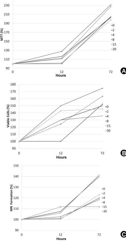
Aiming a better data understanding, Figure 1 shows line graphs based on the median values, for MTT assay (A), Trypan Blue (B) and Alizarin Red (C). The statistical difference between groups, median, 25 percentile and 75 percentile values for each group can be observed on the Tables 1, 2 and 3 for cell viability, cell proliferation and MN formation, respectively.
Cell Viability
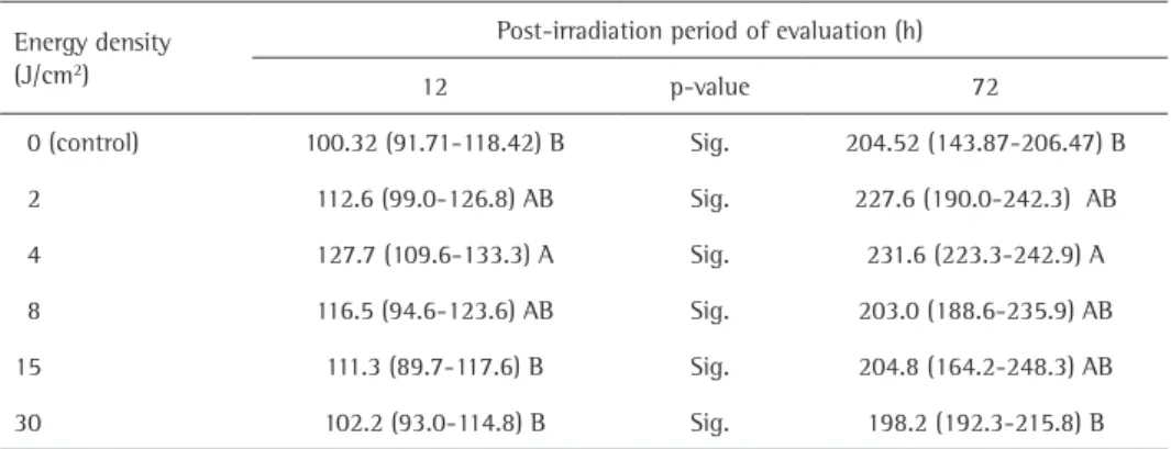
Dose-responses of stem cells to infrared LED in cell viability was observed over time. However, 4 J/
cm2 energy density was the only one to increase cell viability significantly compared with the control group
(p<0.05), by 27.4% and 27.8% for 12 and 72 h, respectively. Other energy densities did not differ from those of the non-irradiated group (p>0.05).
Table 1. Cell viability (% of control) of stem cells from human exfoliated deciduous teeth (SHEDs) 12 and 72 h after irradiation with infrared LED (850 nm) delivering different energy densities
Energy density (J/cm2)
Post-irradiation period of evaluation (h)
12 p-value 72
0 (control) 100.32 (91.71-118.42) B Sig. 204.52 (143.87-206.47) B
2 112.6 (99.0-126.8) AB Sig. 227.6 (190.0-242.3) AB
4 127.7 (109.6-133.3) A Sig. 231.6 (223.3-242.9) A
8 116.5 (94.6-123.6) AB Sig. 203.0 (188.6-235.9) AB
15 111.3 (89.7-117.6) B Sig. 204.8 (164.2-248.3) AB
30 102.2 (93.0-114.8) B Sig. 198.2 (192.3-215.8) B
Values are medians (25th to 75th percentiles), n = 9. p value allows comparisons between post-irradiation
periods (rows). Sig. = statistically significant difference (p<0.05). A Letters allow comparisons among energy
doses (columns). Values identified by the same letter are not statistically different (Mann-Whitney, p>0.05).
Table 2. Cell proliferation (% of control) of stem cells from human exfoliated deciduous teeth (SHEDs) 12 and 72 h after irradiation with infrared LED (850 nm) delivering different energy densities
Energy density (J/cm2)
Period of evaluation (h)
12 p-value 72
0 (control) 100.0 (90.5-111.7) C Sig. 152.2 (143.7-161.7) B
2 124.4 (114.5-150.7) AB N.s. 163.4 (152.2-170.0) B
4 150.0 (144.5-154.0) A Sig. 174.6 (166.7-180.0) A
8 130.0 (105.9-140.4) B Sig. 150.0 (146.7-175.3) AB
15 130.0 (114.7-137.2) AB Sig. 135.8 (135.0- 158.7) B
30 143.0 (117.2-161.2) AB N.s. 145.6 (130.0-155.6) B
Values are medians (25th to 75th percentiles), n = 9. p value allows comparisons between post-irradiation
periods (rows). Sig. = statistically significant difference (p<0.05); N.s. = not statistically different (p>0.05). Capital letters allow comparisons among energy doses (columns). Values identified by the same letter are not statistically different (Mann-Whitney, p>0.05).
Table 3 Mineralized nodule formation (%) by stem cells from human exfoliated deciduous teeth (SHEDs) 12 and 72 h after irradiation with infrared LED (850 nm) delivering different energy densities.
Energy density (J/cm2)
Period of evaluation (h)
12 p-value 72
0 (control) 100.8 (91.1-116.8) AB Sig. 120.0 (99.0-131.0) B
2 111.8 (97.2-116.0) AB N.s. 111.8 (105.5-118.7) B
4 102.5 (100.5-104.3) A N.s. 118.5 (98.8-125.9) B
8 107.5 (102.0-108.5) A Sig. 140.0 (123.5-163.3) A
15 106.0 (102.6-111.9) A Sig. 141.4 (134.8-236.9) A
30 98.4 (91.4-108.2) B Sig. 123.1 (105.0-127.1) B
Values are medians (25th to 75th percentiles), n = 9. p-value Allows comparisons between post-irradiation periods
412
A.P
.S. Turrioni et al.
Cell Proliferation (Trypan Blue)
Analysis of viable cell-counting data (Table 2) demonstrated that, for the 12-hour period, all energy densities promoted an increase on cell proliferation, compared with the control group (p<0.05), and better results for this period were obtained with 4 J/cm2 (50% increase). After 72 h, only the 4 J/cm2 energy density showed statistically higher cell proliferation when compared with
the control group (22.4% increase, p<0.05). When the two evaluation periods were compared, it was observed that, for 4, 8, and 15 J/cm2 energy densities, the period of 72 h was outstanding, showing higher values with statistical significance (p<0.05).
Mineralized Nodule Formation
Quantitative data on mineral nodule formation by cultured pulp cells after LED irradiation are shown in Table 3. Comparison of energy densities at each period demonstrated that, for the 12 h period, there was no statistically significant increase in the irradiated groups when compared with the control group (p>0.05). For the period of 72 h, only the 8 and 15 J/cm2 energy densities were able to increase mineral nodule production (20.0% and 21.4%, respectively) when compared with the control group (p<0.05).
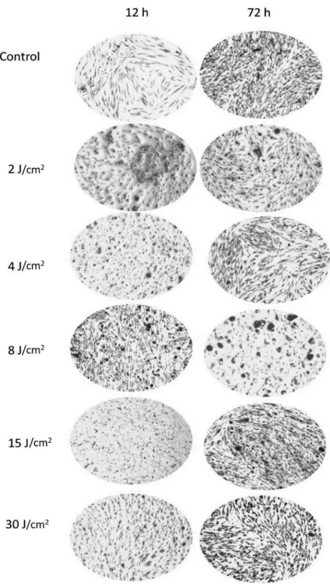
Figure 2 shows the mineral deposition by SHEDs as a function of the different energy densities and post-irradiation periods of evaluation. It was observed that the groups irradiated with 4, 8 and 15 J/cm2 produced more mineralized nodules.
Discussion
The use of phototherapy for transdentinal biostimulation of pulp cells is an interesting adjuvant treatment for pulp healing during clinical procedures in operative dentistry. Based on the analysis of positive data provided by previous studies, researchers have been encouraged to develop more investigations to establish the most beneficial physical parameters of LED or laser irradiation of pulp cells (8-11,20).
Current studies have assessed LED energy densities varying from 0.093 J/cm2 (21) to 162 J/cm2 (22) and power densities from 0.5 mW/cm2 (23) to 140 mW/cm2 (24). According to Alghamdi et al. (12), power values from 1 to 500 mW and energy densities from 0.04 to 50 J/cm2 are considered low-intensity therapy parameters, recommended for the irradiation of cells in culture. Therefore, the irradiation parameters used in this study followed the recommendations of Alghamdi et al. (12) as well as those of Montoro et al. (9), who studied modulation of reactive oxygen species (ROS) and nitric oxide (NO) production by human dental pulp cells
Dose-responses of stem cells to infrared LED
Figure 2. Panel of inverted microscope images representative of mineral deposition by SHEDs, 12 and 72 h after irradiation with infrared LED (850 nm) delivering different energy densities (0, 2, 4, 8, 15 and 30 J/cm2). Original magnification, 4×
subjected to infrared LED therapy.
In the present study, it was shown that infrared LED irradiation can increase the viability and number of pulp cells as well as the formation of mineralized nodules, which play an important role in tertiary dentin formation (8). The
414
A.P
.S. Turrioni et al.
cells was enhanced after exposure to one irradiation session with red LED. Transdentinal biostimulation of odontoblast-like MDPC-23 cells also occurred when the cells were subjected to three irradiation sessions (18). Possibly, a single short irradiation applied to the dentin remaining between the cavity floor and the pulp would be more appropriate for pre-restoration clinical conditions. Therefore, in the present study we decided to evaluate the responses of SHEDs exposed to a single LED irradiation. Since there is no consensus about the best irradiation parameters for pulp biostimulation, we varied only the energy densities from 2 to 30 J/cm2 and assessed the cell responses at 12 and 72 h post-irradiation.
As determined for cell viability, the number of viable SHEDs in the present study was also enhanced after irradiation with infrared LED at the chosen parameters. The most significant increase on cell proliferation was observed at 72 h post-irradiation for the energy density of 4 J/cm2. Holder et al. (8) used the BrdU method to count the number of pulp cells irradiated with LED (653 nm, 3.73 mW/cm2, 448 mJ/cm2). The authors observed a higher number of cells in the irradiated groups than in control groups at 3- and 7-day post-irradiation periods. The increased cell proliferation and viability seem to be related to cytochrome c oxidase activation, which enhances levels in the respiratory chain and adenosine triphosphate (ATP), and these biochemical changes led to macroscopic effects such as increased cell proliferation (25). Analysis of these data confirms that LED therapy may increase the viability and stimulate the proliferation of pulp cells in vitro and in vivo (10,11,20).
The mineral nodule formation by SHEDs was also stimulated by LED irradiation at 8, and 15 J/cm2. Analysis of these data corroborates previous studies in which mesenchymal stem cells (21), odontoblast-like cells (17,18) and rodent pulp cells (8) were subjected to phototherapy. In general, these cells presented increased deposition of mineralized matrix as well as enhanced expression of collagen type I, alkaline phosphatase (ALP), Runx-related transcription factor 2 (Runx2) and osteopontin, which are proteins related to the synthesis and mineralization of the collagen-rich matrix (17,18,21). The molecular pathways that cause increased production of these specific proteins have not been described thus far. However, this macro-effect of phototherapy on pulp cells suggests that light can be an interesting adjuvant treatment to up-regulate the deposition and mineralization of collagen-rich dentin matrix by dental pulp cells during pulp healing. It seems that different energy doses can be the ideal parameter depending on the desired outcome. For number of cells and cell viability, 4 J/cm2 showed better results. On the other hand, for MN formation, doses of 8 and 15 J/cm2
stood out, suggesting that different doses should be used according with different aims.
Overall, in vitro phototherapy with infrared LED biostimulated all SHED functions assessed in the present study. Therefore, the scientific data obtained in the present study can drive future laboratory investigations or even in vivo studies in animal models to establish irradiation parameters for optimal and friendly clinical phototherapy procedures in pulp tissue regeneration.
Resumo
Apesar de diversos estudos envolvendo regeneração tecidual, incluindo o reparo pulpar induzido por diferentes fontes de luz, dados limitados têm sido reportados a respeito dos efeitos da irradiação com diodos emissores de luz (LED) sobre células-tronco de dentes decíduos esfoliados (SHEDs). O objetivo do presente estudo foi avaliar os efeitos de diferentes doses de energia (DE) do LED infravermelho sobre a viabilidade celular, número de células viáveis e produção de nódulos mineralizados (NM) por SHEDs. As células foram obtidas a partir de dentes decíduos próximos ao período de esfoliação (n=3), semeadas em DMEM completo (104 células/
cm2) e irradiadas utilizando um protótipo de LED (LEDTable 850 nm, 40 mW/cm2) com as doses de 0 (controle), 2, 4, 8, 15 ou 30 J/cm2 (n=9). A
viabilidade celular (MTT), o número de células viáveis (trypan blue assay) e a formação de NM (alizarin red stain) foram realizados 12 e 72 h após a irradiação. Os dados foram avaliados utilizando os testes Kruskal-Wallis e Mann-Whitney (α=0,05). As células irradiadas com 2 ou 4 J/cm2 exibiram
uma maior viabilidade em 72 h, e todas as DE aumentaram o número de células viáveis após 12 h. Para a formação de NM, os melhores resultados foram observados 72 h após a irradição das SHEDs, com as doses de 8 e 15 J/cm2. Concluiu-se que a viabilidade celular, o número de células e a
formação de NM por células pulpares são aumentados após exposição ao LED infravermelho. De um modo geral, a melhor bioestimulação celular (SHEDs) foi obtida com 4 e 8 J/cm2.
Acknowledgements
The authors acknowledge the Fundação de Amparo à Pesquisa do Estado de São Paulo – FAPESP (grants: 2011/13895-0 and 2013/17758-3) and the Conselho Nacional de Desenvolvimento Científico e Tecnológico – CNPq (grant: 301291/2010-1) for financial support.
References
1. Smith AJ, Murray PE, Sloan AJ, Matthews JB, Zhao S. Trans-dentinal stimulation of tertiary dentinogenesis. Adv Dent Res 2001;15:51–54. 2. Cohenca N, Paranjpe A, Berg J. Vital pulp therapy. Dent Clin North Am
2013;57:59-73.
3. Carroll JD, Milward MR, Cooper PR, Hadis M, Palin WM. Developments in low level light therapy (LLLT) for dentistry. Dent Mater 2014;30:465-475.
4. Prindeze NJ, Moffatt LT, Shupp JW. Mechanisms of action for light therapy: a review of molecular interactions. Exp Biol Med (Maywood) 2012;237:1241-1248.
5. Tate Y, Yoshiba K, Yoshiba N, Iwaku M. Odontoblast responses to GaAIAs laser irradiation in rat molars: an experimental study using heat-shock protein-25 immunohistochemistry. Eur J Oral Sci 2006;114:50-57. 6. Lizarelli RFZ, Miguel FAC, Villa GEP, Filho EC, Pelino JEP, Bagnato VS.
Clinical effects of low-intensity laser vs light-emitting diode therapy on dentin hypersensitivity. J Oral Laser Appli 2007;7:1–8.
7. Sacono NT, Costa CA, Bagnato VS, Abreu-e-Lima FC. Light-emitting diode therapy in chemotherapy-induced mucositis. Lasers Surg Med 2008;40:625-633.
Dose-responses of stem cells to infrared LED
2012;91:961-966.
9. Montoro LA, Turrioni APS, Basso FG, de Souza Costa CA, Hebling J. Infrared irradiation photobiomodulation of oxidative stress in human dental pulp cells. Int Endod J 2013;47:747-755.
10. Turrioni AP, Basso FG, Montoro LA, Almeida L de F, Costa CA, Hebling J. Phototherapy up-regulates dentin matrix proteins expression and synthesis by stem cells from human exfoliated deciduous teeth. J Dent 2014;42:1292-1299.
11. Turrioni AP, Basso FG, Alonso JR, Oliveira CF, Hebling J, Bagnato VS, Souza Costa CA. Transdentinal cell photobiomodulation using different wavelengths. Oper Dent 2015;40:102-111.
12. Alghamdi KM, Kumar A, Moussa NA. Low-level laser therapy: a useful technique for enhancing the proliferation of various cultured cells. Lasers Med Sci 2012;27:237-249.
13. Turrioni AP, Oliveira CF, Basso FG, Moriyama LT, Kurachi C, Hebling J, et al.. Correlation between light transmission and permeability of human dentin. Lasers Med Sci 2012;27:191-196.
14. Turrioni AP, Alonso JRL, Basso FG, Moriyama LT, Hebling J, Bagnato VS, et al.. LED light attenuation through human dentin: a first step toward pulp photobiomodulation after cavity preparation. Am J Dent 2013;26:319-323.
15. Ablon G. Combination 830-nm and 633-nm light-emitting diode phototherapy shows promise in the treatment of recalcitrant psoriasis: preliminary findings. Photomed Laser Surg 2010;28:141-146. 16. Vinck EM, Cagnie BJ, Cornelissen MJ, Declercq HA, Cambier DC.
Increased fibroblast proliferation induced by light emitting diode and low power laser irradiation. Lasers Med Sci 2003;18:95-99.
17. Oliveira CF, Basso FG, Lins EC, Kurachi C, Hebling J, Bagnato VS, et al.. In vitro effect of low-level laser on odontoblast-like cells. Laser Phys
Lett 2011;8:155-163.
18. Oliveira CF, Basso FG, Reis RI, Parreiras-e-Silva LT, Lins EC, Kurachi C, et al.. In vitro transdentinal effect of low-level laser therapy. Laser Phys 2013;23:1-6.
19. Min JH , Ko SY, Cho YB, Ryu CJ, Jang YJ. Dentinogenic potential of human adult dental pulp cells during the extended primary culture. Human Cell 2011;24:43–50.
20. Ferreira ANS, Silveira S, Genovese WJ, Cavalcante Araújo V, Frigo L, Mesquita RA, et al.. Effect of GaAIAs laser on reactional dentinogenesis induction in human teeth. Photomed Laser Surg 2006;24:358–365. 21. Kim HK, Kim JH, Abbas AA, Kim DO, Park SJ, Chung JY, et al.. Red light
of 647 nm enhances osteogenic differentiation in mesenchymal stem cells. Lasers Med Sci 2009;24:214-222.
22. Taoufik K, Mavrogonatou E, Eliades T, Papagiannoulis L, Eliades G, Kletsas D. Effect of blue light on the proliferation of human gingival fibroblasts. Dent Mater 2008;24:895-900.
23. Vinck EM, Cagnie BJ, Cornelissen MJ, Declercq HA, Cambier DC. Green light emitting diode irradiation enhances fibroblast growth impaired by high glucose level. Photomed Laser Surg 2005;23:167-171. 24. Gritsch K, Ponsonnet L, Schembri C, Farge P, Pourreyron L, Grosgogea
B. Biological behaviour of buccal cells exposed to blue light. Mater Sci Eng C 2008;28:805-810.
25. Gao X, Xing D. Molecular mechanisms of cell proliferation induced by low power laser irradiation. J Biomed Sci 2009;16:4.