Fundação Faculdade Federal de Ciências M édicas de Porto Alegre (FFFCM PA) and Complexo Hospitalar Santa Casa de Porto Alegre (CHSCPA), Porto Alegre RS, Brasil:1Neurologista do CHSCPA, M estre em Clínica M édica pela Universidade Federal do Rio Grande do Sul (UFRGS);2Professora Titular de Anatomia Patológica da FFFCM PA, Livre-Docente em Anatomia Patológica pela FFFCM PA, Coordenadora do Programa de Pós-Graduação em Patologia da FFFCM PA;3Neurologista do CHSCPA, Professora Adjunta de Anatomia Humana da FFFCM PA, M estre e Doutora em Clínica M édica pela UFRGS.
Received 14 M arch 2003, received in final form 29 September 2003. Accepted 7 November 2003.
Dra. Arlete Hilbig - Rua Quintino Bocaiúva 283/401 - 90440-051 Porto Alegre RS - Brasil. E-mail: [email protected]
THE ROLE OF APOPTOSIS,
CELL PROLIFERATION INDEX, BCL-2,
AND P53 IN GLIOBLASTOM A PROGNOSIS
M arlise de Castro Ribeiro
1, Lígia M . Barbosa Coutinho
2, Arlete Hilbig
3ABSTRACT - Glioblastoma is the most common neuroectodermic tumor. It is also the most malignant one. Many genetic changes are found in glioblastomas, among them, the presence of oncoproteins p53 and blc-2, as well as a high mitotic level and the pres-ence of apoptosis. The utility of such findings through immunohistochemistry for the prognosis of patients remains uncertain. Our objectives in this study were to verify the presence of apoptosis, blc-2, p53, and the proliferative index (MIB-1), through immuno-histochemistry, in 30 glioblastomas obtained by surgical resection between August 2000 and August 2001, as well as correlations between those immunohistochemical variables and the patient’s age and survival time. Correlations between immunohistochem-ical variables themselves were also examined. For correlation calculations, Pearson’s and Spermann’s correlations were used and the time of survival was calculated with the Kaplan-Meier method. Results: No correlation was found between immunohisto-chemical variables and survival time.There was also no correlation between those variables and the patients’ age.A moderate inverse correlation was found between the apoptotic index (AI) and the mitotic index (MI) (p = 0.058), besides an inverse correlation between blc-2 and MI. Conclusion: Our study has not demonstrated any of the examined immunohistochemical findings as having a pre-dictive value in the prognosis of glioblastomas.A reverse correlation was found between AI and MI, which has already been demon-strated by a few studies, as well as an inverse correlation between blc-2 and MI. This finding can demonstrate blc-2 as having a pro-apoptotic role in this group of tumors.
KEY WORDS: glioblastoma, apoptosis, p53, bcl-2, MIB-1, prognosis.
O papel da apopt ose, do índice de proliferação celular, bcl-2 e p53 no prognóst ico dos glioblast omas
RESUMO - O glioblastoma é o tumor neuroectodérmico mais comum e também o mais maligno. Muitas são as alterações genéti-cas encontradas nos glioblastomas, entre elas, a presença de oncoproteínas p53 e bcl-2, além de elevado índice mitótico e a pre-sença de apoptose. A utilidade desses achados, através da imuno-histoquímica, no prognóstico dos pacientes ainda permanece incerta. Nossos objetivos neste estudo foram verificar a presença de apoptose, bcl-2, p53 e o índice proliferativo (MIB-1), através de imuno-histoquímica, em 30 glioblastomas obtidos através de ressecção cirúrgica entre agosto de 2000 e agosto de 2001 e tam-bém as correlações entre estas variáveis imuno-histoquímicas e a idade dos pacientes e o tempo de sobrevida.Tamtam-bém pesquisamos as correlações entre as variáveis imuno-histoquímicas entre si. Para os cálculos de correlação utilizamos Correlação de Pearson e Spearmann e a sobrevida foi calculada através do método de Kaplan-Meier. Resultados: Não houve correlação entre as variáveis imuno-histoquímicas e o tempo de sobrevida.Também não houve correlação entre estas variáveis e as idades dos pacientes. Encontramos correlação inversa de grau moderado entre o índice apoptótico (IA) e o índice mitótico (IM) (p= 0,058) e também correlação inver-sa entre bcl-2 e IM. Conclusão: Nosso estudo não demonstrou nenhum dos achados imuno-histoquímico pesquisado como ten-do valor preditivo no prognóstico ten-dos glioblastomas. Houve correlação inversa entre IA e IM, já demonstrada em alguns estuten-dos e também correlação inversa entre bcl-2 e IM, achado que pode demonstrar um papel pró-apoptótico do bcl-2 neste grupo de tumores.
Glioblastoma is the most common malignant neoplasia of the central nervous system, w ith a prognosis of less than 24 months for most patients. Few prognostic factors have been identified in such tumors. Several biological markers are cur-rently under research, in order to verify their predictive fac-tors1,2. Apoptosis, or programmed cell death, plays an
impor-tant role in cell proliferation control in physiological and pathological conditions. Such role has special interest for brain tumor development and progression. The deregulation of ge-netic pathways controlling apoptosis can favor grow th and development of a tumor. Apoptosis is activated and regulat-ed by a number of genes and proteins, such as p53, blc-2, and bax, among others. The apoptotic index (AI) is know n to usu-ally be higher in glioblastomas than in other astrocytic tumors, but its relationship w ith prognosis is still uncertain2-5.
P53, a cell proliferation regulating gene and a pro-apop-totic w hich is altered in as much as half of all astrocytomas, has a controversial role in glioblastoma prognosis6, 7. Bcl-2 and
the blc-2 protein family are mostly considered anti-apoptot-ic, providing cells w ith a longer time of survival by preventing cell death. Bcl-2 expression in some tumors has been related to their ow n prognosis8. How ever, its prognostic correlation
in glioblastomas is still controversial3,9,10.The mitotic index (MI),
assessed through M IB-1 (Ki-67), has proven useful in grading astrocytomas, though its predictive role in glioblastoma patient survival has not been confirmed4,11,12, as it exceeding ranges
from 1 to 22%.
In this study, it was intended to verify the importance of conducting immunohistochemical techniques on glioblas-tomas, noting if the presence of altered genes, such as p53 and blc-2, as w ell as apoptosis, can, in any way, contribute to a better understanding and, consequently, a better treatment for such neoplasia.
M ETHOD
Sample
Thirty patients above 18 years old w ith a diagnosis of glioblas-toma betw een August 2000 and August 2001 w ere retrieved from the Department of Neuropathology of Fundação Faculdade Federal de Ciências M édicas de Porto Alegre files. They w ere all submitted to total surgical resection of the brain tumor at Complexo Hospitalar da Santa Casa de Porto Alegre (Hospital São José), Hospital de Beneficiência Portuguesa, and Hospital São Lucas da PUCRS. Neither patient presented a history of radiotherapy, chemotherapy, and/or pre-vious tumor resection. Slides from all cases w ere review ed to con-firm histopathological diagnosis.
The time of survival was counted from day one - surgery - until 12 months after the surgical procedure had taken place. Death dates w ere know n through death certificates supplied by the responsible state agency.
Immunohistochemistry
Thirty surgical specimens of formalin fixed and paraffin embed-ded tissue were studied.They were cut to 5µm, deparaffined, and
rehy-drat ed for t he immunohist ochemical t echnique, by using t he Estreptoavidin-Biotin-Peroxidase method for visualizing the pres-ence of p53, blc-2 and Ki-67-M ib1. The material was placed in a mi-crowave oven, in order to revert the effect of formaldehyde fixation, w ithin a citrate buffer solution (pH 6.0) until boiling, w hen the tem-perature was reduced and kept moderately boiling for another 15 min-utes, w ith stops for replenishing the lost liquid if necessary. The en-dogenous peroxidase activity was blocked w ith methanol contain-ing 0.3% H2O2for 30 minutes. The tissue was rinsed w ith PBS and the material was then incubated w ith primary monoclonal antibod-ies and rested overnight in the fridge. Primary antibodantibod-ies used w ere: for p53 (D07, DAKO), a 1:500-diluted mouse monoclonal antibody; for blc-2 (Clone 124, DAKO), also a mouse monoclonal antibody, dilut-ed as 1:400, and for Ki-67-M ib1 (DAKO), a mouse monoclonal anti-body diluted as 1:2000. After rinsing w ith PBS, cuts w ere incubated with a secondary mice antibody for 30 minutes at room temperature; and w ith the estreptoavidin-biotin-peroxidase complex for 30 min-utes, undertaking a PBS rinsing between each step.The antigen-antibody complex was visualized with the cromogen DAB and counterstained with hematoxylin.As a positive control, previously positive breast cancer was used for p53 and KI-67/MIB-1 and lymph node for blc-2.
Apoptosis was determined through the TUNEL technique, using the Apoptag plus peroxidase in situ kit (S7101, Intergen). The mate-rial was incubated w ith K proteinase for 10 minutes at room tem-perature and after that, endogenous peroxidase was blocked w ith H2O2for 5 minutes. The reagents in this kit are designated to mark the free DNA 3’ OH terminal in situ with marked nucleotides. Nu-cleotides contained in the buffer reaction are enzymately connect-ed to DNA by deoxynucleotidil transferase (TdT). Incubation with TdT is conducted at 37º Celsius for 60 minutes and the enzyme catalyzes an addition of triphosphate nucleotides to the final 3’ OH of the dou-ble or single helix DNA. Thereafter, incorporated nucleotides form an oligomer randomly by digoxigenin.After that, rinsing with buffer solu-tion is conducted for 10 minutes and then the digoxigenin anti-body is applied and incubated in a damp environment for 30 min-utes at room temperature. Detection of the antigen-antibody link is made through immunoperoxidase follow ed by DAB cromogen. Castrated mice prostate was used as a positive control. The omission of TdT enzyme during the TUNEL technique was used as a negative control and resulted in the non-coloring of the plate.
For counting the tumor cells, 10 fields w ere chosen w ith high-magnification (x 500), selecting the central tumor regions and avoid-ing necrosis areas, through the Sigma Scan Pro 5 imagavoid-ing program. For calculating all the indexes, the following formula was used: num-ber of stained cells/1000 total cells. Immunostained cells can be seen in slides show ed in Figures 1 to 4.
Statistical analysis
survival time, and age, the Pearson’s Correlation Coefficient was used. Due to the observed asymmetrical distributions, the logarith-mic transformation was conducted for several variables. The adopt-ed significance level was α= 0.05. Data were analyzed with the help
of SPSS 10.0 (Statistical Package for the Social Sciences) and SigmaPlot 7.0 programs.
RESULTS
The demographic variables from the w hole group are show n in Table 1. There was a predominance of males (70%)
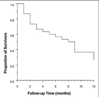
and the patient average age was over 55. Significant differ-ences betw een groups of dead and surviving patients w ere not detected (Table 2). The apoptosis index, as well as the oth-er immunohistochemical markoth-ers, blc-2, MIB-1, and p53, in spite of presenting some oscillation betw een both groups, did not present statistically relevant differences (Table 2).The death curve (Fig 5) showed a relatively constant and regular death occur-rence. After 12 months, it reached a survival-estimated proba-bility of only 25%. Most correlation coefficients between
vari-Fig 1. Glioblastoma with immunohistochemistry for BCL 2 (20 x).
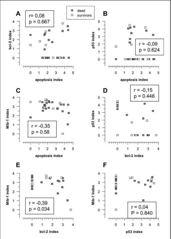
ables (Figs 6,7, and 8) presented a trivial or very small associ-ation power, except for MIB-1, which presented a moderately reverse correlation with the apoptosis index and blc-2.
DISCUSSION
Glioblastomas are a group of neoplasias w ith a broad range of histological and biological findings and, in spite of that, present a homogeneous survival of approximately 1 year.
So far, the age at the time of diagnosis (bellow 45 years) is one of the few relevant criteria for patient prognosis13,14. In our
study, no significant difference was found between patient age and survival time.We did note, however, a tendency of the dead patient group for presenting a higher age average than the survival group (59.5 and 51.9 years, respectively). Such dif-ference w ould probably have statistical relevance if the sam-ple were larger.A significant relationship between age and the Fig 3. Glioblastoma with immunohistochemistry for p53 (20 x).
different expressions in immunohistochemical variables was not verified either, as already demonstrated in a previous
stu-dy by Newcomb et al.1, who did not observe a relationship
between glioblastioma patient survival and p53, blc-2, p16, EGFR, and MDM2 expression, the last three not having been studied by us (Fig 7).
Apoptosis is an active form of cell death, requiring pro-tein and macromolecule synthesis. It is associated w ith dis-tinct morphological changes. Apoptosis is an important phys-iological process for maintaining tissue homeostasis and elim-inating aberrant cells15,16. Some studies have demonstrated that
the AI is higher in glioblastomas than in other astrocytic tumors. Ellison et al.3observed that the AI increased w ith an
increase in astrocytic tumor anaplasia in 81 cases, of w hich 16 w ere fibrillary astrocytomas, 19 w ere anaplasic astrocyto-mas, and 46 w ere glioblastomas. Carrol et al.16studied a total
of 59 cases of astrocytic neoplasias and the presence of apop-tosis in those tumors. They concluded that low -graded astro-cytomas possessed a lower AI than anaplasic ones and glioblas-tomas. Heesters et al.4also observed a higher AI in
glioblas-tomas than in anaplasic astrocyglioblas-tomas, and did not find a prog-nostic role of AI in those patients’ survival. Tew s17, studying
46 gliomas, found a higher AI in glioblastomas than in other astrocytic tumors, and, like Heesters and collaborators4, did not
find a relationship betw een AI and time of survival. How ever, Shiffer et al.10, studying 180 human neuroepithelial tumors,
found a low er AI in glioblastomas than in other gliomas but
as others4,17, could not demonstrate a prognostic role of
apop-tosis in such tumors. Yet, Korshunov et al.2found a significant
direct relationship betw een AI and survival in 168 cases of glioblastomas. Our results have not demonstrated a signifi-cant correlation betw een apoptosis and survival time (Fig 6).
The regulation of apoptosis takes place through a num-ber of mechanisms. As a part of such a complex control, the p53 protein is considered pro-apoptotic. How ever, its muta-tion, verified in neoplasic processes, can nullify its cell-growth-regulating condition18. Loss or mutation in the p53 gene has
been detected in many gliomas and represent an early event in the origin of astrocytomas19. Cunningham et al.6analyzed
120 glial tumors and observed p53 positivity in 85% of the cases, but have not found a relationship between the presence Table 1. Demographic variables in patients diagnosed w ith glioblastoma
(n= 30).
Variable Value
M ale, f (%) 21(70.0)
Age, years 57.5± 11.9
Data are presented as frequency (percentile) and mean ± standard deviation
Table 2. Comparison of demographic and imunohistochemical variables between dead and sur-viving patients diagnosed with glioblastoma.
Variable Dead Survivors P
n= 22 n= 8
M ale, f (%) 15(68.2) 6(75.0) 0.999
Age, yaers 59.5± 12.3 51.9± 9.0 0.122
Apoptosis, % 7.5 (4.2 a 36.0) 5.0 (2.5 a 26.4) 0.344
Bcl-2 0.0 (0.0 a 11.8) 3.0 (0.0 a 13.9) 0.662
P53 0.0 (0.0 a 25.2) 2.2 (0.0 a 49.6) 0.629
M IB-1 20.5 (13.9 a 28.7) 29.7 (15.6 a 33.4) 0.202 Data are presented as frequency (percentile) and median and interquartile amplitude (P25 to P75)
of p53 and survival time. Kraus et al.20have also not
demon-strated a relationship between the presence of p53 in primary glioblastomas and the prognosis of the patients. Jaros et al.21
and Van M eyel et al.22have demonstrated a significant
rela-tionship of the mutation of p53 gene and the accumulation of its protein to a worse prognosis in gliomas. We verified that there was no association betw een the presence of p53 and patients’ survival time (Fig 6), as well as a relationship between p53 and the patients’ age (Fig 7), though the association be-tw een p53 and an age low er than 45 is know n in so-called secondary glioblastomas23. A greater number of studies need
to be conducted in order to clarify the age-stratified role of p53 gene alterations in glioblastomas, as it is know n that the presence of p53 is greater in secondary glioblastomas, w hich are found in younger, and therefore, better-prognosis patients.
It has been suggested that the M I of gliomas can have a predictive value in their ow n prognosis6,24. Ehrmann et al.25
demonstrated a relationship betw een cell proliferation (as measured by the PCNA protein expression) and the progno-sis of 42 astrocytic tumors. In the study by Cunningham et al.6,
there was a prognostic relationship with MIB-1 and PCNA val-ues, which disappeared after an adjustment was made for age and gradation of the 120 astrocytic tumors.
Other studies have demonstrated that, is spite of an increase in cell proliferation along with the increase in the malig-nity degree of the astrocytomas, the prognostic value of M I is not clearly demonstrated in glioblastomas4,10,26. It has been
from previously mentioned studies. There was, how ever, a moderate reverse correlation (p = 0.058) betw een M I and AI, as also demonstrated by Heesters4(Fig 8).
Blc-2, a gene established as programmed cell death sup-pressor, is not found in normal astrocytes w ithin the cortex, w hite matter or cerebellum. How ever, it is found in more than half of all astrocytomas in adults, as demonstrated by Ellison et al.3, w ho found the presence of blc-2 in 44% of the
fibril-lary astrocytomas, 42% of the anaplastic astrocytomas, and 28% of the glioblastomas; by Rieger et al.27, who found a 70%
blc-2 positivity in 20 glioblastomas; and by New comb et al.1,
w ho observed 57% of positivity for blc-2 in astrocytomas in 37 adult patients and in 73% of 21 pediatric cases. The prog-nostic role of the blc-2 expression remains uncertain.We could not demonstrate a significant relationship betw een indexes
of blc-2 and survival, as had already been observed by Nakasu et al.9, New comb et al.8and New comb et al.1(Fig 6). We did
verify, how ever, a statistically significant reverse correlation (p = 0.034) betw een the blc-2 index and the cell proliferation index (Fig 8). Such finding can indicate a greater role for bax, a member of the blc-2 family w ith a pro-apoptotic function, in this group of tumors. How ever, further studies are needed for clarifying such finding.
ing betw een primary and secondary glioblastomas.
CONCLUSION
The reverse correlation betw een AI and M I can be seen in our study, suggesting that the presence of apoptosis can pre-vent tumor growth, though a significant relationship between the presence of apoptosis and time of survival was not demon-strated. A reverse relationship betw een the blc-2 index and
Fig 8. Scatterplots representing correlations found between immunohistochemical variables.
blc-2, since no relationship was established between such genes and the presence of apoptosis.
REFERENCES
1. Newcomb EW, Cohen H, Lee SR, et al. Survival of patients with glioblas-toma is not influenced by altered expression of p16, p53, EGFR, MDM2 or bcl-2 genes. Brain Pathol 1998;8:655-667.
2. Korshunov A, Golanov A, Sycheva R, Pronin I. Prognostic value of tumor associated antigen immunoreactivity and apoptosis in cerebral glioblas-tomas: an analysis of 168 cases. J Clin Pathol 1999;52:574-580. 3. Ellison DW, Steart PV, Gatter KC, Weller RO. Apoptosis in cerebral
astro-cytic rumours and its relationship to expression of bcl-2 and p53 pro-teins. Neuropathol Applied Neurobiol 1995;21:352-361.
4. Heesters MA, Koudstaal J, Go KG, Molenaar WM. Analysis of prolif-eration and apoptosis in brain gliomas: prognostic and clinical value. J Neuro Oncol 1999;44:255-266.
5. Schiffer D, Cavalla A, Migheli A, et al. Apoptosis and cell proliferation in human neuroepithelial tumor. Neurosc Letters 1995;195:81-84. 6. Cunninghan JM, Kimmel DW, Schethauer BW, et al. Analysis of
pro-liferation markers and p53 expression in gliomas of astrocytic origin: relationships and prognostic value. J Neurosurg 1997;86:121-130. 7. Sipos L, Szegedi Z, Fedorscsak I, Afra D, Szende B. Apoptosis and p53
in human giomas. Pathol Oncol Res 1998;4:267-270.
8. Newcomb EW, Bhalla SK, Parrish CL, Hayes RL, Cohen H, Miller DC. bcl-2 protein expression in astrocytomas in relation to patient survival and p53 gene status. Acta Neuropathol 1997;94:369-375.
9. Nakasu S, Nakasu Y, Nioka H. bcl-2 protein expression in tumors of central nervous system. Acta Neuropathol 1994;88:520-526. 10. Schiffer D, Dutto A, Cavala P, et al. Prognostic factor in
oligoden-droglioma. Can J Neurol Sci 1997;24:313-317.
11. Ang LD, Plewes M, Tan L, Begley H, Agranovich A, Shul D. Proliferating cell nuclear antigen expression in the survival of astrocytoma patients. Can J Neurol Sci 1994;21:306-310.
12. Coons SW, Jonhson PC, Pearl DK. Improving interobserver correlation in the classification and gradind of astrocytomas. J Neuropathol Exp Neurol 1993;52:288.
13. Burger PC, Green SB. Patient age, histological features and length of sur-vival in patients with glioblastoma multiforme. Cancer 1987;59:1617-1625. 14. Salmon I, Dewitte O, Pasteels JL, et al. Prognostic scoring in adult astrocytic tumors using patient age, histopathological grade and DNA histogram type. J Neurosurg 1994;80:877-883.
15. Reed JC, Meister L, Tanaka S, et al. Differential expression of bcl-2 pro-tooncogene in neuroblastomas and others tumor cells lines of neural origin. Cancer Res 1991;51:6529-6538.
16. Carrol RS, Zhang J, Chauncey BW, et al. Apoptosis in astrocytic neo-plasms. Acta Neurochir (Wien) 1997;139:845-850.
17. Tews DS. Cell death and oxidative stress in gliomas. Neuropathol Applied Neurobiol 1999;25:272-284.
18. Symonds H, Drall L, Remington L, et al. p53 dependent apoptosis sup-presses tumor growth and progression in vivo. Cell 1994;78:703-711. 19. Van Meir EG, Kikuchi T, Tada M, et al. Analysis of the p53 gene and its
expression in human glioblastomas cell. Cancer Res 1004;54:649-652. 20. Kraus JA, Wengoefer M, Glesmann N. TP53 gene mutations, nuclear p53
accumulation, expression of Waf/p21, bcl-2 and CD95 proteins are not prog-nostic factors in de novo glioblastoma. J Neurooncol 2001;52:263-272. 21. Jaros E, Perry RH, Adam L, et al. Prognostic implications of p53
pro-tein, epidermal growth factor receptor and ki-67 labelling in brain tumors. Cancer 1992;66:373-385.
22. Van Meyel DJ, Ramsay DA, Casson AG, Keenez M, Chamberg AF, Caincross JG. Pattern of mutant p53 expression and DNA ploidy in
evolv-ing gliomas. J Natl Cancer Inst 1994;86:1011-1017.
23. Kleihues P, Ohgaki H. Primary and secondary glioblastoma: from con-cept to clinical diagnosis. Neuro Oncol 1999;1:44-51.
24. Kirkegaard LJ, DeRose PB, Yao B, Cohen C. Image cytometric meas-urement of nuclear proliferation markers (MIB-1, PCNA) in
astrocy-tomas. Prognostic significance. Am J Clin Pathol 1998;109:69-74
25. Erhmann J Jr, Kolar Z, Vojtesek B, Kala M, Komenta S. Prognostic fac-tors in astrocytomas: relationship of p53, MDM-2, bcl-2 and PCNA immunohistochemical expression to tumor grade and overall patient survival. Neoplasma 1997;44:299-304.
26. Hsu DW, Louis DN, Efird JT, Hedley ET. Use of MIB-1 (Ki-67) immunore-activity in diferentiating grade II and grade III gliomas. J Neuropathol Exp Neurobiol 1997;56:857-865.