In vitro
assessment of the effectiveness of
whitening dentifrices for the removal of
extrinsic tooth stains
Abstract: This in vitro study evaluated the effectiveness of whitening dentifrices for the removal of extrinsic tooth stains. Twenty dental blocks (4 x 4 mm), including enamel and dentine, removed from freshly extract-ed bovine incisors, were randomly dividextract-ed into 4 groups: G1 - distillextract-ed water, G2 - Colgate, G3 - Crest Extra Whitening and G4 - Rapid White. In all specimens, the dentin was covered with colorless nail polish, and the enamel was left exposed. Next, the specimens were immersed in a solution of black tea, which was changed every 24 h, for a period of 6 days. After this period, a photo-relectance reading was taken (Time 1) with a spectrometer. The stained specimens were then submitted to lin-ear brushing movements (5,000 cycles) using brushes (Oral B-Soft) cou-pled to an automatic toothbrushing machine, under a static axial load of 200 g and with a speed of 4 movements/second, at 37°C, with the dentifrice or water being injected every 60 s. When toothbrushing ended, a second photo-relectance reading was taken (Time 2). The results were submitted to two-criteria analysis of variance (ANOVA) and to the Tukey test (α = 0.05). When the two times for a same group were compared, Time 2 presented the highest relectance values with statistical difference only for G3 and G4. Among the dentifrices tested, only the Rapid White group differed from the control group, presenting the highest relectance values. Only the whitening dentifrice Rapid White was effective for the removal of extrinsic stains.
Descriptors: Dentifrices; Spectrophotometry; Pigmentation; Toothbrushing.
Débora Alves Nunes Leite Lima(a)
André Luís Faria e Silva(a)
Flávio Henrique Baggio Aguiar(b)
Priscila Christiane Suzy Liporoni(c)
Egberto Munin(d)
Gláucia Maria Bovi Ambrosano(e)
José Roberto Lovadino(f)
(a) PhD Candidates; (b)Assistant Professor;
(f)Chairperson – Department of Restorative Dentistry, Piracicaba Dental School, State University of Campinas, SP, Brazil.
(c) Assistant Professor, Department of Restorative Dentistry, Vale do Paraíba University, SP, Brazil.
(d) Chairperson, Biomedicine Engineering Research Center, Vale do Paraíba University, SP, Brazil.
(e)Assistant Professor, Department of Social Dentistry/Statistics, Piracicaba Dental School, State University of Campinas, SP, Brazil.
Corresponding author:
Débora Alves Nunes Leite Lima
Departamento de Odontologia Restauradora Faculdade de Odontologia de Piracicaba Universidade Estadual de Campinas (Unicamp)
Av. Limeira, 901 Piracicaba - SP - Brazil CEP: 13414-903
E-mail: [email protected]
Introduction
Patients increasingly seek to have an attractive smile, as it is considered to be synonymous with health. This growing demand for enhanced esthetic appearance has led to great development of bleach-ing products.1 However, before any of the bleach-ing products available on the market can be used, it is irst necessary to look into the etiology of dental discoloration.2
Tooth color is inluenced by a combination of intrinsic and extrinsic staining. Extrinsic pigmenta-tions can originate from a smoking habit, from the use of certain cationic agents, such as chlorexidine, and from a diet rich in coloring foods.3 When bev-erages, such as coffee and tea, are frequently con-sumed, colors are deposited on the plaque and bio-logical ilm present on the enamel surface, leading to tooth discoloration.4
These extrinsic stains can be removed by profes-sional prophylaxis. However, the patient’s use of a dentifrice can also contribute to dental stain removal and prevent recurrent discoloration.5 The dentifrice’s stain-removal property is irstly related to the abra-sives present in its composition.6 It is important to point out that if, on one hand, an increased dentifrice abrasiveness leads to improved stain removal eficacy, on the other hand it increases tooth wear.7,8 Thus, dentifrices containing active ingredients speciically designed for tooth stain removal, known as whitening dentifrices, have been introduced on the market.9,10
These dentifrices usually have enzymes in their composition that help to break down the organic components of biological ilm and remove stains and bacterial plaque.11,12 Considering the presence of chromogenic bacteria, bacterial plaque removal also helps whitening teeth.5 Another common dentifrice component is detergent, especially sodium lauryl sul-fate. It acts by lowering the surface tension of stain-producing molecules, thus disrupting their potential to bind to enamel.9 Some whitening dentifrices also contain low peroxide concentrations, with the aim of releasing free oxygen radicals.13 These radicals are responsible for an oxidation reaction, which is the principle of the bleaching technique used for ex-trinsic and inex-trinsic stain removal.9
Considering the fast development of new
whiten-ing products and their growwhiten-ing use by patients, this study aimed to evaluate the effectiveness in vitro of whitening dentifrices for extrinsic tooth stain remov-al. The null hypothesis tested was that there is no difference between the dentifrices used in this study.
Material and Methods
Twenty bovine teeth, stored in a 0.1% thymol solution, were selected for this study in accordance with the color similarity that most of them presented. Very dark or light teeth that stood out against the others were excluded by the visual method. The teeth had their roots removed and, next, the crowns were cleaned with water slurry of pumice lour in a rubber prophylaxis cup at low speed and examined under a light microscope (x4) to exclude those with cracks.
Every tooth crown was set in an acrylic square base, which was ixed to a precision low-speed wa-ter-cooled diamond saw (Imptech PC10, Equilam Lab. Equip., Diadema, SP, Brazil) with two parallel disks, spaced 4 mm from each other and perpendic-ular to the buccal surface of the tooth. Each tooth was cut in the incisogingival and in the mesiodis-tal directions, resulting in a 4 x 4 mm block. The height of each block was also standardized at 3 mm, measured with a digital caliper (727-6/150, Starret, Itu, SP, Brazil). Supericial enamel was wet polished with silicon carbide (SiC) paper #600 and #1,200 grit until the surface was lattened, while trying to maintain the largest possible thickness of enamel.
Afterwards, these dental blocks were randomly distributed into four experimental groups, accord-ing to the dentifrice to be used in this experiment. In one of these experimental groups, the blocks were brushed without dentifrice, with distilled water only, this being considered the control group. The experi-mental groups were as follows: G1 - control (distilled water), G2 - Colgate Total Regular (Colgate-Palmol-ive Indústria e Comércio Ltda., S. B. Campo, SP, Bra-zil), G3 - Crest Extra Whitening (Procter & Gamble, Cincinnati, OH, USA) and G4 - Rapid White (Rapid White Products, Tonawanda, NY, USA). The com-position of each dentifrice is listed in Table 1.
enamel was left exposed. Next, the specimens were immersed in a standard solution of black tea, which was renewed every 24 h, for 6 days (Figure 1). The tea solution was produced by boiling 1.6 g of black tea (black tea leaves, Leão Junior S.A., Curitiba, PR, Brazil) in 100 ml of distilled water, for 5 min and i l-tering it through gauze to remove the tea leaves from the infusion. After this period, a photo-rel ectance reading was taken (Time 1) with a spectrometer.
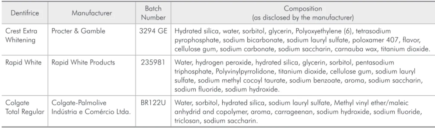
For the rel ectance analysis, a Tel on sphere in the rel ectance coni guration was used. Rel ectance is the luminous radiation portion that is rel ected by the material under study. Before the bleaching procedure, the samples were placed in a sample car-rier that comprises a spectrometer sphere to obtain the initial reading (baseline). The integrating sphere optical signal was captured by an optical i ber with 600 µm in diameter attached to a white light source. The optical potency available in the optical i ber tip was 5 mW, and this i ber was placed 2 mm from the reference pattern (Tel on diffuser) and this distance was kept to the sample (Figure 2).
The rel ectance signal was coni ned inside the in-tegrating sphere and from this a proportional signal
fraction was collected for analysis in the spectrome-ter, where it underwent spectral dispersion through a diffraction grating. The dispersed signal was rel ected to a CCD (Charge Coupled Device) camera that con-verted the optical signal into a digital signal, which was interpreted by the computer and exhibited as in-tensity X wavelength signal. The rel ectance analysis data reading was made with the aid of a microcom-puter, which makes the spectral measurement avail-able to the user in a i le, as a result of the wavelength. The measurements of each sample were dealt with in order to obtain the area given by the graph (%).
After the initial photo-rel ectance reading, the stained specimens were submitted to linear brush-ing. For that, the blocks were allocated to the tooth-brushing machine (Equilabor, Piracicaba, SP, Brazil) and were i xed on the machine support with hot glue so that the vestibular surface of the sample was parallel to this support. The dentifrices used in the experiment were diluted in distilled water at a ratio of 1:3 by weight. This ratio was used in order to al-low the solution to be injected into the toothbrush-ing machine without obstructtoothbrush-ing the syrtoothbrush-inge tip.
The stained specimens were submitted to linear
Table 1 - Dentifrices evaluated in the study.
Dentifrice Manufacturer Batch
Number
Composition (as disclosed by the manufacturer)
Crest Extra Whitening
Procter & Gamble 3294 GE Hydrated silica, water, sorbitol, glycerin, Polyoxyethylene (6), tetrasodium pyrophosphate, sodium bicarbonate, sodium lauryl sulfate, poloxamer 407, flavor, cellulose gum, sodium carbonate, sodium saccharin, carnauba wax, titanium dioxide.
Rapid White Rapid White Products 235981 Water, hydrogen peroxide, hydrated silica, glycerin, sorbitol, pentasodium triphosphate, Polyvinylpyrrolidone, titanium dioxide, cellulose gum, sodium lauryl sulfate, sodium methyl cocoyl taurate, sodium benzoate, aroma, sodium saccharin, sodium fluoride, sodium hydroxide.
Colgate Total Regular
Colgate-Palmolive Indústria e Comércio Ltda.
BR122U Water, sorbitol, hydrated silica, sodium lauryl sulfate, Methyl vinyl ether/maleic anhydrid and copolymer, aroma, carrageenan, sodium hydroxide, sodium fluoride, triclosan, sodium saccharin.
Figure 1 - The enamel of the specimens was stained with a black tea solution.
Dentin covered
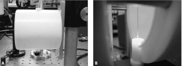
brushing movements using toothbrush heads (Oral-B Indicator 40 Soft, Gillette do Brasil Ltda., Manaus, AM, Brazil) coupled to an automatic toothbrushing machine (Figure 3), under static axial load of 200 g and with a speed of 5 movements/second, at 37°C, the dentifrice or water being injected every 60 s. Five thousand brushing cycles were accomplished, which is equivalent to approximately 6 months of toothbrushing.
When toothbrushing ended, the specimens were removed from the toothbrushing machine and were washed with water spray until the dentifrice residues had been removed. A new photo-relectance reading was taken (Time 2).
The results were submitted to two-criteria analy-sis of variance (ANOVA), the factors being denti-frice and time, and to the Tukey test at a 5% signii-cance level.
Results
The analysis of variance showed no signiicant effect for the dentifrice factor alone (p = 0.28), but for the time factor (p < 0.01) and for the dentifrice and time interaction (p = 0.02) the differences were statistically signiicant. The results for the Tukey test are shown in Table 2.
Before brushing, there was no signiicant dif-ference for the relectance values among any of the studied groups. After brushing, Rapid White pre-sented higher relectance values, although differing signiicantly only from the distilled water group. At this Time, the other two dentifrices used, Regular Colgate and Crest Whitening, did not differ from any of the other groups.
For the same group, there were statistically sig-niicant differences between the values of the read-ing taken after brushread-ing and those of the irst readread-ing only for Rapid White and Crest Whitening. For dis-tilled water and Regular Colgate, there was no sig-niicant difference between the two times of reading.
Discussion
Tooth color is the result of the behavior of inci-dent light on its surface. Part of the inciinci-dent light on
Figure 3 - Automatic toothbrushing machine used in the experiment.
Figure 2 A and B - Positioning of the sample port and optical fiber inside the Teflon sphere in the reflectance analysis set.
the tooth scatters, while the other part is absorbed by pigmented proteins and other pigments present in the tooth. The greater the increase in the amount of these pigments, the greater is the absorption of the incident light and the darker the tooth becomes.14,15
Many methods are currently used to assess tooth color, the objective methods such as relectance spectrophotometer analysis being the most reli-able.16,17 Spectrophotometers and colorimeters are instruments commonly used to measure the color of an object. Spectrophotometers differ from color-imeters in that they measure the relectance of light within the entire visible spectrum, whereas color-imeters measure relected light in only three wave-lengths: red, green and blue.15 In the present study, the color alteration measurements of the specimens were taken by a relectance spectrophotometer. For this, a light was made to fall on the enamel surface of each specimen through an optical iber, the en-tire relected portion of the light being caught by the device and then quantiied. Thus, as the tooth got whiter, a lower amount of light was absorbed and the relectance value was higher.
To standardize the initial color of the specimens, a solution of black tea was used. The method used in this study for simulated extrinsic tooth stain-ing was demonstrated to be effective. At the end of tooth specimen immersion in the black tea solution, the relectance reading disclosed a similarity in the color of dental enamel among the specimens, with-out statistical difference between groups. Sulieman et al.18 (2003) also used black tea for six days to cre-ate staining in dental specimens, which permitted tooth bleaching effectiveness to be assessed. In their
study, the authors also developed an intrinsic dentin pigmentation staining. In the present study, howev-er, contact with the tea solution was restricted to the enamel only.
According to Sexson, Phillips19 (1951), for each toothbrushing session a patient performs, approxi-mately 15 cycles are executed in a determined area. Thus, in two daily toothbrushing sessions, 10,000 cycles are performed at the end of one year. In the present study, 5,000 brushing cycles were per-formed, which is equivalent to approximately 6 months of toothbrushing. The majority of studies that evaluate dentifrice whitening effects are con-ducted in a period that varies from 2 weeks to 6 months.12 Thus, a relatively long time of brushing was simulated in this study, seeking to optimize the whitening effect of the dentifrices used. It is also important that during brushing, dentifrice was injected every 60 s, which allowed the active dentifrice components, among them the abrasives, to be renewed.
When the relectance values for both of the tested times are compared, it was found that the whiten-ing dentifrices, Crest Whitenwhiten-ing and Rapid White, presented higher relectance values than the conven-tional dentifrice (Colgate Regular) after the brush-ing protocol. These whitenbrush-ing dentifrices have en-zymes and detergents in their compositions, which are thought to help with stain removal. Comparing all the dentifrices, the Rapid White was the only one to statistically differ from the control group (dis-tilled water). Rapid White has a small amount of hy-drogen peroxide, which releases free radical oxygen capable of combining with the stain molecule, thus removing it. However, this component stays in con-tact with the tooth for a short period of time, which limits its action.
The whitening effect can be attributed to the ex-trinsic stain removal and also to the change in the relection of the light.14 According to Wulknitz13 (1997), hydrated silica has great cleaning ability and, consequently, stain removal ability. All the den-tifrices tested present hydrated silica in their compo-sitions. The incorporation of abrasives in dentifrices might help physically remove stain, thus some degree of stain removal may be expected even with regular
Table 2 - Reflectance Reading Results.
Group Time
Before brushing After brushing
Distilled water 44.47 (18.65) A a 54.86 (20.22) A b
Colgate Regular 51.94 (17.87) A a 70.52 (33.54) A ab
Crest Whitening 43.58 (14.27) A a 88.89 (33.54) B ab
Rapid White 46.97 (19.29) A a 112.27 (43.65) B a
products.20 The two whitening dentifrices tested in the experiment did not differ from the regular one as regards relectance.
Crest Whitening also has sodium bicarbonate as abrasive. Kleber et al.21 (1998) investigated the whit-ening effect of toothbrushing with different concen-trations of sodium bicarbonate-based dentifrices and concluded that the dentifrices that presented this type of abrasive were more eficient for stain removal. In the present study, Crest Whitening did not signiicantly differ from Rapid White and Col-gate Regular as regards relectance values.
It is important to note that in addition to the type of abrasive, the amount of this component has a di-rect relation to dentifrice abrasiveness. The abrasive
should ideally provide stain removal without caus-ing wear of the tooth.6 Therefore, the results of this study must be viewed with caution, since only the whitening effect of two whitening dentifrices was assessed. The enamel wear caused by these denti-frices still needs to be assessed before they are rec-ommended for routine use.
Conclusion
Only the whitening dentifrice Rapid White was effective for the removal of extrinsic stains.
Acknowledgements
Oral-B (Gillette do Brasil Ltda.) kindly provided the toothbrushes used in this research.
References
1. Gerlach RW, Barker ML. Clinical response of three direct-to-consumer whitening products: strips, paint-on gel, and den-tifrice. Compend Contin Educ Dent. 2003;24(6):458, 461-4, 466 passim.
2. Hattab FN, Qudeimat MA, al-Rimawi HS. Dental discolor-ation: an overview. J Esthet Dent. 1999;11(6):291-310. 3. Matheson JR, Cox TF, Baylor N, Joiner A, Patil R, Karad V et
al. Effect of toothpaste with natural calcium carbonate/perlite on extrinsic tooth stain. Int Dent J. 2004;54(5 Suppl 1):321-5.
4. Watts A, Addy M. Tooth discolouration and staining: a review of the literature. Br Dent J. 2001;190(6):309-16.
5. Claydon NC, Moran J, Bosma ML, Shirodaria S, Addy M, Newcombe R. Clinical study to compare the effectiveness of a test whitening toothpaste with a commercial whiten-ing toothpaste at inhibitwhiten-ing dental stain. J Clin Periodontol. 2004;31(12):1088-91.
6. Meyers IA, McQueen MJ, Harbrow D, Seymour GJ. The sur-face effect of dentifrices. Aust Dent J. 2000;45(2):118-24. 7. Koertge TE. Management of dental staining: can
low-abra-sive dentifrices play a role? Compend Contin Educ Dent. 1997;18(21 Suppl):33-8; quiz 47.
8. Pickles MJ. Tooth wear. Monogr Oral Sci. 2006;19:86-104. 9. Pontefract H, Sheen S, Moran J. The benefits of toothpaste – real or imagined? Review of its role in tooth whitening. Dent Update. 2001;28(2):67-70, 72, 74.
10. Yankell SL, Emling RC, Petrone ME, Rustogi K, Volpe AR, DeVizio W et al. A six-week clinical efficacy study of four commercially available dentifrices for the removal of extrinsic tooth stain. J Clin Dent. 1999;10(3 Spec No):115-8. 11. Lynch E, Samarawickrama DY, Claxson AW, Hawkes JE,
Atherton M, Naughton DP et al. Safety aspects concerning the
therapeutic and cosmetic applications of hydrogen peroxide (H2Ο2)-containing gels, whiteners, oral rinses and dentifrices. J Ir Dent Assoc. 1994;40(3):78-82.
12. Walsh TF, Rawlinson A, Wildgoose D, Marlow I, Haywood J, Ward JM et al. Clinical evaluation of the stain removing ability of a whitening dentifrice and stain controlling system. J Dent. 2005;33(5):413-8.
13. Wulknitz P. Cleaning power and abrasivity of European tooth-pastes. Adv Dent Res. 1997;11(4):576-9.
14. ten Bosch JJ, Coops JC. Tooth color and reflectance as re-lated to light scattering and enamel hardness. J Dent Res. 1995;74(1):374-80.
15. Chu SJ. Use of a reflectance spectrophotometer in evaluat-ing shade change resultevaluat-ing from tooth-whitenevaluat-ing products. J Esthet Restor Dent. 2003;15(Suppl 1):42-8.
16. Horn DJ, Bulan-Brady J, Hicks L. Sphere spectrophotom-eter versus human evaluation of tooth shade. J Endod. 1998;24(12):786-90.
17. Joiner A. Tooth colour: a review of the literature. J Dent. 2004;32(Suppl 1):3-12.
18. Sulieman M, Addy M, Rees JS. Development and evaluation of a method in vitro to study the effectiveness of tooth bleaching. J Dent. 2003;31(6):415-22.
19. Sexson JC, Phillips RW. Studies on the effects of abrasives on acrylic resins. J Prosthet Dent. 1951;1(4):454-71.
20. Amaral CM, Rodrigues JA, Erhardt MC, Araujo MWB, Mar-chi GM, Heymann H et al. Effect of whitening dentifrices on the superficial roughness of esthetic restorative materials. J Esthet Restor Dent. 2006;18(2):111-8.