In Vivo
Characterization of Titanium Implants
Coated with Synthetic Hydroxyapatite by
Electrophoresis
Cristina COSTA de Almeida1
Lídia Ágata SENA1
Marcelo PINTO2
Carlos Alberto MULLER2
José Henrique CAVALCANTI Lima3
Glória de Almeida SOARES1
1Department of Metallurgical and Materials Engineering, COPPE/UFRJ, Rio de Janeiro, RJ, Brazil 2Institute Oswaldo Cruz (FIOCRUZ), Rio de Janeiro, RJ, Brazil
3Brazilian Institute of Implantology (IBI), Rio de Janeiro, RJ, Brazil
This study compared in vivo the performances of commercially pure titanium (cp Ti) screw dental implants either uncoated or coated
with synthetic hydroxyapatite (HA) by electrophoresis. The HA coating was characterized by scanning electron microscopy, energy dispersive spectroscopy (EDS) and Fourier-transform infrared (FT-IR) spectroscopy. Well-adhered carbonated-hydroxyapatite layers
(4- to-8-µm-thick) were obtained. In vivo tests were carried out by insertion of both uncoated and HA-coated implants into rabbit tibiae
for 8 or 12 weeks. Histomorphometric analysis was performed by scanning electron microscopy with the aid of image-processing software. Results showed significantly greater bone-implant contact for HA-coated implants (p<0.05) than cp Ti implants. Comparison of bone content inside the screw implants showed no significant differences (p>0.05) between both types of implants, although cp Ti had numerically higher percentage of bone content than HA-coated implants. In conclusion, the HA-coated implants had better performance regarding the bone-implant contact area than the uncoated implants; coating by electrophoresis proved to be a valuable process to coat metallic implants with an osteoconductive material such as hydroxyapatite.
Key Words: titanium, coating, hydroxyapatite, in vivo study.
Correspondence: Profa. Gloria de Almeida Soares, Programa de Engenharia Metalúrgica e de Materiais, COPPE/UFRJ, Caixa Postal 68505, 21945-970 Rio de Janeiro, RJ, Brasil. e-mail: [email protected]
INTRODUCTION
Due to its biocompatibility, titanium is consid-ered the universal material for permanent implants, such as endosseous dental implants. In other applica-tions requiring higher mechanical strength, titanium-based alloys or Co-Cr alloys are preferred.
Coating metallic implants with biologically ac-tive materials, i.e., hydroxyapatite (Ca10(PO4)6(OH)2;
HA) aims to accelerate bone formation on the initial stages of osseointegration, thus improving implant fixa-tion (1,2). HA-coated dental implants are often recom-mended for the maxillary area where bone is less dense. HA coating is commercially produced by plasma spray techniques, which, in spite of the high productivity,
demand expensive equipment and a robotic system to guarantee a uniform coating on complex shapes. In order to overcome plasma spray disadvantages, alternative processes have been developed (3-6).
phosphate materials are strongly dependent on their composition and crystallographic structure. Among the tricalcium phosphates, amorphous calcium phosphate (ACP) is the most soluble, while sintered stoichiometric HA is the least soluble. Coatings with higher ACP/HA ratios will dissolve or biodegrade to a greater extent than coatings with a low ACP/HA ratio or higher cristallinity (9).
The purpose of this study was to compare the in vivo performances of titanium implants either uncoated or coated with synthetic hydroxyapatite by electro-phoresis.
MATERIAL AND METHODS
Twenty screw-shaped commercially pure tita-nium (cp Ti) implants with an outer diameter of 3.75 mm and 7 mm in length were used in this study. Ten implants were left “as-received” (cp Ti implants) and ten were coated with synthetic hydroxyapatite by elec-trophoresis (HA-coated implants).
A precipitation-wet method was used to synthe-size hydroxyapatite producing a powder with 1.66 ± 0.04 Ca/P ratio. The morphology of HA powder showed acicular nanocrystals and cell parameters compatible with stoichiometric hydroxyapatite (6). The coating methodology was developed by Sena (6), following the Zhitomirsky and Gal-Or procedure (7). Titanium im-plants were used as cathode and platinum was used as anode in an electrophoretic cell with the electrodes positioned 40 mm apart. HA suspension in ethanol was prepared using hydrochloric acid as a dispersing agent, this process being carried out at 24 V for 3 min. After deposition, the coated specimens were calcined in a tubular furnace at 800oC for 2 h.
The characterization of the titanium implants was carried out on 4 specimens (2 HA-coated and 2 uncoated implants) by using a scanning electron micro-scope (DSM 940A model; Carl Zeiss, Jena, GmbH, Germany) on secondary electron and energy dispersive spectroscopy (EDS). A Nicolet-520G spectrometer with diffuse reflectance stage (Thermo Nicolet Corp., Madison, WI, USA) was used for Fourier-transform infrared (FT-IR) analysis.
Eight adult New Zealand rabbits of both sexes, weighing between 3.2 and 4 kg, were used. Each animal received 2 implants, 1 cp Ti (uncoated) and 1 HA-coated, both inserted in the tibial metaphysis. Four
animals received cp Ti implants in the proximal tibial metaphysis and HA-coated implants in the distal tibial metaphysis. After a 4-week interval, the other four ani-mals received the remaining implants following an in-verse design, i.e., the HA-coated implants were inserted in the proximal tibial metaphysis while the cp Ti implants were inserted in the distal tibial metaphysis. All im-plants were supposed to remain in place for 12 weeks. The animals were fasted 12 h before surgery and no prophylactic medication was prescribed. Ketamine (Francotar, Virbac, Carros, French; 50 mg/kg body weight) and Acepromazine (Acepran, Univet, São Paulo, SP, Brazil; 2 mg/kg body weight) were administered intramuscularly. After sedation, the left tibia of each animal was shaved and thereafter washed with Extran MA02 detergent (Merck, Rio de Janeiro, RJ, Brazil)
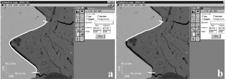
magnifi-cation) of both sides (A and B) of each section were obtained by SEM. The digital images were processed using image-analysis software (Global Lab Image, Data Translation, Marlboro, MA, USA). The percentage of direct bone contact was calculated as being the length of the implant in contact with bone divided by the total length of the screw (Fig. 1). In addition, the bone area inside the threads, expressed as a percentage of the total area, was also determined.
For statistical analysis, the variables were con-sidered to have normal or about normal distribution. Bone-implant contact means for the three best
consecu-tive threads and for all threads, as well as the means of bone area inside the HA-coated and uncoated implant threads were compared using the F test (α=0.05%). Student’s t-test for two samples supposing equivalent variances or different variances was also done.
RESULTS
Coating Characterization
SEM micrographs of HA-coated implants (Fig. 2) showed homogenous layers of hydroxyapatite with a
Figure 1. Calculation of the percentage of direct bone-implant contact - length of the implant in contact with bone (white line in b) divided by the total length of the screw (white line in a).
“crackled” appearance due to the volumetric contrac-tion after calcinacontrac-tion (Fig. 2). EDS spectra (Fig. 3) showed peaks of titanium, calcium and phosphorus. On FT-IR spectra (Fig. 3), the characteristic bands of the group PO4-3 (566, 570, 605, 1030 and 1100 cm-1) of
hydroxyapatite were identified. The band at 872 cm-1
correspondent to CO3-2 in HA A sites and the bands at
1422 and 1449 cm-1 related to HA A and B sites were
also identified, indicating that the coatings were com-posed by carbonated-hydroxyapatite.
Histologic Analysis
SEM images of bone sections showed an almost
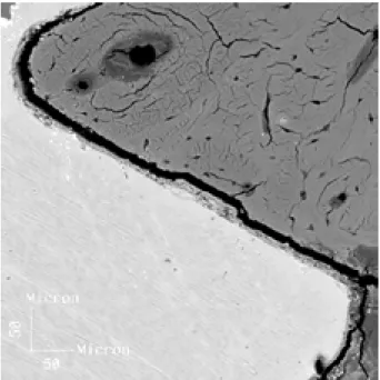
continuous Ca-P-rich layer, with thickness ranging from 4 to 8 µm, well adhered to titanium implant (10). In Figure 4, a fracture line can be seen along the HA-bone interface, probably caused by the rapid polymerization of acrylic resin. Regions where the fracture gap was thicker than 10 µm were not considered as osseointegrated. For all HA-coated implants, this fracture occurred at the bone-hydroxyapatite interface, the coating layer remaining adhered to the metal.
Histomorphometric Analysis
Table 1 shows the means (±SD) of the percent-age of bone-implant contact obtained after 8 and 12
Figure 4. SEM micrograph showing a fracture line along the HA-bone interface, probably caused by the rapid polymerization of acrylic resin (original magnification X1000).
Figure 3. EDS spectrum of HA-coated implant showing peaks of titanium, calcium and phosphorus (top). FT-IR spectrum of the coating layer (bottom).
Table 1. Bone-implant contact (%) for cp Ti implants and HA-coated implants.
After 8 weeks After 12 weeks
All screw threads
Cp Ti 43.82 ± 0.95 36.49 ± 4.50
HA-coated 63.20 ± 10.60 60.27 ± 8.20
3 best consecutive screw threads
Cp Ti 59.47 ± 3.81 55.98 ± 21.70
weeks for the three best consecutive threads and for all threads in the cortical area. No statistically significant differences (p>0.05) were observed when the 8- and 12-week results were compared. Data were grouped, regardless of the osseointegration period, and the per-centage of bone-implant contact of cp Ti and HA-coated implants was compared. Student’s t-test showed significant differences (p<0.05) between uncoated and HA-coated implants for the three best consecutive threads and for all threads (Fig. 5). Means (±SD) for the bone area inside the three best titanium threads and for all threads are given in Figure 6. Student’s t-test showed no statistically significant difference (p>0.05) between the results obtained for both types of surfaces.
DISCUSSION
In this study, titanium implants were satisfac-torily coated with hydroxyapatite by using electro-phoresis. According to Sena et al. (6) and Lacefield (11), calcination is of paramount importance to en-hance adhesion and promote hydroxyapatite densifica-tion. HA deposition by electrophoresis was described by Ducheyne and Qiu (12) as the best technique to coat irregular surfaces, such as those of the screw-shaped dental implants. The “crackled” appearance of HA coatings (Fig. 2) is characteristic of metal-ceramic systems and is attributed to the development of thermal tensions due to the different expansion coefficients between the metal and ceramic layers during heating and cooling cycles (6,8).
Carbonate bands in HA A and B sites were
identified in the FTIR spectra (Fig. 3), suggesting that the HA coating actually is a carbonated-apatite coating. These results are justified by hydroxyapatite trend to incorporate carbonate ions from air, during synthesis and/or coating process. Although carbonate ions act to turn hydroxyapatite more soluble, this appears to be positive for the osseointegration process. The partial dissolution of hydroxyapatite makes the surrounding fluids rich in calcium and phosphate ions, which seems to induce the precipitation of “bone-like” apatite on implant surface (12-14). The release of calcium and phosphate ions followed by precipitation of “bone-like” apatite can trigger cellular differentiation and conse-quent bone formation (apatite plus osteoblast cells), and would perhaps explain why HA-coated implants tend to exhibit faster osseointegration than uncoated implants.
Several authors (9,12,15) have stated that the role of coating ends after the initial stages of osseointegration. Therefore, the coating layer has not to be necessarily insoluble or thick, like plasma spray coatings usually are. Indeed, thick layers tend to exfoliate or delaminate (16) because the interfacial resistance decreases with increasing coating thickness. In a previous investigation, HA coatings deposited by plasma spray with thickness ranging from 75-150 µm showed delami-nation, while 25 to 50-µm-thick coatings were found to have no effect on fatigue strength (17). By adjusting the process parameters, electrophoresis can produce coat-ing as thin as 5 µm.
Gotfredsen et al. (18) and Vidigal et al. (19) have proposed the use of SEM images obtained with backscattered electrons for histomorphometric analy-sis. This methodology allows for use of thick sections and acquisition of high-contrast images because BSE contrast is highly dependent on atomic number of chemi-cal specimens. Consequently, bone and chemi-calcium phos-phate appears darker than titanium.
The non-significant differences observed after 2 or 3 months of osseointegration in this study are in agreement with the findings of other investigations (18,20). The better performance of HA-coated implants regarding bone-implant contact percentage (Fig. 5) is also consistent with the results of previous reports (2,18). However, the number of implants and animals used in this study cannot unequivocally support such comparisons.
Determination of the bone area inside the threads
Figure 5. Percentage of bone-implant contact (means and SD) for the three best consecutive threads and for all threads of both HA-coated and unHA-coated implants.
0 10 20 30 40 50 60 70 80 90
3 BEST THREADS ALL THREADS
% CONTACT BONE-IMPLANT
(Fig. 6) showed that the cp Ti implants presented slightly higher numerical values than the HA-coated implants, but this difference was not statistically sig-nificant. Likewise, Gottlander et al. (20) reported higher values for bone area inside titanium implant threads (60.6%) than inside HA-coated implant threads (41.8%) placed on tibia bone for 6 months. This behavior sug-gests that the efficacy of the coating layer is somehow restricted to the surroundings of the implant surface, without any long-distance effects.
The discrepancies between in vivo investigations and clinical applications must be appreciated and in vivo results cannot be directly extrapolated to the clinical reality. Moreover, the major limitation of our study was the small number of animals, which was further re-duced with the loss of two animals during the experi-mental phase. The findings of this study led to the conclusion that the HA-coated implants had better per-formance regarding the bone-implant contact area than the uncoated implants. Coating by electrophoresis proved to be a valuable process to coat metallic im-plants with an osteoconductive material such as hy-droxyapatite, for small or medium scale production. Further investigations should be carried out before this process can be rendered commercial.
RESUMO
Este estudo comparou in vivo a performance de implantes orais
rosqueáveis fabricados em titânio comercialmente puro (Ti cp) e implantes recobertos com hidroxiapatita (HA) estequiométrica pelo processo de eletroforese. A camada recoberta foi caracterizada por microscopia eletrônica de varredura, espectroscopia de energia dispersiva e espectroscopia de infravermelho com transformada de Fourier. Foram obtidas camadas de carbonato-apatita bem
aderidas ao substrato metálico, com espessura variando de 4 a 8
µm. Testes in vivo foram realizados por meio da inserção de
implantes em tíbias de coelho por períodos de 8 ou 12 semanas. A análise histomorfométrica foi realizada quantificando-se imagens obtidas em microscopia eletrônica de varredura e com o auxílio de um software de processamento de imagens. Os resultados mostraram que a porcentagem de contato direto osso-implante foi significativamente maior para os osso-implantes recobertos (p<0,05) do que para os implantes de titânio cp. A comparação da quantidade de osso no interior das roscas não mostrou diferença significativa para os dois tipos de implantes (p>0,05), embora os implantes de titânio cp tenham apresentado valores de porcentagem numericamente maiores que os implantes recobertos. Concluiu-se que os implantes recobertos com hidroxiapatita tiveram melhor performance em relação à área de contato osso-implante que os osso-implantes de titânio cp. O processo de eletroforese parece ser uma alternativa simples e viável para se recobrir implantes de titânio com material osteocondutivo, como a hidroxiapatita.
ACKNOWLEDGEMENTS
This research was supported by CNPq, CAPES, FAPERJ and FUJB. The authors are grateful to Neodent (Curitiba, PR, Brazil) for providing the titanium implants.
REFERENCES
1. Strnad Z, Strnad J, Povysil C, Urban K. Effect of plasma-sprayed hydroxyapatite coating on the osteoconductivity of commercially pure titanium implants. Int J Oral Maxillofac Implants 2000;15:483-490.
2. Vidigal GM Jr, Aragones LC, Campos Jr A, Groisman M. Histomorphometric analyses of hydroxyapatite-coated and un-coated titanium dental implants in rabbit cortical bone. Implant Dent1999;8:295-302.
3. Kokubo T, Miyaji F, Min-Kim H, Nakamura T. Spontaneous formation of bone-like apatite layer on chemically treated tita-nium metal. J Amer Ceram Soc 1996;79:1127-1129.
4. De Groot K. Calcium phosphate coatings: alternatives to plasma spray. Proceedings of the 11th International Symposium on Ceramics in Medicine. Bioceramics 1998;11:41-43.
5. Prado da Silva MH, Lima JHC, Soares GA, Elias CN, De Andrade MC, Best SM, Gibson IR. Transformation of monetite to hydroxyapatite in bioactive coatings on titanium. Surface Coatings Technol 2001;137:270-276.
6. Sena LA, Andrade MC, Rossi AM, Soares GA. Hydroxyapatite deposition by electrophoresis on titanium sheets with different surface finishing. J Biomed Mater Res2002;60:1-7.
7. Zhitomirsky I and Gal-Or L. Electrophoretic deposition of hydroxyapatite. J Mater Sci Mater Med 1997;8:213-219. 8. Wei M, Ruys A, Miltphorpe BK, Sorrel CC. Mechanical
testing of electrophoretically deposited hydroxyapatite. Bioceramics 1999;12:463-466.
9. LeGeros RZ, LeGeros JP, Daculsi G, Kijkowska R. Calcium phosphate biomaterials: preparation, properties and biodegrada-tion. In: Encyclopedic Handbook of Biomaterials and Bioengi-neering. Wise DL, Trantolo DJ, Altobelli DE, et al. (Editors). Part A: Materials. vol 2. New York: Marcel Dekker; 1995. p.
1429-Figure 6. Percentage of bone area (means and SD) inside the titanium threads of both HA-coated and uncoated implants.
0 1 0 2 0 3 0 4 0 5 0 6 0 7 0 8 0 9 0 100
3 BEST THREADS
% AREA INSIDE THREADS
ALL THREADS
1463.
10. Costa CA. Caracterização in vivo de implantes de titânio recobertos com hidroxiapatita estequiométrica por eletroforese. [Master’s thesis]. Rio de Janeiro: COPPE, Universidade Federal do Rio de Janeiro; 2002. 112 p.
11. Lacefield WR. Current status of ceramic coatings for dental implants. Implant Dent 1998;7:315-320.
12. Ducheyne P, Qiu, Q. Bioactive Ceramics: The effect of surface reactivity on bone formation and bone cell function. Biomaterials 1999;20:2287-2303.
13. Kokubo T, Kim HM, Kawashita M, Nakamura T. What kinds of materials exhibit bone-bonding? In: Bone Engineering. Davies JE (Editor). Toronto: EM2 Corporation; 2000. p. 190-194. 14. Monteiro MM, Rocha NCC, Rossi AM, Soares GA. Dissolution
properties of calcium phosphate granules with different composi-tions in simulated body fluid. J Biomed Mater Res 2003;1:299-305.
15. De Groot K, Wolke JCK, Jansen JA. State of art: hydroxyapatite coatings for dental implants. J Oral Implantol
1992;20:232-234.
16. Gross KA, Berndt CC, Iacono VJ. Variability of hydroxyapatite-coated dental implants. Int J Oral Maxillofac Implants 1998;13:601-610.
17. Lynn AK, DuQuesnay DL. Hydroxyapatite-coated Ti-6Al-4V. Part 1: the effect of coating thickness on mechanical fatigue behaviour. Biomaterials 2001;23:1-10.
18. Gotfredsen K, Wennerberg A, Johansson C, Skovgaard LT, Hjorting-Hansen E. Anchorage of TiO2-blasted, HA-coated and machined implants: an experimental study with rabbits. J Biomed Mater Res 1995;29:1223-1231.
19. Vidigal Jr GM, Sader MS, Soares GA. Osseointegration evalua-tion through histomorphometry on SEM images. Proceedings of the XVIII Meeting of the Brazilian Society for Microscopy and Microanalysis (SBMM), Águas de Lindóia, SP 2001;28-31. 20. Gottlander M, Johansson CB, Wennerberg A, Albrektsson T,
Radin S, Ducheyne P. Bone tissue reactions to an electrophoreti-cally applied calcium phosphate coating. Biomaterials 1997;18:551-557.