INTRODUCTION
In endodontic practice, special emphasis is given to biomechanical preparation in order to obtained clean, well shaped and disinfected root canals. However, varia-tions in the internal anatomy of the root canal system, such as the cross-sectional shape, can interfere with cleaning and allow the persistence of tissue remnants in isthmuses, re-entrances and ramifications (1-3).
Although the use of rotary nickel-titanium instru-ments in endodontics brought significant contributions to clinical practice in terms of safety, speed, cleanliness and shaping of root canals (3-4),it is currently well estab-lished in the literature that their use during preparation of middle and coronal cross-sections of ovoid-shaped or flattened root canals frequently shows circular bulges,
Cleaning of Flattened Root Canals with Different
Irrigating Solutions and Nickel-Titanium
Rotary Instrumentation
Marcos Pôrto de ArrudA1,2
Jacy ribeiro de CArvAlHo JunIor2
Carlos Eduardo Saraiva MIrAndA1
Cristina PASCHoAlAto1
Silvio rocha C. SIlvA1
1DentalSchool, University of Ribeirão Preto,Ribeirão Preto, SP, Brazil 2DentalSchool, Catholic University of Brasília, Brasília, DF, Brazil
the aim of this study was to evaluate the cleaning capacity of Profile .04 files combined with different irrigating solutions in flattened root canals using histological, morphometrical and SEM analyses. Eighty human mandibular incisors were prepared with Profile .04 instruments and randomly divided into 4 groups according to the irrigating solutions used (n=20): G1: distilled water (control); G2: 1% sodium hypochlorite (naoCl); G3: 1% naoCl alternated with 17% EdtA, and G4: 1% naoCl with rCPrep cream. ten teeth of each group were evaluated with an optical microscope to determine the percentage of root canal debris. the remaining teeth were evaluated under scanning electron microscopy (SEM). Data were analyzed statistically by ANOVA and Tukey's test (α=0.01).There was a significant difference (p<0.001) among the groups regarding the percentage of debris left in the canals (distilled water: 18.82 ±
5.55; 1% naoCl: 6.29 ± 5.55; 1% naoCl + 17% EdtA: 12.47 ± 6.92; 1% naoCl + rCPrep: 7.82 ± 1.91). the SEM analysis showed the best results for 1% naoCl + 17% EdtA on smear layer removal. It may be concluded that the combination of Profile .04 rotary instrumentation and the tested solutions was not able to totally remove debris and smear layer from flattened root canals.
Key Words:Endodontics, root canals, irrigating solutions, rotary instruments.
whereas the buccal and lingual extensions of the ovoid root canals, often remained unprepared (1-3,5-7). In addition, the deposit of dentinal debris resulting from biomechanical preparation contributes to the formation of an amorphous structure (smear layer), which adheres to the root canal walls (1-3,6).
In this way, the use of an irrigating solution is essential for cleaning and removing the smear layer and debris from the root canals (5,8). Among these substances are sodium hypochlorite (naoCl) (9), EdtA solution (10), rC-Prep (11,12), naoCl associated with EdtA (13), chlorhexidine gluconate (14) and MtAd (15). naoCl solutions at different concentrations (9,16) are the most commonly used irrigants and accepted due to important physicochemical properties including clarifi-cation, organic tissue-solving capacity, saponificlarifi-cation,
transformation of amines into chloramines, deodoriza-tion, and antimicrobial action.
the most commonly used methods to evaluate the cleanliness of root canals are scanning electron mi-croscopy (SEM) (4,16), optical mimi-croscopy (1-3,5,6), before and after instrumentation analyses and, more recently, computed tomography (17). Studies using these methodologies have shown that endodontic researchers and practitioners has not yet been successful in produc-ing root canals completely free of organic and inorganic debris (1,2).
the aim of this in vitro study was to evaluate, by optical microscopy and SEM, the effectiveness of 1% naoCl used either alone or alternated with 17% EdtA or rC-Prep used during rotary instrumentation with ni-ti files in cleaning flattened root canals.
MATERIAL AND METHODS
Human mandibular central incisors, obtained from laboratory stock and stored in thymol solution, were washed in running water for 24 h to eliminate possible residues of thymol and were radiographed. Eighty teeth with single canals and no calcifications were selected for the study.
Conventional endodontic access was done and a size 10 K-type file (dentsply/Maillefer, Ballaigues, Switzerland) was introduced into the canal until its tip appeared at the apical foramen. the working length was established by subtracting 1.0 mm from this mea-surement. the teeth were prepared by the crown-down technique, using rotary instrumentation with Profile .04 ni-ti files (dentsply/Maillefer) as follows: after initial enlargement with a stainless steel size 15 file, sequential preparation with Profile instruments (sizes 15 to 40) was performed to the working length.
the teeth were randomly assigned to 4 groups (n=20) according to the irrigating substance used. In Group 1, root canals were irrigated between each instru-ment with 2 ml of distilled water and with 10 ml after final instrumentation (control). In Group 2, root canals were irrigated between each instrument with 2 ml of 1% naoCl and 10 ml after final instrumentation. In Group 3, root canals were irrigated with 1 ml of 1% naoCl followed by aspiration, irrigation with 1 ml of 17% EdtA and final flush with 10 ml of 1% naoCl. In Group 4, the canals were irrigated with 2 ml of 1% naoCl, followed by filling of the pulp chamber with
rC-Prep cream, which was continuously replenished as the effervescence decreased in order to keep the chamber filled with the cream, proper instrumentation, and final irrigation with 10 ml of 1% naoCl.
Histological and Morphometric Analyses
the apical thirds of 40 roots (10 teeth from each group) were sectioned and removed for histological pro-cessing. Canals were immersed in 10% buffered formalin and stored for 12 h in the same solution until histological processing. the teeth were then washed, decalcified in 10% glycoacetic acid and embedded in paraffin. Serial 5-μm-thick cross-sections were stained with hematoxylin and eosin, and examined with an optical microscope at ×40 maginification (Eclipse E600; nikon, tokyo, Japan) coupled to a computer where the images were recorded. A grid was placed over these images to evaluate the total canal area and the area with debris. the percentage of debris was calculated. AnovA and tukey's test were used for statistical analysis (α=0.01).
SEM Analysis
two parallel longitudinal grooves were made with a diamond disc in low-speed rotation on the root outer surface, without penetrating into the canal. Following the orientation grooves, the roots were separated using a cleaver. the specimens were dehydrated using a series of graded ethanol solutions (70, 80, 90, and 100%) and were sputter-coated with platinum after drying. the apical thirds of the root canals were then viewed for evaluation.
representative areas for each group were pho-tographed with a scanning electron microscope (JSM-t220A; JEol, tokyo, Japan) at ×1000 magnification. the SEM micrographs were evaluated for the presence of smear layer. the rating system used was proposed by takeda et al. (18) and was modified for this study as follows: 0 = no smear layer, open dentinal tubules; 1 = moderate smear layer, visible entrances of dentinal tubules, lack of smear layer in some areas; 2 = thin smear layer covering the entrances of dentinal tubules, which were not discernible; tubule location was indicated by a crack; 3 = heavy smear layer, entrances of tubules obliterated.
RESULTS
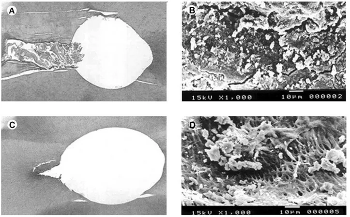
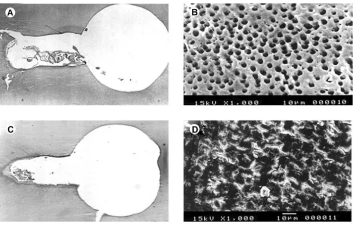
Figures 1 and 2 show a panel of photomicrographs and SEM micrographs of the groups treated with distilled water (control), 1% naoCl, 1% naoCl + 17% EdtA and 1% naoCl + rCPrep.
the results of the histological and morphomet-ric analysis showed statistically significant difference (p<0.001) among the groups regarding the percentage of debris left in the canals after instrumentation and irrigation protocols (table 1) (Figs. 1A, 1C, 2A, 2C).
In the SEM analysis, statistically significant dif-ference (p<0.001) was found in the cleaning provided by the studied solutions. there was no statistically significant difference (p>0.001) among distilled wa-ter (Fig. 1B), 1% naoCl (Fig. 1d) and 1% naoCl + rCPrep (Fig. 2d), which presented a great amount of smear layer, whereas 1% naoCl + 17% EdtA, showed
dentin walls free of a smear layer and exposed a large number of tubules (table 1) (Fig. 2B).
DISCUSSION
there is general agreement among endodontic researchers and practitioners regarding importance of biomechanical preparation. However, there is still discussion on which instrumentation technique and ir-rigating solution is more effective.
this study used optical microscopy because it reveals by histological and morphometric analyses, the amount of debris presented in the root canal system (1,2,5,6), and used SEM to analyze the presence of smear layer on the internal walls of root canal after longitudinal section (4,16). these are two complementary methods to evaluate root canal cleaning.
In the optical microscopic analysis, irrigation with
1% naoCl showed the best results for debris removal, which are due to the capacity of this solution to dissolve organic tissue, whether necrotic or not, turning solid pulp particles into liquid (9). However, the SEM analysis showed that naoCl was not able to completely remove smear layer from dentin walls during biomechanical preparation. the presence of sodium hydroxide and hypochlorous acid, formed by the dissociation of naoCl, dissolves only organic compounds due to its selectively on organic matrix (9), thus not totally removing the smear layer from the canal walls.
the specimens irrigated with 1% naoCl alter-nated with 17% EdtA presented intermediate values of debris when compared to those irrigated only with 1% naoCl. nevertheless, the SEM values show absence of smear layer and smear plug in this group probably
because naoCl acts selectively on organic matrices, and EdtA on inorganic matrices, leading to a root canal.
Figure 2. A = Photomicrograph of the apical region (×40) irrigated with 1% naoCl alternated with 17% EdtA showing areas not touched by the instrument and presence of debris; B = SEM micrograph (×1000) showing dentin surface with open dentinal tubules and no smear layer after biomechanical preparation using 1% naoCl alternated with EdtA solution for irrigation; C = Photomicrograph of the apical region (×40) treated with 1% naoCl with rCPrep cream showing areas not touched by the instrument and presence of debris; d = SEM micrograph (×1000) showing smear-covered dentin surface after biomechanical preparation using 1% naoCl with rCPrepcream solution for irrigation.
table 1. Mean percentage of debris (Sd) in flattened root canals for each group tested.
H2o naoCl naoCl
+ EdtA
naoCl + rCPrep
debris 18.82 A (5.55)
6.29 B (5.55)
12.47 C (6.92)
7.82 d (1.91)
Smear layer 3.00 a (0.00)
2.20 a (0.44)
0.60 b (0.54)
2.60 a (0.54)
surface with no smear layer and smear plug (19). there are controversies in the literature about the effects of naoCl alternated with EdtA. Saquy et al. (13) observed that naoCl increases the action of the EdtA. Pécora et al. (19) showed that EdtA may inhibit the action of the naoCl solution in dissolving tissue. the findings of the present study are in agreement with previous findings (14) since there was substantially less debris when naoCl was used alone rather than associ-ated to EdtA.
the specimens irrigated with 1% naoCl and rCPrep cream showed similar results on debris removal when compared to 1% naoCl solution alternated with 17% EdtA. debris removal can be explained by the reaction between naoCl and the urea peroxide presented in the rCPrep cream that produces an effervescent exothermic reaction, which can interfere with naoCl’s tissue-solving property (9). Anderson et al. (12) reported that using rCPrep cream as a lubricant when preparing curved canals leds to less instrument stress. the SEM images of this group showed the presence of smear layer. rCPrep cream remaining on the root canal walls hinders the action of hypochlorite solution on the organic matrices in the smear layer, resulting in obliteration of the dentinal tubules (20).
Each irrigating solution must be evaluated regard-ing their physicochemical properties and used selectively during biomechanical preparation in order to enhance their chemical properties and produce cleaner root canals (8). In the present study, the combination of Profile .04 rotary instrumentation and the tested irrigating solutions was not able to totally remove debris and smear layer from flattened root canals. Although naoCl used alone is the solution that best aids the removal of debris, a bet-ter way of using it associated with chelating solutions needs to be found.
RESUMO
o objetivo deste estudo foi avaliar a capacidade de limpeza dos instrumentos Profile .04 associados a diferentes soluções ir-rigantes, em canais radiculares achatados, por meio de análises histológica, morfométrica e MEv. oitenta incisivos inferiores humanos foram submetidos ao preparo biomecânico com in-strumentos Profile .04 e aleatoriamente distribuídos em 4 grupos de acordo com as soluções irrigantes usadas (n=20): G1 - água destilada (controle); G2 - naoCl 1%; G3 - naoCl 1% alternado com EdtA 17% e G4 - naoCl 1% com rCPrep creme. dez dentes de cada grupo foram avaliados em microscopia óptica para determinação da porcentagem de debris do canal radicular.
os demais dentes foram avaliados em microscopia eletrônica de varredura. A análise estatística mostrou diferença significante (p<0,001) entre os grupos ao considerar a porcentagem de debris remanescente nos canais (água destilada: 18,82 ± 5,55; naoCl: 6,29 ± 5,55; naoCl + EdtA: 12,47 ± 6,92; naoCl + rCPrep: 7,82 ± 1,91). A análise em MEv evidenciou melhores resultados na remoção da camada de smear com o naoCl 1% alternado com EdtA 17%. Pode-se concluir que a associação entre a instrumentação rotatória com Profile .04 e as soluções testadas não foi capaz de remover totalmente debris e camada de smear
de canais radiculares achatados.
REFERENCES
1. Barbizam JvB, Fariniuk lF, Marchesan MA, Pécora Jd, Sousa-neto Md. Effectiveness of manual and rotatory instrumenta-tion techniques for cleaning flattened root canals. J Endod 2002;28:365-366.
2. Marchesan AM, Arruda MP, Silva-Sousa YtC, Saquy PC, Sousa-neto Md. Morphometrical analysis of cleaning capacity using nickel-titanium rotary instrumentation associated with irrigating solutions in mesio-distal flattened root canals. J Appl oral Sci 2003;11:55-59.
3. Weiger r, Elayouti A, löst C. Efficiency of hand and rotary instru-ments in shaping oval root canals. J Endod 2002;28:580-583. 4. Schäfer E, Zapke KA. Comparative scanning electron microscopic
investigation of the efficacy of manual and automated instrumenta-tion of root canal. J Endod 2000;26:660-664.
5. nadalin Mr, Perez dE, vansan lP, Paschoalato C, Souza-neto Md, Saquy PC. Effectiveness of different final irrigation pro-tocols in removing debris in flattened root canals. Braz dent J 2009;20:211-214.
6. Baratto-Filho F, Carvalho Jr Jr, Fariniuk lF, Sousa-neto Md, Pé-cora Jd, Cruz-Filho AM. Morphometric analysis of the effective-ness of different concentrations of sodium hypochlorite associated with rotary instrumentation for root canal cleaning. Braz dent J 2004;15:36-40.
7. Wu MK, van der Sluis lW, Wesselink Pr. the capability of two hand instrumentation techniques to remove the inner layer of dentine in oval canals. Int Endod J 2003;36:218-224.
8. de-deus G, Garcia-Filho P. Influence of the niti rotary system
on the debridement quality of the root canal space. oral Surg oral
Med oral Pathol oral radiol Endod 2009;108:e71-e76. 9. Spanó JCE, Barbin El, Santos tC, Guimarães lF, Pécora Jd.
Sol-vent action of sodium hypochlorite on bovine pulp and physico-chemical properties of resulting liquid.Braz dent J 2001;12:154-170.
10. Østby nB. Chelation in root canal therapy. Ethylenediamine tetra-acetic acid for cleansing and widening of root canals. odont tidskrift 1957;65:3-11.
11. Stewart GG, Kapsimalis P, rappaport H. EdtA and urea perox-ide for root canal preparation. J Am dent Assoc 1969;78:335-338.
12. Anderson dn, Joyce AP, roberts S, runner r. A comparative pho-toelastic stress analysis of internal root stresses between rC Prep and saline when applied to the Profile/Gt rotary instrumentation system. J Endod 2006;32:222-224.
13. Saquy PC, Campos GM, Sousa-neto Md, Guimarães lF, Pécora Jd. Evaluation of chelating action of EdtA in association with dakin’s solution.Braz dent J 1994;5:65-70.
Endod 2004;1:3-7.
15. torabinejad M, Cho Y, Khademi AA, Bakland lK, Shabahang S. the effect of various concentrations of sodium hypochlorite on the ability of MtAd to remove the smear layer.J Endod 2003;29:233-239.
16. Scelza MFZ, Antoniazzi JH, Scelza P. Efficacy of final irrigation - a scanning electron microscopic evaluation. J Endod 2000;6:355-358.
17. limongi o, Albuquerque dS, Baratto-Filho F, vanni Jr, oliveira EPM, Barletta FB. In vitro comparative study of manual and mechanical rotary instrumentation of root canals using computed tomography. Braz dent J 2007;18:289-293.
18. takeda FH, Harashima t, Kimura Y, Matsumoto K. A comparative study of the removal of smear layer by three endodontic irrigants and two types of laser. Int Endod J 1999;32:32-39.
19. Pécora Jd, Sousa-neto Md, Saquy PC, Silva rG, Cruz-Filho AM. Effect of dakin’s and EdtA solutions on dentin permeability of root canals. Braz dent J 1993;4:79-84.
20. Cameron JA. the use of ultrasound and EdtA-urea peroxide compound in the cleansing of root canals. An SEM study. Aust dent J 1984;29:80-85.