Radiol Bras. 2015 Mar/Abr;48(2):69–73 69
Effects of iodinated contrast agent, xylocaine and gadolinium
concentration on the signal emitted in magnetic resonance
arthrography: a samples study
*
Efeitos do contraste iodado, da xilocaína e da concentração de gadolínio no sinal emitido em exames de artrorressonância magnética: estudo por amostras
Pinheiro YLS, Costa RZV, Pinho KEP, Ferreira RR, Schuindt SM. Effects of iodinated contrast agent, xylocaine and gadolinium concentration on the signal emitted in magnetic resonance arthrography: a samples study. Radiol Bras. 2015 Mar/Abr;48(2):69–73.
Abstract
R e s u m o
Objective: To investigate the effects of dilution of paramagnetic contrast agent with iodinated contrast and xylocaine on the signal intensity during magnetic resonance arthrography, and to improve the paramagnetic contrast agent concentration utilized in this imaging modality. Materials and Methods: Samples specially prepared for the study with three different concentrations of paramagnetic contrast agent diluted in saline, iodinated contrast agent and xylocaine were imaged with fast spin echo T1-weighted sequences with fat saturation. The samples were placed into flasks and graphical analysis of the signal intensity was performed as a function of the paramagnetic contrast concentration.
Results: As compared with samples of equal concentrations diluted only with saline, the authors have observed an average signal intensity decrease of 20.67% for iodinated contrast agent, and of 28.34% for xylocaine. However, the increased gadolinium concentration in the samples caused decrease in signal intensity with all the dilutions.
Conclusion: Minimizing the use of iodinated contrast media and xylocaine and/or the use of a gadolinium concentration of 2.5 mmol/L diluted in saline will improve the sensitivity of magnetic resonance arthrography.
Keywords: Magnetic resonance arthrography; Contrast media; Magnetic resonance imaging; Xylocaine.
Objetivo: Investigar, mediante quantificação da intensidade do sinal emitido em amostras, se a diluição do agente de contraste para-magnético com contraste iodado e xilocaína alteram o sinal emitido pelo meio de contraste parapara-magnético durante o exame de artrorres-sonância magnética, e aperfeiçoar a concentração de contraste paramagnético utilizada no exame.
Materiais e Métodos: Foi realizada sequência de pulso fast spin eco ponderada em T1 com saturação de gordura, utilizando três diferentes concentrações de contraste paramagnético diluídas em solução salina, contraste iodado e xilocaína. As amostras foram co-locadas em frascos e a análise gráfica da intensidade do sinal em função da concentração de contraste paramagnético foi realizada. Resultados: Constatou-se que as diluições de contraste paramagnético em contraste iodado e xilocaína diminuíram, em média, a intensidade do sinal em 20,67% para o contraste iodado e 28,34% para a xilocaína, em comparação com as amostras de concentração idêntica diluídas apenas em solução salina. Porém, o aumento da concentração de gadolínio nas amostras ocasionou a diminuição da intensidade do sinal emitido pelo gadolínio, para todas as diluições.
Conclusão: Minimizar o uso do meio de contraste iodado e da xilocaína e/ou a utilização de uma concentração de gadolínio com 2,5 mmol/L, diluída em solução salina, irá aperfeiçoar a sensibilidade do exame de artrorressonância magnética.
Unitermos: Artrorressonância magnética; Meios de contraste; Ressonância magnética; Xilocaína.
* Study developed at Centro Diagnóstico Água Verde (Cedav), Curitiba, PR, Brazil. 1. Radiology Technologist, Universidade Tecnológica Federal do Paraná (UTFPR), Curitiba, PR, Brazil.
2. PhD in Sciences, Professor at Universidade Tecnológica Federal do Paraná (UTFPR), Curitiba, PR, Brazil.
3.Fellow PhD degree in Sciences, Professor at Universidade Tecnológica Federal do Paraná (UTFPR), Curitiba, PR, Brazil.
4. MD, Radiology and Ultrasonography Specialist at Centro Diagnóstico Água Verde (Cedav), Curitiba, PR, Brazil.
5. Radiology Technologist, Graduate Student of Medicine, Universidade Federal do Paraná (UFPR), Curitiba, PR, Brazil.
Mailing Address: Yvana Lopes Pinheiro da Silva. Rua Pedro Collere, 699, Vila Izabel, Curitiba, PR, 80320-320, Brazil. E-mail: [email protected].
Received October 3, 2013. Accepted after revision September 3, 2014.
INTRODUCTION
The use of magnetic resonance imaging (MRI) has in-creased in the last years, both for the greater availability of apparatuses and its wide application in different clinical
set-tings(1). This method is considered as the gold standard for
acquisition of gadolinium-enhanced abdominal images(2),
cardiac images for cardiac mass and volume quantification(3),
and is also widely used in the evaluation of joints, allowing for direct visualization of relevant anatomical structures, including ligaments, menisci and periarticular soft tissues. Studies show that the delimitation of many intra-articu-lar structures, the visualization of the normal articuintra-articu-lar
Yvana Lopes Pinheiro da Silva1, Rita Zanlorensi Visneck Costa2, Kátia Elisa Prus Pinho3, Ricardo Rabello
anatomy, and the description of abnormalities are reinforced by the presence of articular effusion. At magnetic resonance arthrography (MRA), such an articular effusion is iatrogeni-cally obtained, combining the advantages of the joint dis-tension, the high-contrast resolution and the multiplanar
acquisition capability of the method(4).
Before the MR images acquisition, a needle is inserted into the joint to be studied and the intra-articular location is confirmed by means of either computed tomography (CT)-or flu(CT)-oroscopically-guided injection of a small volume of iodinated contrast agent. Then, the diluted paramagnetic contrast/saline solution is injected into the intra-articular region and the images acquisition is performed. The tech-nique might include a simultaneous injection of xylocaine without vasoconstrictor combined with the contrast agent in order to relieve the pain, and to help the patient to remain
still during the scanning(5).
According to the literature, the paramagnetic contrast dilution with iodinated contrast agent might have a
signifi-cant effect on the quality of the MRA scan(6). Thus, the
present study was aimed at investigating if the presence of iodinated contrast agent and xylocaine without vasoconstrictor in the solution injected into the joint affects the image qual-ity and the signal emitted by the paramagnetic contrast agent, besides evaluating which is the most appropriate paramag-netic contrast concentration in MRA.
MATERIALS AND METHODS
An in vitro study was developed with three different
gadodiamide (gadolinium) concentrations, as follows: 2.5 mmol/L, 5.0 mmol/L and 10.0 mmol/L, and each concen-tration was diluted in 100 mL saline solution (0.9% sodium chloride). Subsequently, nine 10 mL syringes received 8 mL of those solutions and were added as follows: syringes num-bers 1, 2 and 3 received 1.5 mL saline solution; syringes 4, 5 and 6 received 1.5 mL non-ionic iodinated contrast agent; and syringes 7, 8 and 9 received 1.5 mL xylocaine without vasoconstrictor. Thus, the final volume in each syringe was 9.5 mL.
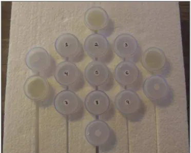
Each syringe contents was transferred to standard poly-styrene 15 mL flasks which were inserted into an expanded polystyrene block for immobilization during the images ac-quisition (Figure 1). Additionally, six control tubes were utilized – three out of them filled with 9.5 mL saline solu-tion and the other three flasks filled with vegetal soya oil to aid in the samples orientation during the study (Figure 2). Neither color plastic materials, glues with high proton concentration, nor other materials with magnetic suscepti-bility very different from the solutions were utilized in the making of the samples, since these types of materials could
produce local inhomogeneity and susceptibility artifacts(7).
Images acquisition
The samples’ images were acquired with a 1.5 T MRI apparatus utilizing a skull coil for acquisition of T1-weighted
fast spin echo pulse sequences with fat saturation, TR = 416.7 ms, TE = 7.9 ms, matrix = 256 × 256 pixels, slice thickness = 3.0 mm and FOV = 21.
The flasks were kept closed during the images acquisi-tion in order to minimize possible changes in the soluacquisi-tions (for example, evaporation). The air conditioning tempera-ture was set at 18°C, providing the same room temperatempera-ture and, consequently the same temperature of the samples dur-ing the process of images acquisition.
Figure 2. Test samples.
Figure 1. Representation of the test samples. The numbers represent the gado-linium concentration (in mmol/L) utilized in each of the flasks.
Saline solution Vegetal soya oil
Saline solution + gadolinium
Saline solution + gadolinium + iodinated contrast agent
Images analysis
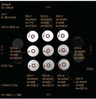
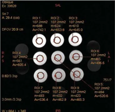
The images were transferred and analyzed with the soft-ware Advantage Workstation GE 2010. The region of inter-est (ROI) was utilized to define the central region of the sample and to quantify the signal emitted by the different gadolinium dilutions and concentrations at the images, determining the average value of pixels in different areas of grey level in the ROI, i.e. the mean brightness of the ROI. The ROIs were identified in the center of the flasks where the signal inten-sities were measured and amplitude values were recorded. Three different sizes of ellipse-shaped ROI (6 mm, 9 mm and 12 mm in diameter), were utilized to calculate the mean signal intensity as measured on these three ROI sizes.
The charts were designed in accordance with the ampli-tude values (signal intensity on the ROI) versus gadolinium concentration for each one of the three sample mixtures (sa-line solution, iodinated contrast agent and xylocaine).
RESULTS
With T1-weighted, fast spin eco sequence (TR/TE = 416.7/7.9) with fat saturation (Figure 3), the peak signal amplitude was produced at the gadodiamide 2.5 mmol/L con-centration diluted in regular saline solution for all the ROI sizes. Gadodiamide dilution with iodinated contrast agent and xylocaine resulted in decreased signal amplitude for all the ROI sizes, as compared with gadodiamide dilution with saline solution.
For ROI with 6 mm in diameter (Figure 4), the signal intensity with gadolinium diluted in iodinated contrast agent decreased 18.56% at 2.5 mmol/L concentration, 20.13% at 5.0 mmol/L concentration, and 23.21% at 10.0 mmol/L concentration, as compared with their respective
concentra-tions diluted in saline solution only. For dilution with xylocaine, the decrease in the gadolinium signal intensity was of 27.80% at 2.5 mmol/L concentration, 29.21% at 5.0 mmol/ L concentration, and 27.68% at 10.0 mmol/L concentration, also compared with their respective concentrations diluted in saline solution only.
For ROI with 9 mm in diameter (Figure 5), the signal intensity with gadolinium diluted in iodinated contrast agent decreased 18.57% at 2.5 mmol/L concentration, 20.20% at 5.0 mmol/L concentration, and 23.48% at 10.0 mmol/L
Figure 4. Image of the test object analyzed with a ROI with 6 mm in diameter.
Figure 5. Image of the test object analyzed with a ROI with 9 mm in diameter.
concentration, as compared with their respective concentra-tions diluted in saline solution only. For dilution with xylocaine, the decrease in the gadolinium signal intensity was of 27.97% at 2.5 mmol/L concentration, 29.37% at 5.0 mmol/L concentration, and 27.87% at 10.0 mmol/L concen-tration, also compared with their respective concentrations diluted in saline solution only.
For ROI with 12 mm in diameter (Figure 6), the signal intensity with gadolinium diluted in iodinated contrast agent decreased 18.42% at 2.5 mmol/L concentration, 20.38% at 5.0 mmol/L concentration, and 23.01% at 10.0 mmol/L concentration, as compared with their respective concentra-tions diluted in saline solution only. For dilution with xylocaine, the decrease in the gadolinium signal intensity was of 27.72% at 2.5 mmol/L concentration, 29.20% at 5.0 mmol/L concentration, and 27.87% at 10.0 mmol/L concen-tration, also compared with their respective concentrations diluted in saline solution only.
Figure 7 shows the decrease in gadodiamide signal in-tensity after dilution with iodinated contrast agent and xylocaine. One has considered that the signal emitted by these three gadodiamide solutions diluted in saline solution only represent the maximum intensities (100%) emitted by gado-linium for each of the different concentrations, because the saline solution is utilized only as a diluent for the solution to be injected into the joint. The signal intensity with dilu-tion in iodinated contrast agent was 81.48% for gadodiamide 2.5 mmol/L concentration, 79.76% for 5.0 mmol/L tration, and 76.76% for gadodiamide 10.0 mmol/L concen-tration. On the other hand, the signal intensity with dilution in xylocaine was 72.17% for gadodiamide 2.5 mmol/L con-centration, 70.74% for 5.0 mmol/L concon-centration, and 72.09% for gadodiamide 10.0 mmol/L concentration.
DISCUSSION
Greater intrinsic soft tissue contrast resolution in asso-ciation with non-exposure to ionizing radiation have made
MRI an excellent choice for screening purposes(8), as well
as in the evaluation of joints. MRA has been utilized for a detailed evaluation of internal joint derangements because of its capacity to describe small anatomical details, increas-ing the diagnostic accuracy. In many circumstances, MRA is superior to conventional non-contrast-enhanced MRI and to CT arthrography in the evaluation of several diseases
af-fecting shoulders, knees, hip and other joints(9).
Brown et al.(5) have developed an in vitro study, where
three iodinated contrast agents were mixed and incubated with paramagnetic contrast agent. They observed that no ga-dolinium ion dissociated from the complex, even after addi-tion of saline soluaddi-tion, xylocaine or epinephrine. Such re-sults demonstrate that the mixture of contrast material, ga-dolinium and iodinated contrast agent is safe, and xylocaine and/or epinephrine might also be added for clinical purposes.
According to Montgomery et al.(6), the literature reports
inconsistency both in the gadolinium concentration utilized in MRA and in the amount of injected iodinated contrast agent and xylocaine.
In the present study, the gadolinium concentrations were different (2.5 mmol/L, 5.0 mmol/L and 10.0 mmol/L) and the amounts of iodinated contrast agent (1.5 mL) and xylocaine (1.5 mL) remained equal, and are the same uti-lized by physicians who perform MRA in the diagnostic cen-ter where the present study was developed.
The authors observed that the increased gadolinium con-centration in the samples causes decrease in the gadolinium
signal intensity for all the dilutions. According to Bushong(10),
this occurs because if the gadolinium concentration in a Figure 6. Image of the test object analyzed with a ROI with 12 mm in diameter.
Figure 7. Comparison of the decrease in signal intensity considering the gado-diamide dilution in saline solution as maximum intensity.
Gadodiamide concentration (mmol/L)
S
ig
n
a
l
in
te
n
si
ty
Saline solution Iodinated contrast agent
determined area becomes extremely high, it is possible that the T2-weighting effect predominates even on a T2-weighted image, causing signal intensity loss on both image types.
According to Montgomery et al.(6), this may be harmful,
particularly in scans performed in joints with small volume of synovial fluid, as in the region of the wrist, since in such joints there is less contrast dilution. Therefore, the utiliza-tion of a gadolinium 2.5 mmol/L concentrautiliza-tion allows for some dilution in the synovial fluid, which may even increase the signal intensity, according to the results reported by
Montgomery et al.(6).
The improvement in the gadolinium concentration and in the amount of iodinated contrast agent and xylocaine re-sults in visually perceptible differences which may signifi-cantly affect the MRA diagnostic quality. Another implica-tion of such data is that previous reports evaluating the MRA efficacy may have not utilized appropriate gadolinium con-centrations and/or minimized the use of iodinated contrast agent and xylocaine. As a result, the MRA diagnostic rel-evance in relation to the other imaging modalities might have
been underestimated(6).
The present study demonstrates that the addition of io-dinated contrast agent or xylocaine leads to a decrease in the signal emitted by the gadolinium, as suggested by Kopka et
al.(11), who say that the iodinated contrast agent reduces the
gadolinium T1 effect, although the exact mechanism of this action is still unknown. As regards xylocaine, up to this moment there is no comparative study demonstrating its effect on MRA images, probably because xylocaine is not as frequently utilized as iodinated contrast agent in this type of procedure.
CONCLUSION
The present study results demonstrate that the peak sig-nal intensity was obtained with a gadodiamide concentration of about 2.5 mmol/L diluted in regular saline solution (Fig-ures 4, 5 and 6). Thus, on the basis of the results and con-sidering the presence of iodinated contrast agent and xylocaine without vasoconstrictor in the solution injected into the joint, a gadolinium concentration = 2.5 mmol/L is recommended. Gadolinium dilution in iodinated contrast agent and xylocaine has led to a reduction of respectively 20.76% and
28.34% in the signal intensity as compared with the samples with equal concentrations diluted in saline solution only. Such percentage values correspond to the calculated means for the three different gadolinium concentrations, diluted in iodinated contrast agent and xylocaine and to the different ROI sizes. Therefore, according to the present results, minimizing the use of iodinated contrast agent and xylocaine and/or the utilization of a gadolinium 2.5 mmol/L concentration diluted in saline solution will improve the sensitivity and specificity of MRA in the evaluation of internal joint derangements.
REFERENCES
1. Galvão BVT, Torres LR, Cardia PP, et al. Prevalência de cistos sim-ples e hemangiomas hepáticos em pacientes cirróticos e não cirróti-cos submetidos a exames de ressonância magnética. Radiol Bras. 2013;46:203–8.
2. Kim YH, Shin SS, Burke LMB, et al. Hemangioma hepático sub-capsular com realce perilesional: achados de RM. Radiol Bras. 2010; 43:384–8.
3. Barranhas AD, Santos AASMD, Coelho-Filho OR, et al. Cardiac magnetic resonance imaging in clinical practice. Radiol Bras. 2014; 47:1–8.
4. Hajek PC, Sartoris DJ, Neumann CH, et al. Potential contrast agents for MR arthrography: in vitro evaluation and practical observations. AJR Am J Roentgenol. 1987;149:97–104.
5. Brown RR, Clarke DW, Daffner RH. Is a mixture of gadolinium and iodinated contrast material safe during MR arthrography? AJR Am J Roentgenol. 2000;175:1087–90.
6. Montgomery DD, Morrison WB, Schweitzer ME, et al. Effects of iodinated contrast and field strength on gadolinium enhancement: implications for direct MR arthrography. J Magn Reson Imaging. 2002;15:334–43.
7. Price RR, Axel L, Morgan T, et al. Quality assurance methods and phantoms for magnetic resonance imaging: report of AAPM Nuclear Magnetic Resonance Task Group No. 1. Med Phys. 1990;17:287– 95.
8. Hernandes MA, Semelka RC, Elias Jr, et al. Whole-body MRI: com-prehensive evaluation on a 48-channel 3T MRI system in less than 40 minutes. Preliminary results. Radiol Bras. 2012;45:319–25. 9. Choi JY, Kang HS, Hong SH, et al. Optimization of the contrast
mixture ratio for simultaneous direct MR and CT arthrography: an in vitro study. Korean J Radiol. 2008;9:520–5.
10. Bushong SC. Magnetic resonance imaging: physical and biological principles. 3rd ed. Philadelphia, PA: Mosby; 2003.