Letters to the Editor
Radiol Bras. 2016 Mai/Jun;49(3):199–204
199
0100-3984 © Colégio Brasileiro de Radiologia e Diagnóstico por Imagem
Letters to the Editor
PET/CT used in the evaluation of pulmonary nodules suspicious for lung cancer in regions where infectious lung disease is endemic: to be or not to be?
http://dx.doi.org/10.1590/0100-3984.2016.49.3.ce1 Dear Editor,
The assessment of patients with suspected lung malig-nances(1–4) has routinely included morphological imaging
evalu-ation, with either chest X-rays or chest computed tomography (CT). In addition—although not diagnostic in character—18
F-fluorodeoxyglucose positron emission tomography (FDG-PET), bone scintigraphy, and (occasionally) somatostatin receptor scin-tigraphy have been increasingly incorporated into daily practice in recent decades, providing physicians with useful and complemen-tary information on the functional characteristics of lesions(5,6).
More recently, the emergence of combined PET/CT imaging has greatly aided the investigation of lung cancer by allowing even better delineation of areas with increased tracer uptake. This modality has helped radiologists avoid the technical difficulties that arose from the independent combination of PET and CT exami-nations, which resulted in substantial artifacts.
Many patients with early stage lung cancer will present with a solitary pulmonary nodule (SPN), defined as a single spherical or oval lesion that is less than 3 cm in diameter and is completely surrounded by pulmonary parenchyma without accompanying atelectasis or lymph node enlargement(5,6). A very important step
in investigating the etiology of an SPN is to determine whether it is benign or malignant in nature. In addition, PET/CT has been shown to be an accurate tool for the work-up of SPNs and for lung cancer staging, by improving the detection of metastatic disease, guiding therapy, and allowing clinical outcomes to be predicted(5– 7). However, there are a number of pitfalls to be considered during
the assessment of SPNs with PET. In patients with inflamma-tory conditions or infections—such as bacterial or fungal infec-tions; granulomatous diseases (tuberculosis, sarcoidosis, histoplas-mosis, etc.); and pyogenic abscesses—there is a greater likelihood of higher metabolic activity due to increased granulocyte or mac-rophage activity, and such comorbidities have become a cause for great concern in some regions of Brazil(8–10).
In a recent study published in Radiologia Brasileira, Mos-mann et al.(11) reviewed the evaluation of SPNs, in order to
dis-cuss the current role of FDG-PET (addressing its accuracy and cost-effectiveness) and to detail the current recommendations for the examination in this scenario. However, the authors did not focus on the applicability of FDG-PET in areas endemic for in-fectious granulomatous diseases. Deppen et al.(12) performed the
most recent and biggest meta-analysis about the diagnostic ac-curacy of FDG-PET for pulmonary nodules suspicious for lung cancer, comparing the accuracy of the test in regions where in-fectious lung disease is endemic with that reported for regions where such disease is rare(8). The pooled (unadjusted) sensitivity
and specificity were 89% (95% CI: 86–91%) and 75% (95% CI: 71–79%), respectively. The adjusted specificity was 16% lower for regions where infectious lung disease is endemic than for those where it is not—61% (95% CI, 49–72%) versus 77% (95% CI, 73–80%). The specificity was also lower when the analysis was limited to rigorously conducted and well-controlled studies. The conclusion is that the data do not support the use of FDG-PET to diagnose lung cancer in areas where infectious lung disease is endemic unless an institution achieves test performance accu-racy similar to that found in areas where it is not(12). Because
Mosmann et al.(11) did not include these data in their review, is
important to highlight that fact.
REFERENCES
1. Franco RM, Guimaraes MD, Moreira BL, et al. Enhancing survival with early surgical resection of endobronchial metastasis in a follow-up of ovarian carcinoma. Radiol Bras. 2015;48:130.
2. Guimaraes MD, Hochhegger B, Koenigkam-Santos M, et al. Magnetic resonance imaging of the chest in the evaluation of cancer patients: state of the art. Radiol Bras. 2015;48:33–42.
3. Batista MN, Barreto MM, Cavaguti RF, et al. Pulmonary artery sar-coma mimicking chronic pulmonary thromboembolism. Radiol Bras. 2015;48:333–4.
4. Silva Junior GM, Zanetti GMR, Barillo JL, et al. Peripheral primitive neuroectodermal tumor of chest wall in young adult. Radiol Bras. 2015;48:59–60.
5. Hochhegger B, Alves GR, Irion KL, et al. PET/CT imaging in lung cancer: indications and findings. J Bras Pneumol. 2015;41:264–74. 6. Sharma P, Singh H, Basu S, et al. Positron emission
tomography-com-puted tomography in the management of lung cancer: an update. South Asian J Cancer. 2013;2:171–8.
7. Ambrosini V, Nicolini S, Caroli P, et al. PET/CT imaging in different types of lung cancer: an overview. Eur J Radiol. 2012;81:988–1001. 8. Truong MT, Viswanathan C, Erasmus JJ. Positron emission tomogra-phy/computed tomography in lung cancer staging, prognosis, and as-sessment of therapeutic response. J Thorac Imaging. 2011;26:132– 46.
9. Opoka L, Kunikowska J, Podgajny Z, et al. Accuracy of FDG PET/CT in the evaluation of solitary pulmonary lesions – own experience. Pneumonol Alergol Pol. 2014;82:198–205.
10. Allen TL, Kendi AT, Mitiek MO, et al. Combined contrast-enhanced computed tomography and 18-fluoro-2-deoxy-D-glucose-positron emis-sion tomography in the diagnosis and staging of non-small cell lung cancer. Semin Thorac Cardiovasc Surg. 2011;23:43–50.
11. Mosmann MP, Borba MA, Macedo FP, et al. Solitary pulmonary nodule and 18F-FDG PET/CT. Part 2: accuracy, cost-effectiveness, and cur-rent recommendations. Radiol Bras. 2016;49:104–11.
12. Deppen S, Putnam JB Jr, Andrade G, et al. Accuracy of FDG-PET to diagnose lung cancer in a region of endemic granulomatous disease. Ann Thorac Surg. 2011;92:428–32.
Bruno Hochhegger1
1. Universidade Federal de Ciências da Saúde de Porto Alegre (UFCSPA), Porto Alegre, RS, Brazil. E-mail: [email protected].
Neurological symptoms in a case of acute aortic dissection
Dear Editor,
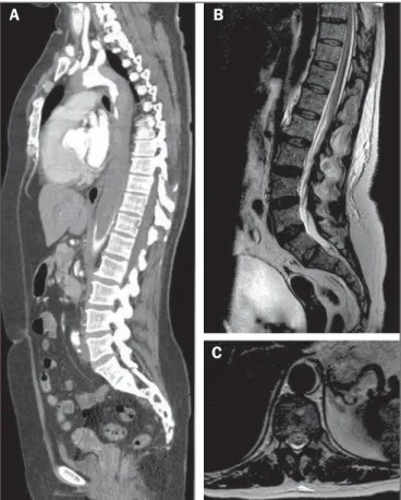
A 52-year-old female with aortic dissection presented with neurological symptoms and signs, in a markedly acute presenta-tion, of flaccid paraplegia and painful hypoesthesia of the lower limbs. She also presented postoperative monoplegia of the left arm. Computed tomography angiography of the chest confirmed the
diagnosis of type A dissection (Stanford classification), with ex-tension to the infrarenal abdominal aorta, associated with exten-sive suboccluexten-sive thrombus in the thoracoabdominal transition of the aorta (Figure 1A). On T2-weighted magnetic resonance imaging (MRI) sequences, hyperintensity was observed in the anterior horns of the spinal cord (Figures 1B and 1C), featuring an “owl eye” sign in axial images(1), together with enhancement
Letters to the Editor
Radiol Bras. 2016 Mai/Jun;49(3):199–204
200
http://dx.doi.org/10.1590/0100-3984.2015.0070
anterior two thirds of the spinal cord (mainly by the artery of Adamkiewicz) is more susceptible to ischemia than is the poste-rior segment, which has several levels of vascular supply(6). A high
degree of clinical suspicion of neurological involvement of the spinal cord is indicative of the diagnosis. Symptoms vary depend-ing on the extent of the affected area and the level of spinal injury. Cerebral ischemic lesion is also a possible complication of aortic dissection and can result from reduced blood flow to the brain caused by the surgical procedure or even from carotid involvement caused by dissection or embolism from the thrombus in the aorta. In addition, data in the literature indicate that there is a right-side dominance of lesions, which is explained by different mechanical dynamics in the progression of the dissecting hematoma.
MRI is particularly sensitive in the detection of aortic dissec-tion and can reveal signal abnormality in the anterior horns of the spinal cord, which can be associated with enhancement after contrast agent injection. The spinal segment most often affected is the thoracic segment, due to the border arterial supply(6).
Diffu-sion sequences can show restriction in the ischemic area. In fact, diffusion sequences can provide early detection(7), although this
technique is not always applied in routine MRI scans of the spinal cord. Therefore, we have presented a case of aortic dissection with a rare combination of neurological complications of brain and spi-nal cord ischemia.
REFERENCES
1. Udiya AK, Shetty GS, Singh V, et al. “Owl eye sign”: anterior spinal ar-tery syndrome. Neurol India. 2015;63:459.
2. Amaral RH, Souza VV, Nin CS, et al. Aortic lesion simulating pulmonary disease: a case report. Radiol Bras. 2014;47:320–2.
3. Metzger PB, Novero ER, Rossi FH, et al. Evaluation of preoperative com-puted tomography angiography in association with conventional angiog-raphy versus computed tomogangiog-raphy angiogangiog-raphy only, in the endovascular treatment of aortic diseases. Radiol Bras. 2013;46:265–72.
4. Netto OS, Hasselmann CL, Osterne ECV, et al. Detection of abdominal aortic calcification by densitometry. Radiol Bras. 2013;46:35–8. 5. Moulakakis KG, Mylonas SN, Dalainas I, et al. Management of
compli-cated and uncomplicompli-cated acute type B dissection. A systematic review and meta-analysis. Ann Cardiothorac Surg. 2014;3:234–46.
6. Lee SJ, Kim JH, Na CY, et al. Eleven years of experience with the neuro-logic complications in Korean patients with acute aortic dissection: a retrospective study. BMC Neurol. 2013;13:46.
7. Fujikawa A, Tsuchiya K, Takeuchi S, et al. Diffusion-weighted MR im-aging in acute spinal cord ischemia. Eur Radiol. 2004;14:2076–8. Figure 1.A: Maximum intensity projection reconstruction of computed
tomogra-phy angiogratomogra-phy in the sagittal plane showing an extensive mural thrombus in the thoracic aorta, extending to the infrarenal aorta. B: Sagittal T2-weighted MRI sequence showing areas of hyperintensity within the anterior spinal cord. C: Axial T2-weighted MRI at the level of the T10 spinal segment demonstrating anterior areas of hyperintensity, with the “owl eye” sign.
A
B
C
diffusion of water at the levels studied. Cranial MRI revealed acute lesions (also with restricted diffusion) in the right middle cerebral artery. The patient underwent surgery to treat the aortic dissec-tion, and her neurological function was monitored.
The evaluation of the aorta by imaging methods has been the subject of a series of recent publications in the Brazilian radiology literature(2–4). In the case presented here, neurological findings
were associated with aortic dissection, and the MRI findings were consistent with the diagnosis of spinal cord infarction with ischemic stroke in the right middle cerebral artery. Although spinal cord infarction is not a rare event(5), the subtlety of the findings and
the wide range of differential diagnoses can make its diagnosis difficult. Spinal cord ischemia can be attributed to several causes, including aortic dissection (as in the case presented) and thora-columbar sympathectomy, or can even occur as a postpartum complication. The single anastomotic segment that irrigates the
Igor Aloísio Garcez Zamilute1, Fabiano Reis1, Nivaldo Adolfo
Silva Junior1, Tania Aparecida Marchiori de Oliveira Cardoso1,
Wendy Caroline de Souza Costa França1
1. Faculdade de Ciências Médicas da Universidade Estadual de Campinas (FCM-Unicamp), Campinas, SP, Brazil. Mailing address: Dr. Igor Aloísio Garcez Zamilute. Faculdade de Ciências Médicas, Departamento de Radiologia, Unicamp. Rua Tessália Vieira de Camargo, 126, Cidade Universitária Zeferino Vaz. Campinas, SP, Brazil, 13083-887. E-mail: [email protected].
Use of multislice computed tomography in the diagnosis of annular constrictive pericarditis
Dear Editor,
A 65-year-old man with a history of pleural tuberculosis was referred to our outpatient clinic due to respiratory difficulty. He presented with worsening dyspnea on minimal exertion. Exami-nation confirmed that the patient was experiencing mild respira-tory difficulty; his respiration rate was 25 breaths per minute, and his heart rate was 98 beats per minute. Cyanosis, jaundice, and
signs of heart failure were absent, and other systems appeared normal. Multislice computed tomography showed a calcified peri-cardial band encircling the left ventricular cavity at the level of the atrioventricular groove (Figure 1).