Radiol Bras. 2017 Jan/Fev;50(1):55–59
55
Abstract
R e s u m o
PET/CT is widely used for the evaluation of patients with thoracic malignancies. Although the levels of 18F-fluorodeoxyglucose (FDG) uptake
are usually high in neoplastic diseases, they can also be physiological, due to artifacts. In addition, FDG uptake can occur in benign conditions such as infectious, inflammatory, and iatrogenic lesions. Furthermore, some malignant tumors, such as adenocarcinoma in situ (formerly known as bronchoalveolar carcinoma) and carcinoid tumors, may not show FDG uptake. Here, we illustrate the main pitfalls and artifacts in the interpretation of the results of oncologic PET/CT of the chest, outlining strategies for avoiding misinterpretation.
Keywords: Pitfalls; Chest; PET/CT; Oncology.
PET/CT é amplamente utilizada para avaliação de pacientes com neoplasias torácicas. Altos níveis de captação de 18F-fluordesoxiglicose
(FDG) são geralmente vistos em neoplasias, mas também podem ser fisiológicos, decorrentes de artefatos ou ocorrerem em condições benignas, como lesões infecciosas, inflamatórias e iatrogênicas. Por outro lado, alguns tumores malignos podem não captar FDG, como o adenocarcinoma in situ (anteriormente denominado de carcinoma bronquioloalveolar) e tumores carcinoides. Os autores ilustram as principais armadilhas e artefatos na interpretação dos exames torácicos de PET/CT oncológicos, com estratégias para evitar erros de interpretação.
Unitermos: Armadilhas; Tórax; PET/CT; Oncologia.
Study conducted by the Grupo Fleury, São Paulo, SP, Brazil.
1. MD, PhD, Postdoctoral work in PET/CT, Medical Coordinator of the Grupo Fleury, São Paulo, SP, Brazil.
2. MD, Specialist in PET/CT, Radiologist for the Grupo Fleury, São Paulo, SP, Brazil.
3. Nuclear Medicine Physician for the Grupo Fleury, São Paulo, SP, Brazil.
Mailing address: Dr. Gustavo de Souza Portes Meirelles. Rua Cincinato Braga, 282, Bela Vista. São Paulo, SP, Brazil, 01333-010. E-mail: gustavo.meirelles@ grupofleury.com.br.
Received October 10, 2015. Accepted after revision February 16, 2016.
of FDG typically occurs in the brain, lymphoid tissue, liver,
spleen, kidneys, and urinary tract
(11). In the thorax, normal
metabolic activity can be seen in the myocardium
(12), great
vessels, esophagus, thymus, breast (especially of lactating
fe-males), bone marrow, muscles, and brown fat
(Figure 1)
(13).
FALSE-POSITIVE FDG UPTAKE
False-positive FDG uptake can occur in infections,
in-flammatory lesions, benign tumors, and iatrogenic
condi-tions such as surgical manipulation, pleurodesis, radiation
therapy, and granulocyte-colony-stimulating factor
adminis-tration, as well as after chemotherapy (Figures 2–10)
(13–17).
FALSE-NEGATIVE STUDIES
False-negative FDG uptake (Figure 11) can be seen in
adenocarcinomas
in situ
(formerly known as bronchoalveolar
carcinomas), carcinoid tumors
(18), ground-glass nodules, and
small lesions
(19).
ARTIFACTS
Artifacts (Figures 12 and 13) can be induced by PET
attenuation correction, misregistration related to free
breath-ing, truncation, FDG extravasation, and FDG embolism
(20).
CONCLUSION
Awareness of normal FDG distribution, physiological
FDG uptake, and their variants is mandatory for
interpret-ing oncologic PET/CT examinations of the chest.
False-INTRODUCTION
Positron emission tomography/computed tomography
(PET/CT) imaging has been the subject of a series of recent
publications in the radiology literature in Brazil
(1–10). PET/
CT is an integral part of the management of patients with
thoracic neoplasms, improving staging, monitoring of therapy,
and prognostic assessment. However, many artifacts and
pitfalls can be seen on the examination, such as normal
vari-ants, physiological areas of
18F-fluorodeoxyglucose (FDG)
uptake, acquisition or reconstruction artifacts, and
false-positive or false-negative results.
The purpose of this essay was to describe and illustrate
the main pitfalls and artifacts in the interpretation of
onco-logic PET/CT examinations of the chest and to present
strat-egies for avoiding their ‘misinterpretation.
PHYSIOLOGICAL FDG UPTAKE
positive and false-negative results can be avoided if the reader
has knowledge of their main aspects and interprets the CT
and PET findings carefully. When these points are well
known by the radiologist, PET/CT is a powerful imaging
technique for characterizing pulmonary lesions, providing
accurate staging for lung neoplasms and contributing to
better evaluation of the effectiveness of therapy and
prog-nostic assessment.
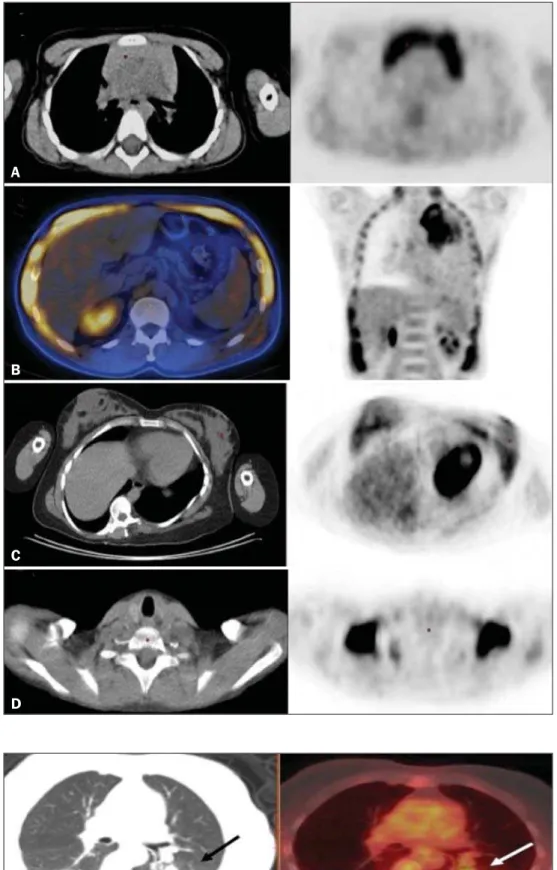
Figure 1.Causes of physiological FDG up-take in the thorax. A: A 6-year-old boy, with normal metabolic activity in the thymus. The strategy for differentiating between focal up-take in the anterior mediastinum and a le-sion is to look at the normal aspect of the thymus on CT images. B: A 56-year-old man with lung cancer and a forceful cough dur-ing the PET/CT examination. FDG PET/CT shows increased metabolic activity in the thoracic muscles, due to vigorous cough-ing. C: A 31-year-old lactating woman, with normal FDG uptake in the breast. Lactation induces higher metabolic activity in the breasts, which should not be interpreted as disease. D: A 28-year-old man with testicu-lar cancer. Multiple areas of FDG uptake in the supraclavicular regions and in the upper mediastinum, consistent with increased metabolic activity in brown fat, which regu-lates body weight and temperature and can be activated by satiety or cold environments.
A
B
C
D
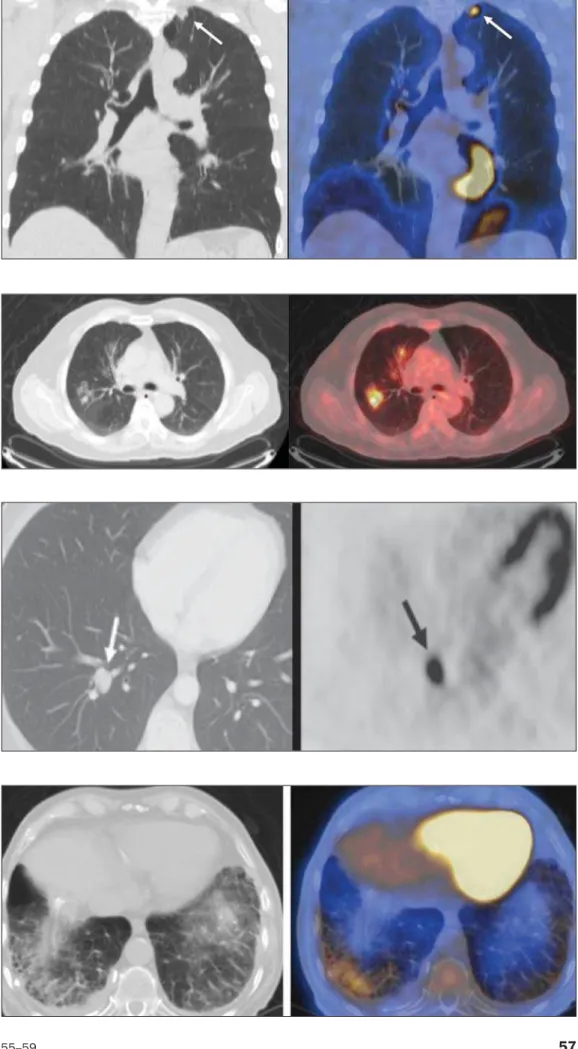
Figure 6. A 81-year-old man with an occupational history of asbestos ex-posure. PET/CT images demonstrate high metabolic activity in the left lower lobes, especially in the cortical regions. CT images depict interstitial lung ab-normalities, with ground-glass opaci-ties, reticulation, and mild bronchiolec-tasis.
Figure 5. A 42-year-old man with a lobulated pulmonary nodule in the right lower lobe (arrows), indetermi-nate on CT. PET/CT was performed and showed increased metabolic activity (arrow) in the lung nodule, which was surgically resected and was found to be consistent with pulmonary menin-gioma.
Figure 7. A 78-year-old woman sub-mitted to PET/CT for the staging of a cervical carcinoma, with increased FDG uptake in bilateral subscapular masses (arrows), consistent with elastofibroma dorsi.
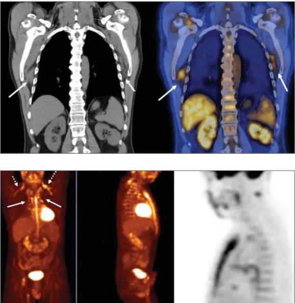
Figure 8. Postoperative PET/CT of a patient with history of cardiac tumor. Note the diffuse FDG uptake in the sternum, consistent with inflamma-tory changes induced by the surgical procedure (arrows). Also note the in-creased metabolic activity in areas of brown fat in the supraclavicular and paravertebral regions (dashed ar-rows).
Figure 10. A 41-year-old woman with a history of Hodgkin lymphoma and paramediastinal opacities with FDG uptake (arrows), consistent with in-flammatory lesions induced by radia-tion therapy.
9. Mosmann MP, Borba MA, Macedo FPN, et al. Solitary pulmo-nary nodule and 18F-FDG PET/CT. Part 1: epidemiology, mor-phological evaluation and cancer probability. Radiol Bras. 2016; 49:35–42.
10. Mosmann MP, Borba MA, Macedo FPN, et al. Solitary pulmo-nary nodule and 18F-FDG PET/CT. Part 2: accuracy, cost-ef-fectiveness, and current recommendations. Radiol Bras. 2016;49: 104–11.
11. Shreve PD, Anzai Y, Wahl RL. Pitfalls in oncologic diagnosis with FDG PET imaging: physiologic and benign variants. Radiographics. 1999;19:61–77.
12. Nose H, Otsuka H, Otomi Y, et al. The physiological uptake pat-tern of (18)F-FDG in the left ventricular myocardium of patients without heart disease. J Med Invest. 2014;61:53–8.
13. Abouzied MM, Crawford ES, Nabi HA. 18F-FDG imaging: pit-falls and artifacts. J Nucl Med Technol. 2005;33:145–55. 14. Bakheet SM, Powe J, Ezzat A, et al. F-18-FDG uptake in
tubercu-losis. Clin Nucl Med. 1998;23:739–42.
5. Guimarães JB, Rigo L, Lewin F, et al. The importance of PET/CT in the evaluation of patients with Ewing tumors. Radiol Bras. 2015; 48:175–80.
6. Valadares AA, Duarte PS, Carvalho G, et al. Receiver operating char-acteristic (ROC) curve for classification of 18F-NaF uptake on PET/ CT. Radiol Bras. 2016;49:12–6.
7. Barbosa FG. PET/CT in the evaluation of pulmonary solitary nod-ule. Radiol Bras. 2016;49(2):xi.
8. Hochhegger B. PET/CT used in the evaluation of pulmonary nod-ules suspicious for lung cancer in regions where infectious lung dis-ease is endemic: to be or not to be? Radiol Bras. 2016;49:199.
15. Yasuda S, Shoht0su A, Ide M, et al. High fluorine-18 labeled deoxy-glucose uptake in sarcoidosis. Clin Nucl Med. 1996;21:983–4 . 16. Blodgett TM, Ames JT, Torok FS, et al. Diffuse bone marrow
up-take on whole-body F-18 fluorodeoxyglucose positron emission to-mography in a patient taking recombinant erythropoietin. Clin Nucl Med. 2004;29:161–3.
17. Peek H, van der Bruggen W, Limonard G. Pleural FDG uptake more than a decade after talc pleurodesis. Case Rep Med. 2009; 2009:650864.
18. Cheran SK, Nielsen ND, Patz EF Jr. False-negative findings for primary lung tumors on FDG positron emission tomography: stag-ing and prognostic implications. AJR Am J Roentgenol. 2004;182: 1129–32.
19. Kostakoglu L, Agress H Jr, Goldsmith SJ. Clinical role of FDG PET in evaluation of cancer patients. Radiographics. 2003;23:315-40; quiz 533.
20. Sureshbabu W, Mawlawi O. PET/CT imaging artifacts. J Nucl Med Technol. 2005;33:156–61; quiz 163–4.
Figure 11. A 63-year-old male, current smoker, with a ground-glass nodule in the right lung (arrow), which had consistently increased in size over the years. PET/CT was performed and was negative. Surgical resection confirmed the diagnosis of adenocarcinoma in situ (formerly known as bronchoalveolar carcinoma). PET/CT is limited for these conditions and should not be used for small nodules with ground-glass attenuation, due to the high pretest probability of false-negative results.
Figure 12. A 57-year-old woman with breast cancer and focal FDG uptake in a left axillary lymph node (arrow). That uptake is consistent with extravasation of FDG in the left arm, draining to the ipsilateral axillary region.