ABSTRACT
http://dx.doi.org/10.1590/1678-775720130174
A microleakage study of gutta-percha/AH Plus
and Resilon/Real self-etch systems after different
irrigation protocols
Maíra PRADO1,2, Renata Antoun SIMÃO2, Brenda Paula Figueiredo de Almeida GOMES1
1- Department of Restorative Dentistry, Endodontics Division, State University of Campinas - UNICAMP, Piracicaba, SP, Brazil. 2- Department of Metallurgic and Materials Engineering, Federal University of Rio de Janeiro, Rio de Janeiro, RJ, Brazil.
Corresponding address: Brenda Paula F. A. Gomes - Departamento de Odontologia Restauradora, Área de Endodontia - Faculdade de Odontologia de Piracicaba - Universidade Estadual de Campinas - UNICAMP - Avenida Limeira, 901 - Piracicaba - SP - Brazil - 13414-018 - Phone: (55) 19 2106-5215 - Fax: (55) 19 2106-5218 - E-mail: [email protected]
Submitted: April 8, 2013 - Modiication: February 10, 2014 - Accepted: February 19, 2014
T
he development and maintenance of the sealing of the root canal system is the key to the success of root canal treatment. The resin-based adhesive material has the potential to reduce the microleakage of the root canal because of its adhesive propertiesand penetration into dentinal walls. Moreover, the irrigation protocols may have an inluence
on the adhesiveness of resin-based sealers to root dentin. Objective: The objective of the present study was to evaluate the effect of different irrigant protocols on coronal bacterial microleakage of gutta-percha/AH Plus and Resilon/Real Seal Self-etch systems. Material and Methods: One hundred ninety pre-molars were used. The teeth were divided into 18
experimental groups according to the irrigation protocols and illing materials used. The
protocols used were: distilled water; sodium hypochlorite (NaOCl)+eDTA; NaOCl+H3PO4; NaOCl+eDTA+chlorhexidine (CHX); NaOCl+H3PO4+CHX; CHX+eDTA; CHX+ H3PO4; CHX+eDTA+CHX and CHX+H3PO4+CHX. Gutta-percha/AH Plus or Resilon/Real Seal Se
were used as root-illing materials. The coronal microleakage was evaluated for 90 days
against Enterococcus faecalis. Data were statistically analyzed using Kaplan-Meier survival
test, Kruskal-Wallis and Mann-Whitney tests. Results: No signiicant difference was veriied
in the groups using chlorhexidine or sodium hypochlorite during the chemo-mechanical preparation followed by eDTA or phosphoric acid for smear layer removal. The same results
were found for illing materials. However, the statistical analyses revealed that a inal lush with 2% chlorhexidine reduced signiicantly the coronal microleakage. Conclusion: A inal lush with 2% chlorhexidine after smear layer removal reduces coronal microleakage of teeth illed with gutta-percha/AH Plus or Resilon/Real Seal SE.
Keywords: Chlorhexidine. Dental leakage. Root canal irrigants. Root canal obturation.
INTRODUCTION
The major aim of root canal therapy is to prevent
and treat periradicular inlammation by eliminating
microorganisms from the root canal system. The methods commonly used for this purpose include root canal preparation using different instruments and irrigants, adequate filling, and coronal restoration1,14.
Chemical irrigants are essential for successful debridement of root canals during cleaning and shaping procedures11. They are used during
chemo-mechanical procedures not only as antimicrobial agents, but also to lubricate the dentinal walls,
lush out debris and dissolve organic and inorganic
components of the smear layer, thus cleaning the dentin surface2,17. Different irrigants have been
proposed and used, including: 5.25% sodium hypochlorite, 2% chlorhexidine, 17% eDTA, 10% citric acid and 37% phosphoric acid solution17,18,20.
to dissolve the tissue. Additionally, chlorhexidine
has been suggested as a inal irrigant30. Regarding
its use as final irrigant, a final flush with 2% chlorhexidine favors the wettability of AH Plus and Real Seal Se sealers on the dentin surface.
Furthermore, it was veriied that the bond strength
of ActiV GP, a glass ionomer based system, was
improved by using 2% chlorhexidine in the inal
irrigation after 17% eDTA.
The development and maintenance of the sealing of the root canal system is the key to the success of root-canal treatment. The resin-based adhesive material has the potential to reduce the microleakage of the root canal because of its adhesive properties and penetration into dentinal walls25. Moreover, the irrigation protocols may have an inluence on the adhesiveness of resin-based
sealers to root dentin8.
A variety of experimental models are used to detect and measure any leakage along endodontic
illings, such as dye penetration, clearing of the
teeth, radioisotope tests, bacterial penetration,
electrochemical tests, luid iltration, and glucose
penetration model12,24.
The aim of the present study was to evaluate the effect of different irrigant protocols on coronal bacterial microleakage of gutta-percha/AH Plus and Resilon/Real Seal Self-etch systems.
MATERIAL AND METhODS
Sample preparation
One hundred ninety single-rooted pre-molars with straight roots, mature root apices and similar anatomical characteristics were used in this study. All instruments used in the root canal preparation were sterilized previously to the procedure. The teeth were positioned on a metallic apparatus that allowed for all procedures to be carried out without manual contact with the roots. Conventional access was performed using high-speed diamond burs. A
size 10 K-ile (Dentsply Maillefer, Petrópolis, Rio
de Janeiro, Brazil) was used to verify the patency of the canals and to determine the total length of the root canal, i.e. the work length. This was observed when the instrument reached the apical foramen. Next, the foramina were standardized by
using a size 20 K-ile and root canals were shaped
by using MTwo NiTi rotary system (VDW, Münich, Bavaria, Germany). The sequence employed was the following: 10/.04, 15/.05, 20/.06, 25/.06, 30/.05, 35/.04, 40/.04, and 25/.07. The teeth were divided into groups of ten according to the irrigation
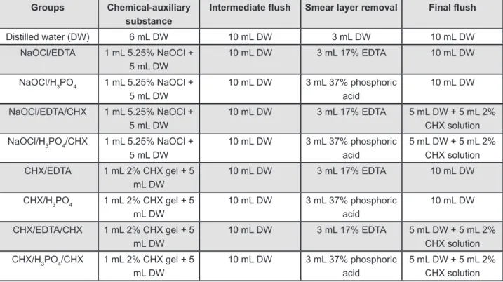
regimen (Figure 1) and root canal illing.
Before the insertion of each ile, 2% chlorhexidine
(CHX) gel (Drogal, Piracicaba, São Paulo, Brazil) or 5.25% sodium hypochlorite (NaOCl) (Drogal, Piracicaba, São Paulo, Brazil) were used as chemical-auxiliary substance. Once the preparation
was inished, 10 mL of distilled water (DW) was
used to remove the chemical-auxiliary substance.
Groups Chemical-auxiliary
substance
Intermediate lush Smear layer removal Final lush
Distilled water (DW) 6 mL DW 10 mL DW 3 mL DW 10 mL DW
NaOCl/EDTA 1 mL 5.25% NaOCl + 5 mL DW
10 mL DW 3 mL 17% EDTA 10 mL DW
NaOCl/H3PO4 1 mL 5.25% NaOCl + 5 mL DW
10 mL DW 3 mL 37% phosphoric acid
10 mL DW
NaOCl/EDTA/CHX 1 mL 5.25% NaOCl + 5 mL DW
10 mL DW 3 mL 17% EDTA 5 mL DW + 5 mL 2% CHX solution
NaOCl/H3PO4/CHX 1 mL 5.25% NaOCl +
5 mL DW
10 mL DW 3 mL 37% phosphoric acid
5 mL DW + 5 mL 2% CHX solution
CHX/EDTA 1 mL 2% CHX gel + 5 mL DW
10 mL DW 3 mL 17% EDTA 10 mL DW
CHX/H3PO4 1 mL 2% CHX gel + 5
mL DW
10 mL DW 3 mL 37% phosphoric acid
10 mL DW
CHX/EDTA/CHX 1 mL 2% CHX gel + 5 mL DW
10 mL DW 3 mL 17% EDTA 5 mL DW + 5 mL 2% CHX solution
CHX/H3PO4/CHX 1 mL 2% CHX gel + 5 mL DW
10 mL DW 3 mL 37% phosphoric acid
5 mL DW + 5 mL 2% CHX solution
*CHX - chlorhexidine
Next, 17% eDTA or 37% phosphoric acid solution (Drogal, Piracicaba, São Paulo, Brazil) was used for 3 minutes to remove the smear layer, with changes every 1 minute (1 mL per minute). Again, DW was used to remove the remaining solution. Finally, 2% chlorhexidine solution (Drogal, Piracicaba,
São Paulo, Brazil) was used for inal lush. During
the chemo-mechanical preparation, all teeth had their apices sealed with utility wax (Technew, Rio
de Janeiro, Rio de Janeiro, Brazil) to prevent low
through them.
The root canals were dried with sterilized medium-sized paper points (endopoints, Paraíba do Sul, Rio de Janeiro, Brazil). Groups 1 to 9 had
the canals illed with gutta-percha cones (Odous,
Belo Horizonte, Minas Gerais, Brazil) associated with AH Plus sealer (Dentsply, Petrópolis, Rio de Janeiro, Brazil), whereas Groups 10 to 18 had the
canals illed with Resilon associated with Real Seal
Se (Sybronendo, Orange, California, USA). The cones packages were opened and they were used immediately, and the sealers were manipulated in sterile plates according to the manufacturer’s recommendations.
A System-B endodontic heat source unit (Sybronendo, Orange, California, USA) was used to down-pack and Obtura System (J Morita, São Paulo, São Paulo, Brazil) to backfill. All
procedures were conducted at the laminar low
cabinet. Subsequently, teeth were radiographed mesiodistally and buccolingually to assess the
quality of the illing.
All roots were kept on gauzes at 37°C and 100% humidity for 2 weeks before leakage measurement in order to allow the materials to set properly.
Next, the external root surface of all specimens was sealed with two layers of red nail varnish (Revlon, New York, New York, USA), except the last
1 mm of the apex.
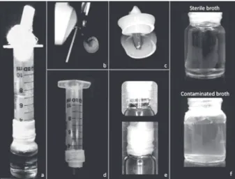
Analysis of coronal bacterial microleakage Figure 2a illustrates the apparatus used to evaluate coronal leakage10. Glass vials with rubber
stoppers were adjusted for use. By using a shear, a hole was made at the center of each rubber stopper (Figure 2b), through which each tooth was inserted under pressure up to the cementoenamel junction, so that its crown was outside the vial and its root inside (Figure 2c). Cyanoacrylate glue (CG) was applied at the interface between tooth and stopper for sealing15. Cylinders prepared from 10 mL plastic
syringes were adapted to the outer surface of the stoppers to create a chamber around the crown of the tooth (Figure 2d). Again, CG was used at the interface between syringes and stoppers, followed
by a Parailm layer (American National CanTM,
Menasha, Wisconsin, USA) to help in the sealing. The syringe/stopper/tooth sets were submitted to sterilization by gamma-rays (embrarad, São Paulo,
São Paulo, Brazil). The glass lasks were autoclaved
at 121°C for 15 minutes.
The sterilized glass lasks were then illed with
sterile Brain Heart Infusion broth (BHI; Oxoid, São Paulo, São Paulo, Brazil) so that a 2-mm length of root apex was immersed in the broth. CG and
Parailm were used to seal the interface between stopper and lask (Figure 2e). In all samples, in order to ensure the eficiency of the seal, 2 mL of
1% sterile methylene blue dye was placed into the tube until the coronal portion of the sample was reached15. The lasks were then incubated at 37°C
for 3 days to ensure sterilization. After the third day, the methylene blue was removed with sterile distilled water by using a pipette. When a green medium was observed the specimen was discarded. The green color was due to the blue dye association
with the yellow medium.
For preparation of microbial medium28,
Enterococcus faecalis (ATCC 29212) was grown on BHI agar plates (Brain Heart Infusion agar; Oxoid, São Paulo, São Paulo, Brazil) and supplemented with 5% sheep blood for 24 hours at 37°C in CO2. Then, the Enterococcus faecalis was inoculated into tubes containing 5 mL sterile BHI suspension, which were adjusted spectrophotometrically at 800 nm (OD800) to a turbidity of 1.5x10⁸ colony-forming
units (CFU)/mL. With the aid of a pipette, 5 mL of the suspensions were placed into the syringe apparatuses (in the upper region, removing the gauze stop), which were left at 37°C for 90 days in CO2 and checked daily for turbidity in the BHI broth. When turbidity (Figure 2f) was observed, the day was recorded.
every 2 days, 3 mL of the suspension (BHI+microorganisms) were removed from the chamber and replaced by 3 mL of BHI to
avoid saturation and to conirm the growth of Enterococcus faecalis5.
After this period, all apparatuses were opened to evaluate the sterile hood. Positive cultures were
conirmed by using Gram staining (gram-positive),
colony morphology on blood agar plates (cocci) and
biochemical identiication kits (Rapid ID 32 Strep,
BioMérieux SA, Marcyl’etoile, Charbonnieres-les-Bains, France).
Ten samples were used as positive (n=5) and negative (n=5) controls. The positive controls consisted of instrumented teeth without obturation, while negative controls consisted of sound teeth, both with no contamination.
The results were analyzed with Kaplan-Meier survival test, Kruskal-Wallis and Mann-Whitney
tests (p<0.05).
RESULTS
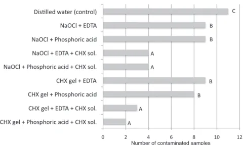
No differences were found following the use of chlorhexidine or NaOCl associated with eDTA or phosphoric acid for smear layer removal. However,
it was clearly observed that groups receiving a inal lush with chlorhexidine showed a lower number of
contaminated samples.
Figure 3 shows the number of contaminated samples according to the irrigation protocol.
Regarding the root canal illing system
(gutta-percha/AH Plus and Resilon/Real Seal Se), there
was no statistically signiicant differences in relation
to the coronal microleakage.
Figure 4 shows the number of contaminated samples in relation to time. Statistic analysis of the contamination days revealed difference in the
groups receiving the inal lush with chlorhexidine.
Chlorhexidine groups started to contaminate only in the 6th week, while in the others the microbial
growth was veriied in the 1st or 2nd week. The
control apparatuses showed broth turbidity within 1 day of incubation in all samples, whereas no microbial growth was found in the negative control throughout the experiment.
DISCUSSION
Leakage of the root canal has been deined as the passage of bacteria, luids, and chemical
substances between the dentinal wall and the root
canal illing material, and results from the presence of space at the interface of the illing material and
the root canal wall. This space can result from
deicient adaptation of the illing material to the root
dentin, solubility of the sealer, or sealer expansion or shrinkage. There are 2 possibilities of leakage: at
the interface between the main illing material and
sealer, or between the sealer and root canal wall26.
*Different letters indicate statistically signiicant values (P<0.05)
Figure 3- Graph showing the number of contaminated samples according to the irrigation protocol
In the present study, the coronal bacterial microleakage was used. This methodology is reproducible and has clinical relevance, presenting reliable data and simulating clinical conditions7,13,16.
This methodology allows the observation of the exact day of the sample contamination, showed by the broth turbidity.
The 37% phosphoric acid was evaluated in comparison with eDTA because this solution is effective for smear layer removal, showing better results than eDTA in the apical third during 3 minutes19. Additionally, the protocols associating
NaOCl with phosphoric acid showed higher bond strength values when compared with NaOCl associated with eDTA20. Although phosphoric acid
had showed better performance in removing the smear layer from the apical third19, it did not inluence the results of coronal leakage.
The 2% chlorhexidine, a cationic bisbiguanide,
was used here for the inal lush after smear layer
removal. This substance has a broad-spectrum MMP-inhibitory effect that improves the integrity of the hybrid layer6 and the resin-dentin bond
stability23. Additionally, the use of CHX increases
the wettability of endodontic sealers on dentin3,
which can be explained by the presence of surface surfactant in CHX, increasing the surface energy and promoting higher wetting ability to dentin. Our
results showed that a inal lush with chlorhexidine signiicantly reduced the coronal microleakage,
when compared to the other experimental groups. It might be explained by the fact that chlorhexidine is adsorbed onto dentin and prevent microbial colonization21, increasing the time required for
recontamination of illed root23 up to 12 weeks due
to its substantivity21.
Regarding the effect of a final flush with chlorhexidine on adhesion, previous studies showed that this solution did not affect the bond strength of resin-based sealers17,20 and improved the adhesion
of hydrophilic bonded materials such as ActiV GP and epiphany23.
NaOCl was not used as a inal irrigant because a
previous study showed that this solution decreased the bond strength between epoxy resin and dentin and increased the leakage17.
Regarding the root canal illing system
(gutta-percha/AH Plus and Resilon/ Real Seal Se), there
was no statistically signiicant differences in relation
to the coronal microleakage, agreeing with previous
indings4,9,26. However, some studies reported that
Resilon/Epiphany sealer was more eficient than
gutta-percha/AH Plus12,29, whereas others found
the opposite22,27.
GROUPS WEEK 1 WEEK 2 WEEK 3 WEEK 4 WEEK 5 WEEK 6 WEEK 7 WEEK 8 WEEK 9 WEEK 10 WEEK 1 1 WEEK 12 WEEK 13 DW/GP AH 0/10 2/10 0/10 2/10 0/10 0/10 0/10 0/10 0/10 0/10 0/10 0/10 1/10 NaOCl/EDT A/GP AH 0/10 1/10 1/10 0/10 0/10 1/10 0/10 0/10 1/10 0/10 0/10 0/10 1/10 NaOCl/H 3 PO 4 /GP AH 0/10 2/10 1/10 0/10 0/10 0/10 0/10 0/10 0/10 0/10 0/10 0/10 1/10 NaOCl/EDT A/CHX/GP AH 0/10 0/10 0/10 0/10 0/10 0/10 0/10 0/10 1/10 0/10 0/10 0/10 1/10 NaOCl/H 3 PO 4 /CHX/GP AH 0/10 0/10 0/10 0/10 0/10 1/10 0/10 0/10 0/10 0/10 0/10 0/10 1/10 CHX/EDT A/GP AH 0/10 1/10 0/10 0/10 0/10 0/10 0/10 0/10 0/10 0/10 1/10 1/10 2/10 CHX/H 3 PO 4 /GP AH 0/10 1/10 0/10 0/10 0/10 0/10 0/10 0/10 0/10 0/10 1/10 0/10 1/10 CHX/EDT A/CHX/GP AH 0/10 0/10 0/10 0/10 0/10 0/10 0/10 0/10 0/10 0/10 0/10 0/10 2/10 CHX/H 3 PO 4 /CHX/GP AH 0/10 0/10 0/10 0/10 0/10 0/10 0/10 0/10 0/10 0/10 0/10 0/10 1/10 DW/RRS 2/10 2/10 0/10 0/10 0/10 0/10 0/10 0/10 1/10 0/10 0/10 0/10 1/10 NaOCl/EDT A/RRS 1/10 1/10 0/10 0/10 0/10 0/10 0/10 0/10 0/10 0/10 0/10 1/10 1/10 NaOCl/H 3 PO 4 /RRS 1/10 1/10 0/10 0/10 1/10 1/10 0/10 0/10 0/10 0/10 0/10 0/10 1/10 NaOCl/EDT A/CHX/RRS 0/10 0/10 0/10 0/10 0/10 1/10 0/10 0/10 0/10 0/10 0/10 0/10 1/10 NaOCl/H 3 PO 4 /CHX/RRS 0/10 0/10 0/10 0/10 0/10 0/10 0/10 0/10 0/10 1/10 0/10 0/10 1/10 CHX/EDT A/RRS 0/10 1/10 1/10 0/10 0/10 0/10 0/10 0/10 0/10 0/10 0/10 1/10 1/10 CHX/H 3 PO 4 /RRS 0/10 1/10 1/10 0/10 0/10 0/10 0/10 0/10 0/10 1/10 1/10 0/10 1/10 CHX/EDT A/CHX/RRS 0/10 0/10 0/10 0/10 0/10 0/10 0/10 0/10 0/10 0/10 0/10 0/10 1/10 CHX/H 3 PO 4 /CHX/RRS 0/10 0/10 0/10 0/10 0/10 0/10 0/10 0/10 0/10 0/10 0/10 0/10 1/10 DW
- Distilled water; CHX- chlorhexidine; GP
AH- gutta-percha/AH Plus; RRS- Resilon/Real Seal SE
Figure
Number of contaminated samples
per
CONCLUSION
In conclusion, a inal lush with 2% chlorhexidine
after smear layer removal reduces coronal
microleakage of teeth illed with gutta-percha/AH
Plus or Resilon/Real Seal Self-etch. The bacterial leakage methodology used made possible the
veriication of this behavior.
ACKNOWLEDgEMENTS
This work was supported by the Brazilian agencies FAPeSP – São Paulo Research Foundation (2009/53976-0; 2010/50817-5), CAPeS – Coordination for the Improvement of Higher education Personnel, and CNPq – National Council for Scientific and Technological Development (302575/2009-0).
REFERENCES
1- Adamo HL, Buruiana R, Schertzer L, Boylan RJ. A comparison
of MTA, Super EBA, composite and amalgam as root end illing
materials using a bacterial microleakage model. Int endod J. 1999;32:197-203.
2- Akisue e, Tomita VS, Gavini G, Poli de Figueiredo JA. effect of the combination of sodium hypochlorite and chlorhexidine on dentinal permeability and scanning electron microscopy precipitate observation. J endod. 2010;36:847-50.
3- Assis DF, Prado M, Simão RA. evaluation of the interaction between endodontic sealers and dentin treated with different irrigant solutions. J endod. 2011;37:1550-2.
4- Baumgartner G, Zehnder M, Paqué F. Enterococcus faecalis
type strain leakage through root canals illed with Gutta-Percha/
AH Plus or Resilon/epiphany. J endod. 2007;33:45-7.
5- Berber VB, Gomes BP, Sena NT, Vianna Me, Ferraz CC,
Zaia AA, et al. Eficacy of various concentrations of NaOCl and
instrumentation techniques in reducing Enterococcus faecalis
within root canals and dentinal tubules. Int endod J. 2006;39:10-7. 6- Carrilho MR, Carvalho RM, Goes MF, di Hipólito V, Geraldeli S,
Tay FR, et al. Chlorhexidine preserves dentin bond in vitro. J Dent
Res. 2007;86:90-4.
7- Chailertvanitkul P, Saunders WP, MacKenzie D. Coronal leakage of obturated root canals after long-term storage using a polymicrobial marker. J endod. 1997;23:610-3.
8- De-Deus G, Namen F, Galan J Jr, Zehnder M. Soft chelating irrigation protocol optimizes bonding quality of Resilon/epiphany
root illings. J Endod. 2008;34:703-5.
9- Fransen JN, He J, Glickman GN, Rios A, Shulman JD, Honeyman A. Comparative assessment of ActiV GP/glass ionomer sealer, Resilon/epiphany, and gutta-percha/AH plus obturation: a bacterial leakage study. J endod. 2008;34:725-7.
10- Gomes BP, Sato e, Ferraz CC, Teixeira FB, Zaia AA, Souza-Filho FJ. evaluation of time required for recontamination of coronally sealed canals medicated with calcium hydroxide and chlorhexidine. Int endod J. 2003;36:604-9.
11- Hashem AA, Ghoneim AG, Lutfy RA, Fouda MY. The effect of different irrigating solutions on bond strength of two root
canal-illing systems. J Endod. 2009;35:537-40.
12- Hirai VH, Silva Neto UX, Westphalen VP, Perin CP, Carneiro e,
Fariniuk LF. Comparative analysis of leakage in root canal illings
performed with gutta-percha and Resilon cones with AH Plus and epiphany sealers. Oral Surg Oral Med Oral Pathol Oral Radiol endod. 2010;109:e131-5.
13- Imura N, Otani SM, Campos MJ, Jardim Júnior eG, Zuolo ML. Bacterial penetration through temporary restorative materials
in root-canal-treated teeth in vitro. Int endod J. 1997;23:1-5.
14- Jacobovitz M, Vianna Me, Pandolfelli VC, Oliveira IR, Rossetto
HL, Gomes BP. Root canal illing with cements based on mineral
aggregates: an in vitro analysis of bacterial microleakage. Oral
Surg Oral Med Oral Pathol Oral Radiol endod. 2009;108:140-4.
15- Malone KH 3rd, Donnelly JC. In vitro evaluation of coronal
microleakage in obturated root canals without restorations. J endod. 1997;23:35-8.
16- Nair U, Ghattas S, Saber M, Natera M, Walker C, Pileggi R. A
comparative evaluation of the sealing ability of 2 root-end illing
materials: an in vitro leakage study using Enterococcus faecalis.
Oral Surg Oral Med Oral Pathol Oral Radiol endod. 2011;112:e74-7. 17- Neelakantan P, Subbarao C, Subbarao CV, De-Deus G, Zehnder M. The impact of root dentine conditioning on sealing ability and push-out bond strength of an epoxy resin root canal sealer. Int endod J. 2011;44:491-8
18- Park DS, Torabinejad M, Shabahang S. The effect of MTAD on the coronal leakage of obturated root canals. J endod. 2004;30:890-2.
19- Prado M, Gusman H, Gomes BP, Simão RA. Scanning electron microscopic investigation of the effectiveness of phosphoric acid in smear layer removal when compared with eDTA and citric acid. J endod. 2011;37:255-8.
20- Prado M, Simão RA, Gomes BP. effect of different irrigation protocols on resin sealer bond strength to dentin. J endod. 2013;39:689-92.
21- Rosenthal S, Spångberg L, Safavi K. Chlorhexidine substantivity in root canal dentin. Oral Surg Oral Med Oral Pathol Oral Radiol endod. 2004;98:488-92.
22- Saleh IM, Ruyter Ie, Haapasalo M, Ørstavik D. Bacterial
penetration along different root canal illing materials in the
presence or absence of smear layer. Int endod J. 2008;41:32-40.
23- Shariian MR, Shokouhinejad N, Aligholi M, Jafari Z. Effect of
chlorhexidine on coronal microleakage from root canals obturated with Resilon/epiphany Self-etch. J Oral Sci. 2010;52:83-7. 24- Shemesh H, Wu MK, Wesselink PR. Leakage along apical root
illings with and without smear layer using two different leakage
models: a two-month longitudinal ex vivo study. Int endod J.
2006;39:968-76.
25- Shipper G, Ørstavik D, Teixeira FB, Trope M. An evaluation
of microbial leakage in roots illed with a thermoplastic synthetic polymer-based root canal illing material (Resilon). J Endod
2004;30:342-7.
26- Shokouhinejad N, Shariian MR, Aligholi M, Assadian H, Tabor
RK, Nekoofar MH. The sealing ability of resilon and gutta-parcha
following different smear layer removal methods: an ex vivo study.
Oral Surg Oral Med Oral Pathol Oral Radiol endod. 2010;110:e45-9.
27- Stratton RK, Apicella MJ, Mines P. A luid iltration comparison
of gutta-percha versus Resilon, a new soft resin endodontic obturation system. J endod. 2006;32:642-5.
28- Vianna Me, Gomes BP, Sena NT, Zaia AA, Ferraz CCRF,
Souza-Filho FJ. In vitro evaluation of the susceptibility of endodontic
pathogens to calcium hydroxide combined with different vehicles. Braz Dent J. 2005;16:175-80.
29- Wedding JR, Brown Ce, Legan JJ, Moore BK, Vail MM. An in
vitro comparison of microleakage between Resilon and
gutta-percha with a luid iltration model. J Endod. 2007;33:1447-9.