ABSTRACT
http://dx.doi.org/10.1590/1678-775720130528
Physical and chemical properties of orthodontic
brackets after 12 and 24 months:
in situ
study
Bernardo de Azevedo Bahia MENDES1, Ricardo Alberto Neto FERREIRA2, Matheus Melo PITHON3, Martinho Campolina Rebello HORTA1, Dauro Douglas OLIVEIRA1
1- Graduate Program in Orthodontics, Pontiical Catholic University of Minas Gerais, Belo Horizonte, MG, Brazil.
2- Center for Development of Nuclear Technology, National Nuclear Energy Commission (CDTN/CNEN), Belo Horizonte, MG, Brazil. 3- Department of Orthodontics, Southwest Bahia State University - UESB, Jequié, BA, Brazil.
Corresponding address: Dauro Douglas Oliveira - Pontifícia Universidade Católica de Minas Gerais - Mestrado em Odontologia - Av. Dom José Gaspar, 500 - Prédio 46 - Sala 106 - Belo Horizonte - MG - Brazil - 30535-610 - Phone: 55 - 31 - 3319-4414 - Fax: 55 31 3319 4415 - e-mail: [email protected]
Submitted: September 22, 2013 - Modiication: December 13, 2013 - Accepted: February 4, 2014
O
bjective: The aim of this article was to assess how intraoral biodegradation inluencedthe surface characteristics and friction levels of metallic brackets used during 12 and 24 months of orthodontic treatment and also to compare the static friction generated in these brackets with four different methods of the ligation of orthodontic wires. Material and Methods: Seventy premolar brackets as received from the manufacturer and 224 brackets that were used in previous orthodontic treatments were evaluated in this experiment. The surface morphology and the composition of the deposits found in the brackets were evaluated with rugosimetry, scanning electron microscopy, and energy dispersive X-ray spectroscopy. Friction was analyzed by applying tensile tests simulating sliding mechanics with a 0.019x0.025” steel wire. The static friction levels produced by the following ligation methods were evaluated: loosely attached steel ligature around all four bracket wings, steel ligature attached to only two wings, conventional elastomeric ligation around all 4 bracket wings, and non-conventional Slide® elastomeric ligature. Results: The results demonstrated
the presence of biodegradation effects such as corrosion pits, plastic deformation, cracks, and material deposits. The main chemical elements found on these deposits were Carbon and Oxygen. The maximum friction produced by each ligation method changed according to the time of intraoral use. The steel ligature loosely attached to all four bracket wings produced the lowest friction levels in the new brackets. The conventional elastic ligatures
generated the highest friction levels. The metallic brackets underwent signiicant degradation
during orthodontic treatment, showing an increase in surface roughness and the deposit of chemical elements on the surface. Conclusion: The levels of static friction decreased with use. The non-conventional elastic ligatures were the best alternative to reduce friction.
Keywords: Orthodontic brackets. Friction. Biodegradation.
INTRODUCTION
Orthodontic tooth movement (OTM) eficiency is inluenced by both biological and mechanical
variables, and friction is one of the most studied due to its complexity and importance. OTM occurs when the orthodontic forces applied to the teeth are higher than the static friction22. Some physical
variables interfere in the friction magnitude, such as the type of bracket and wire material14, the angle of
insertion of the wires2, the surface characteristics11,
geometry and dimensions of these materials22, as
well as the type of ligature12 and method of ligating
the wire to the brackets13. It is impossible to fully
eliminate friction in sliding mechanics. Therefore, orthodontists must understand the factors that
inluence friction and how they can effectively
control them, according to the demands of the different stages of treatment22.
In recent years, the so-called low friction systems22, such as self-ligating brackets22, have
been gaining ever-greater space in the orthodontic product market18, despite their higher costs22.
suggested, such as modiications on the surface
and geometry of the bracket slot3,5,22, coating the
external layer of speciic orthodontic wires19, the
use of loosely tied steel ligature wires13, or elastic
ligatures with new designs24.
A very important aspect directly related to friction that has not been adequately studied is the mid and long-term changes that take place in orthodontic materials after their intraoral use. The few studies available in the literature have reported signs of corrosion, structural wear, plastic deformation, and
deposition of bioilm after the use of stainless steel
brackets and wires in orthodontic treatments20.
These indings were considered co-responsible for
the increased friction levels registered after their biodegradation in the oral cavity15,16,21,23.
In addition to the limited number of studies that have evaluated the biodegradation of orthodontic materials22, none have analyzed if different
methods of ligating the wire to the brackets may reduce some of the negative effects generated from exposure of these materials to the oral environment. Thus, the aim of the present study was to: (1) evaluate how the biodegradation
inluenced the surface characteristics and friction
levels of metallic brackets used during 12 and 24 months of orthodontic treatment; (2) compare the static friction generated in these brackets with four ligation methods for orthodontic wires; and (3) test the hypothesis that brackets as received from the manufacturer present lower friction levels than those that were used for 12 or 24 months and that different methods of ligation produce different amounts of friction.
MATERIAL AND METhODS
Sample and groups
The present study evaluated the static friction of metallic brackets as received from the manufacturer and after their intraoral use during 12 or 24 months
of orthodontic treatment in a private ofice. The
sample consisted of stainless steel premolar brackets (edgewise Standard 0.022x0.028”, Morelli Ortodontia, Sorocaba, SP, Brazil). After completing the intraoral exposure time, the brackets were carefully removed with debonding orthodontic pliers especially designed to apply force only to the bracket base to avoid slot and wing deformation. Immediately after debonding, all brackets were cleaned with a dental water jet, placed in an ultrasound device for 5 minutes, and maintained in closed recipients until they were tested. Before friction evaluation, the brackets were brushed with an electric toothbrush (model 3744, Oral-B/Braun, Queimados, RJ, Brazil) with a round head for 10 seconds, without placing pressure onto the slot.
A total of 294 brackets were tested, 224 were
used in the orthodontic treatment of 25 patients from a private practice (average age of 31 years and 6 months) and 70 were new, tested as received from the manufacturer. During orthodontic treatment, stainless steel and nickel-titanium wires were used, which were tied to the brackets with elastomeric and/or steel ligatures.
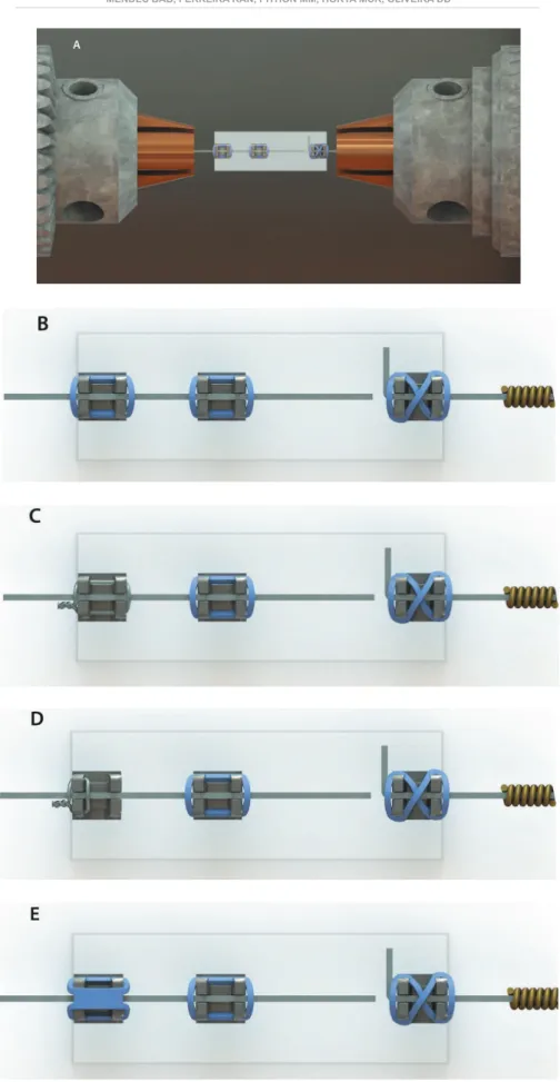
Friction was evaluated by simulating sliding mechanics with a 0.019x0.025” stainless steel orthodontic wire (Morelli Ortodontia, Sorocaba, SP, Brazil). Four types of ligation were tested: (1) conventional gray elastic ligatures (Morelli Ortodontia, Sorocaba, SP, Brazil); (2) 0.010” steel ligatures (Morelli Ortodontia, Sorocaba, SP, Brazil) tied around all four bracket wings; (3) steel ligatures tied only to the two bracket wings closest to the load-cell; (4) low friction gray elastic ligatures (Slide®, Leone Orthodontics and Implantology,
Florence, Italy) (Figure 1).
The new brackets (control group), ligatures, and wires were used as received from the manufacturer. Thus, they were removed from their original packaging and immediately submitted to the tests. Four test groups with seven new brackets each, of the same brand, model, and ligation method, were used as controls. According to the combinations of bracket and types of ligations tested, 12 groups were formed, as shown in Figure 2.
Scanning Electron Microscopy (SEM) analysis
S e M w a s p e r f o r m e d w i t h a J S M - 5 3 1 0 microscope (JeOL, Tokyo, Japan) from the Center for Development of Nuclear Technology (CDTN/ CNeN). It was used to obtain the images of the brackets surface and analyze the possible damages generated after the exposure to oral cavity during the orthodontic treatment. Twelve orthodontic brackets were randomly selected for this analysis, 4 as received from the manufacturer (Group 0), 4 from the 12-month treatment samples (Group 12), and 4 from the 24-month treatment specimens (Group 24).
For each sample, 5 photographic images were
taken, in the following order: (1) 15x magniication
at a 90° angle; (2) 15x at 45°; (3) 50x at 90°; (4) between 100x; and (5) 350x of the slot region near the tie-wings.
Semi-quantitative analysis through Energy Dispersive X-ray Spectroscopy
Groups Time of use (month) Type of ligation
0-LE 0 Elastomeric ligatures
0-LM 0 Metallic ligatures 4/4*
0-2/4LM 0 Metallic ligatures 2/4**
0-LS 0 Slide®
12-LE 12 Elastomeric ligatures
12-LM 12 Metallic ligatures 4/4*
12-2/4LM 12 Metallic ligatures 2/4**
12-LS 12 Slide®
24-LE 24 Elastomeric ligatures
24-LM 24 Metallic ligatures 4/4*
24-2/4LM 24 Metallic ligatures 2/4**
24-LS 24 Slide®
Figure 2- Distribution of brackets and types of wire ligation evaluated * Metallic ligation tied to 4 bracket tie-wings
** Metallic ligature tied to only 2 and 4 bracket tie-wings impurities were analyzed to identify the composition of the bracket.
Bracket surface roughness analysis
Surface roughness was measured on 5 brackets of each group to verify possible differences on the effects of their exposure to the oral environment. The images were obtained with 2,000x of vertical
magniication and 100x horizontally. Magniications
of 2,000x or 5,000x and a cut-off of 0.25 mm were used to obtain Ra values. The values of the ive
largest distances between the highest peak and the
lowest valley in each of the ive sample lengths (R1, R2, R3, R4, and R5), as well as the average of these values (Rtm) were also registered. The images were obtained at 1 mm from the central region of the brackets slots. Five measurements were performed for each bracket, resulting in a total of 75 analyses of Ra andRtm.
Evaluation of the friction levels
The static friction was evaluated with an AN8032 testing machine (Analógica Instrumentação e Controle Ltda., Belo Horizonte, MG, Brazil) at room temperature, in a dry environment and using the tensile test in a straight line. The load cell had a maximum capacity of 222.4 N and the machine was operated at 0.075 mm/seg. each trial had its
initial (500 µm) and inal (1,400 µm) positions set
by the machine.
In order to perform the friction tests, the brackets were positioned on an acrylic plate that remained in a horizontal position and was hooked to the lower claw of the universal testing machine, while the wire was hooked to the upper claw and pulled to slide through the bracket slots (Figure 1). This positioning was carefully set to allow the test to
be performed through the long axis of the machine. The load cell registered the levels of force necessary to move the wire through the brackets for 9 mm. However, only the measurements registered
along the irst 7 mm were taken into consideration
because this distance is equivalent to the average space available when premolars are extracted in an orthodontic treatment. The data was transmitted to a computer connected to the machine, which plotted a graph with XY coordinates. The X-axis registered the displacement of the wire in mm, while the Y-axis registered the friction in Newtons at the bracket/wire interface.
The same operator prepared the specimens and performed all tests of this study. During the preparation of the brackets, the operator carefully standardized the insertion of the elastic ligatures, stretching them with Mathieu tweezers (Quinelato, São Paulo, SP, Brazil) to the minimum needed to place them around the bracket wings. The non-conventional Slide® elastic ligatures were inserted
according to the manufacturer’s instructions.
Statistical procedures
The Kolmogorov-Smirnov normality test showed that the data presented normal distribution. The two-way analysis of variance (two-way ANOVA) followed by the Bonferroni’s post hoc test was used to compare static friction registered with the different types of ligation and various duration of intraoral use of the brackets.
The one-way analysis of variance (One-way ANOVA) followed by the Tukey post hoc test was used to compare the surface roughness (Ra) and the largest distance between peaks and valleys (Rtm) between different times of intraoral exposure.
Type of ligation Force (N)
New 12 months 24 months P value*
Metallic ligatures 2/4 2.64±0.43 1.72±0.35 1.91±0.47 n.s.3/p<0.051,2
Metallic ligatures 4/4 1.98±0.26 1.95±0.26 2.42±0.36 n.s.1,2,3
Elastomeric 4.31±0.27 3.70±0.29 4.18±0.59
n.s.
2,3/p<0.051Slide®
2.74±0.35
1.67±0.19
1.51±0.16
n.s.
3/p<0.051,2P value*
n.s.
6n.s.
4,6,8n.s.
6p<0.05
4,5,7,8,9p<0.05
5,7,9p<0.05
4,5,7,8,9Table 1- Mean and standard deviations of the static friction during sliding tests. Comparison between different types of ligation and duration of intraoral bracket use
*P values were obtained by two way analysis of variance (one way ANOVA) followed by the Bonferroni’s post hoc test n.s.=not signiicant (p>0.05)
1 New versus 12 months
2 New versus 24 months
3 12 months versus 24 months
4 2/4 versus 4/4
5 2/4 versus Elastomeric
6 2/4 versus Slide®
7 4/4 versus Elastomeric
8 4/4 versus Slide®
9 Elastomeric versus Slide®
Condition
New 12 months 24 months P value *
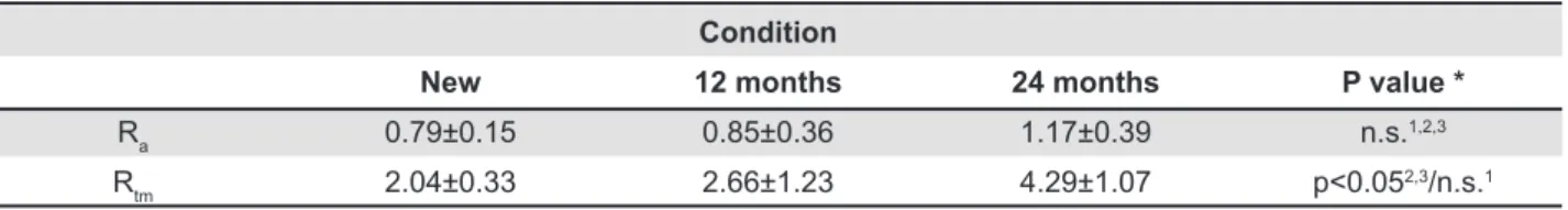
Ra 0.79±0.15 0.85±0.36 1.17±0.39 n.s.
1,2,3
Rtm 2.04±0.33 2.66±1.23 4.29±1.07 p<0.052,3/n.s.1
*P values were obtained by one way analysis of variance (one way ANOVA) followed by the Tukey post hoc test n.s.=not signiicant (p>0.05)
1New versus 12 months
2New versus 24 months
312 months versus 24 months
Table 2- Mean and standard deviation of surface roughness (Ra) and of the largest distance between peaks and valleys (Rtm) found on the brackets’ surfaces
tests that were performed with the GraphPad Prism®
software (GraphPad Software, San Diego, CA, USA).
RESULTS
Friction evaluation
The descriptive analyses of the mean static friction values, registered for all combinations of time of exposure to the oral environment and wire ligation methods, are presented in Figure 2. Table 1 shows the comparison of the static friction levels registered among all groups and the comparison of static friction levels produced with the different wire ligation methods at each time interval, according to the two-way ANOVA followed by the Bonferroni’s post hoc test.
Bracket surface roughness analysis
even tough the brackets as received from the manufacturer presented the lowest surface roughness (Ra) mean values, no statistically difference was observed when the new, the 12- and the 24-month brackets were compared (Table 2). The largest distance between the peaks and valleys on the slot surface (Rtm) was higher in the 24-month brackets when compared to the new and to the 12-month brackets (Table 2).
SEM analysis
The SeM evaluation of the brackets as received from the manufacturer showed presence of machining marks and some corrosion pits. Some of
Figure 3- Cleft found on the external surface of the base of the tie-wing on a new bracket with 75x magniication (A). Marks of machining, corrosion cavities (which appear as dark spots), some illed with impurities; 15x magniication (B)
Figure 4- Changes stemming from use and consequent exposure to the intraoral medium. New bracket (A), after 12 months of use (B), and after 24 months of use (C), all with 15x magniication
as shiny white spots in the SeM images (Figure 3A). There were also clefts of different sizes on the external surface of the bracket wings base (Figure 3B).
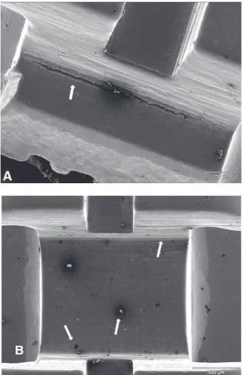
The SeM qualitative evaluation of the brackets used for 12 and 24 months revealed important differences in relation to the evaluation of the brackets as received from the manufacturer. These changes were signs of corrosion and wear, plastic deformation, presence of clefts, cracks, and grooves. Furthermore, the used brackets presented
bioilm deposits and accumulations of materials of
various sizes on their surface (Figure 4). Figure 5 illustrates the bracket slots after 12 and 24 months of orthodontic treatment.
Semi-quantitative analysis through Energy Dispersive X-ray Spectroscopy
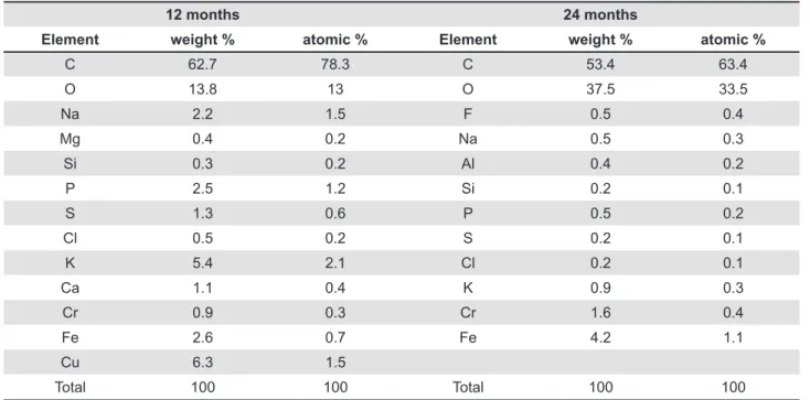
Semi-quantitative eDS chemical analysis of the deposits found on the slot surfaces of the orthodontic brackets that were used for 12 or 24 months are shown in Tables 2 and 3, respectively.
Carbon and oxygen were the elements detected in the highest concentrations. However, signs of sodium, phosphorus, potassium, calcium, iron,
lead, and aluminum could also be identiied. In
12 months 24 months
Element weight % atomic % Element weight % atomic %
C 62.7 78.3 C 53.4 63.4
O 13.8 13 O 37.5 33.5
Na 2.2 1.5 F 0.5 0.4
Mg 0.4 0.2 Na 0.5 0.3
Si 0.3 0.2 Al 0.4 0.2
P 2.5 1.2 Si 0.2 0.1
S 1.3 0.6 P 0.5 0.2
Cl 0.5 0.2 S 0.2 0.1
K 5.4 2.1 Cl 0.2 0.1
Ca 1.1 0.4 K 0.9 0.3
Cr 0.9 0.3 Cr 1.6 0.4
Fe 2.6 0.7 Fe 4.2 1.1
Cu 6.3 1.5
Total 100 100 Total 100 100
Table 3- EDS semi-quantitative chemical analysis on the deposits found in an orthodontic bracket slot used for 12 or 24 months
Figure 5- Bioilm deposit over the bracket slot after exposure to the intraoral medium for 12 (A) and 24 (B) months; 150x magniication
DISCUSSION
The physical properties of orthodontic materials as received from the manufacturer have been widely studied over the years16. However, the knowledge
about the clinical performance of orthodontic materials over the duration of treatment, thus after they are exposed to the oral environment is also very important. Variations in temperature and pH as registered intraorally22 may cause the
biodegradation of these materials, changing some of their properties22, which may compromise their
clinical performance9.
The combined action of these biological factors
can signiicantly alter the integrity of the orthodontic
materials surface, especially metallic brackets8,9.
However, the extension of these changes and how they actually affect the clinical performance of these materials remain unclear. Thus, the purpose of this study was to evaluate physical and chemical changes on metallic brackets surfaces after different intervals of clinical use. The present study also analyzed the static friction generated on these biodegraded brackets with 4 different wire ligation methods. Furthermore, the surface roughness of the brackets was also evaluated. SeM and eDS were the methods of evaluation used to quantify the
chemical elements present in the bioilm generated
during the intraoral use of the materials tested.
Friction evaluation
brackets as received from the manufacturer combined to different methods of wire ligation showed that loose ligation with steel ligatures around the four bracket wings presented the lowest mean friction values, while the maximum friction was registered when the brackets had the wire ligated with conventional elastic ligatures. These
indings are compatible with what has previously
been reported in the literature4. However, our
results differ from others regarding the use of the non-conventional Slide® elastic ligatures. In
the present study, no signiicant differences were identiied between ligation with metallic ligatures
on two bracket wings and the Slide® ligatures.
Conversely, Chen, et al.6 (2010) found the lowest
friction values using Slide® also combined with
stainless steel brackets and wires.
The comparison of the maximum friction generated among the new brackets and brackets used over a 12-month period revealed a reduction for all different methods of wire ligation, except for loose ligation with steel ligatures around the four bracket wings. The comparison of the maximum friction generated among the new brackets and brackets used over a 24-month period revealed a reduction for all different methods of wire ligation, except for loose ligation with steel ligatures around the four bracket wings and for elastomeric ligatures. These results are in accordance with previously reported results21 and are partially in agreement
with the indings from the evaluation of friction
levels produced with three bracket models after an average period of intraoral use of 41 months. The Mini Standard edgewise bracket (American Orthodontics, Sheboygan, WI, USA) presented a reduction of 3.10% in friction levels, while the Kirium edgewise brackets (3M/Abzil, Sumaré, SP, Brazil) and the Nuedge Roth Prescription brackets (TP Orthodontics, LaPorte, IN, USA) presented a
signiicant increase in relation to those observed
with brackets as received from the manufacturer9.
All friction tests of this study were performed under dry conditions. The effects of lubricating
agents such as artiicial saliva in the results of
friction tests are controversial. Some studies suggest the use of a lubrication medium because it would better simulated the intraoral conditions1,
whereas others attained controversial results10.
The use of brackets exposed to the oral environment for 12 or 24 months positively contributes to the external validity of our results, since it resembled and evaluated some of the challenges that any orthodontist may face upon dealing with materials that age in the oral cavity during treatment. Another precaution taken to increase the reliability of our results was the development of a mechanism that eliminated any tipping between the wire and the brackets attached
to the lower portion of the load cell4, preventing
the existence of binding effect which would be
a signiicant confounder, increasing the friction
levels11. Moreover, the conventional elastomeric
ligatures were inserted prior to performing the tests in each group. This alternative was chosen to standardize any possible alteration of the ligature’s modules of elasticity, an effect which has previously been described1.
However, all the limitations that involve in vitro studies must be taken into consideration when attempting to apply the present study’s results to a clinical reality. Factors such as second and third order sloping, the binding effect between the wire and the bracket, the presence of masticatory forces, and the
thin bioilm were not reproduced9. Nevertheless,
the present study presents evidence of the effects caused by the clinical use of metallic brackets and represents an improvement in understanding how biodegradation may compromise the clinical performance of orthodontic metallic brackets.
Bracket surface roughness analysis
The analysis of Rtm demonstrated the lower surface roughness values in the brackets as received from the manufacturer. The group of brackets after 24 hours of intraoral use presented the highest Rtm values, when compared to both other groups. These results are in accordance with reports in the literature that evaluated surface roughness changes on metallic orthodontic wires after use in the oral environment. The authors concluded such exposure caused an increase in the variety, type, and number of surface irregularities1, and our results support
these indings.
Scanning electron microscopy analysis
In the present study, the new brackets presented marks of machining in the form of parallel lines,
various corrosion cavities (pits) (some illed with
impurities), clefts of various sizes on the external surface of the base of the bracket tie-wings, in addition to containing an accentuated curvature in relation to the axis of the bracket slots. These
indings are in accordance with prior reports that
evaluated the surface of stainless steel brackets as received from the manufacturer and those after
exposure to the oral environment or artiicial saliva1.
Therefore, the results of the present study showed that the manufacturing processes of the metallic brackets are not perfect and may produce brackets presenting grooves and marks in the bracket slot,
which could compromise the ideal itting of the
orthodontic wire within the slot walls1.
plastic deformation, clefts, cracks, and grooves.
These indings are in accordance with previous
reports that have also evaluated the performance of orthodontic materials after their exposure to the oral environment or to different mediums that simulated in vivo conditions7,13,16,17,23,24.
Furthermore, the used brackets presented bioilm
deposits and accumulations of a wide range of materials of various sizes, with results similar to those previously described in the literature9, and
that were also present in orthodontic wires10 and
components of extra-oral devices9. However, a
variation in the bioilm levels could be observed
among brackets with the same time of use, which
reinforces the inluence of individual variation and
of methods of personal hygiene9.
The signs of wear and corrosion observed in the bracket slots, slightly below the bracket tie-wings, are compatible with galvanic corrosion which occurs when two materials made of different metals are used to construct an orthodontic bracket9,16. Finally,
the signs of plastic deformation after clinical use, found in the region corresponding to the end of the bracket tie-wings, were also registered in previous investigations9.
Semi-quantitative analysis through Energy Dispersive X-ray Spectroscopy
The semi-quantitative chemical analysis of the bracket wing top after 12 months of intraoral use revealed the presence of Fe (70.2%), Cr (19.5%), Mo (0.8%), and Ni (8.8%) (Table 3). These values correspond to those used in the manufacturing of ABNT 316 steel. These results were expected and are coherent, since this alloy is commonly used to manufacture orthodontic brackets1.
The eDS analysis of metallic brackets after 12 and 24 months of intraoral use detected deposits of C, O, Na, P, S, K, and Ca. There were also traces of Cl and Si, as well as small quantities of Fe and Cr. The presence of some of these chemical elements (C, O, Ca, P, S, and K) as products of the biodegradation of orthodontic brackets has been previously reported in the literature9. Other studies also found bioilm
deposits with the presence of the same elements in NiTi orthodontic wires12 and in extra-oral devices
after their clinical use12, indicating similar indings
but in different proportions than those found in the present study.
The eDS analysis of deposits found on the base
of the used brackets identiied similar results to
those observed in the slot region of the brackets, except for the presence of relatively large quantities of Cu and Ag in the brackets used for 12 months and of Al in the brackets used for 24 months. However, it was impossible to determine the origin of these elements.
Clinical application
The results of this investigation have signiicant
clinical application because they showed that different types of wire ligation generate different levels of friction even after the biodegradation of orthodontic brackets in the oral environment. Based
on these indings, the orthodontist may use a type
of wire ligation to generate less friction at the early stages of the orthodontic treatment and implement
a different ligation protocol at the inishing stages,
when higher levels of friction are desired.
CONCLUSIONS
Based on the results of the present study, the conclusions are as follows:
The largest distance between peaks and valleys (Rtm) of metallic brackets increased after 24 months of exposure in the oral environment;
The maximum friction levels produced during the sliding mechanics decreased with use;
The ligation method that presented the lowest friction levels for new brackets was the steel ligature loosely attached around all four bracket wings;
The assays using brackets after 12 or 24 months of exposure to the oral environment presented the lowest static friction levels;
For all assays, the wire ligation using conventional elastic ligatures presented the highest static friction levels.
The hypothesis that different types of wire ligation generates different levels of static friction
was conirmed.
REFERENCES
1- Alcock JP, Barbour Me, Sandy JR, Ireland AJ. Nanoindentation of orthodontic archwires: the effect of decontamination and clinical use on hardness, elastic modulus and surface roughness. Dent Mater. 2009;25:1039-43.
2- Articolo LC, Kusy RP. Inluence of angulation on the resistance to sliding in ixed appliances. Am J Orthod Dentofacial Orthop.
1999;115:39-51.
3- Baccetti T, Franchi L, Camporesi M, Defraia e, Barbato e. Forces produced by different nonconventional bracket or ligature systems during alignment of apically displaced teeth. Angle Orthod. 2009;79:533-9.
4- Burrow SJ. Friction and resistance to sliding in orthodontics: a critical review. Am J Orthod Dentofacial Orthop. 2009;135:442-7. 5- Castro RM, Neto PS, Horta MC, Pithon MM, Oliveira DD.
Comparison of static friction with self-ligating, modiied slot design
and conventional brackets. J Appl Oral Sci. 2013;21:314-9. 6- Chen SS, Greenlee GM, Kim Je, Smith CL, Huang GJ. Systematic review of self-ligating brackets. Am J Orthod Dentofacial Orthop. 2010;137:726.e1-726.e718.
7- Dolci GS, Menezes LM, Souza RM, Dedavid BA. Biodegradtion of orthodontics brackets: in vitro ion release. Rev Dent Press Ortodon Ortopedi Facial. 2008;13:77-84.
9- eliades T, Bourauel C. Intraoral aging of orthodontic materials: the picture we miss and its clinical relevance. Am J Orthod Dentofacial Orthop. 2005;127:403-12.
10- eliades T, Zinelis S, eliades G, Athanasiou Ae. Characterization of as-received, retrieved, and recycled stainless steel brackets. J Orofac Orthop. 2003;64:80-7.
11- Fidalgo TK, Pithon MM, Maciel JV, Bolognese AM. Friction
between different wire bracket combinations in artiicial saliva - an
in vitro evaluation. J Appl Oral Sci. 2011;19:57-62.
12- Grifiths HS, Sherriff M, Ireland AJ. Resistance to sliding with
3 types of elastomeric modules. Am J Orthod Dentofacial Orthop. 2005;127:670-5; quiz 754.
13- Hain M, Dhopatkar A, Rock P. The effect of ligation method on friction in sliding mechanics. Am J Orthod Dentofacial Orthop. 2003;123:416-22.
14- Kapila S, Angolkar PV, Duncanson MG Jr, Nanda RS. evaluation of friction between edgewise stainless steel brackets and orthodontic wires of four alloys. Am J Orthod Dentofacial Orthop. 1990;98:117-26.
15- Lima VN, Coimbra Me, Derech CD, Ruellas AC. Frictional forces in stainless steel and plastic brackets using four types of wire ligation. Dental Press J Orthod. 2010;15:82-6.
16- Marques IS, Araújo AM, Gurgel JA, Normando D. Debris, roughness and friction of stainless steel archwires following clinical use. Angle Orthod. 2010;80:521-7.
17- Menezes LM, Souza RM, Dolci GS, Dedavid BA. Analysis of biodegradation of orthodontic brackets using scanning electron microscopy. Dental Press J Orthod. 2010;15:48-51.
18. Miles PG. Self-ligating brackets in orthodontics: do they deliver what they claim? Aust Dent J. 2009;54:9-11.
19- Muguruma T, Iijima M, Brantley WA, Mizoguchi I. effects of a diamond-like carbon coating on the frictional properties of orthodontic wires. Angle Orthod. 2011;81:141-8.
20- Normando D, Araújo AM, Marques Ida S, Barroso Tavares Dias CG, Miguel JA. Archwire cleaning after intraoral ageing: the effects on debris, roughness, and friction. eur J Orthod. 2013;35:223-9. 21- Pacheco MR, Oliveira DD, Smith Neto P, Jansen WC. evaluation of friction in self-ligating brackets subjected to sliding mechanics: an in vitro study. Dental Press J Orthod. 2011;16:107-15. 22- Redlich M, Mayer Y, Harari D, Lewinstein I. In vitro study of frictional forces during sliding mechanics of "reduced-friction" brackets. Am J Orthod Dentofacial Orthop. 2003;124:69-73. 23- Sathler R, Silva GR, Janson G, Castello Branco NC, Zanda M. Demystifying self-ligating brackets. Dental Press J Orthod. 2011;16:e1-8.