Arnica montana does not afect mast cell populations in
experimentally induced oral ulcers in rats
Leandro Boutin PerussoloI, Ana Maria Trindade GrégioI, Luciana Reis Azevedo-AlanisI, Maria Ângela Naval MachadoII, Aline Cristina Batista Rodrigues JohannI, Antonio Adilson Soares de LimaII
DOI: 10.5935/MedicalExpress.2015.03.06
I Pontifícia Universidade Católica do Paraná, Faculdade de Odontologia, Curitiba, Paraná, Brazil
II Universidade Federal do Paraná, Faculdade de Odontologia, Departamento de Estomatologia, Curitiba, Paraná, Brazil
BACKGROUND: Studies have shown that Arnica montana shows anti-inlammatory and antioxidant activities. It has been used in traditional medicine for the treatment of several disorders. The aim of this study was to investigate the efect of Arnica montana on mast cells during the wound healing of oral ulcers.
METHOD: An ulcerated lesion was chemically induced on the tongue of 75 male albino rats and, then, treated topically for seven days using saline solution (control), Arnica montana gel or tincture. The animals were killed after 2nd, 7th, 14th, 21th and 42th day of treatment. The tongues were removed and subjected to routine laboratory (0.2%
toluidine blue staining). The numbers of mast cell were determined in two regions: supericial and submucosa.
RESULTS: The numbers of mast cells were signiicantly increased for all groups in the region of the deeper tissue when compared to the supericial region. No statistical diference was observed in mast cell numbers for each group.
CONCLUSION: This study revealed that Arnica montana tincture and gel were unable to change mast cell population during wound healing of oral ulcer of rats. According to these results, the anti-inlammatory efects of Arnica montana were not related to inhibition of mast cell degranulation.
KEYWORDS: Arnica montana, Mast cell, Oral mucosa, Oral ulcer, Inlammation.
Perussolo LB, Grégio AMT, Azevedo-Alanis LR, Machado MAN, Johann ACBR, Lima AAS. Arnica Montana does not afect mast cell populations in experimentally induced oral ulcers in rats. MedicalExpress (São Paulo, online). 2015;2(3):M150306
Received for Publication on March 14, 2015; First review on April 26, 2015; Accepted for publication on May 22, 2015
E-mail: lbperussolo@hotmail.com
■
INTRODUCTIONOral ulcer is the name for the appearance of an open sore inside the mouth caused by a break in the continuity of the mucous membrane or the epithelium on the lips or surrounding the mouth. The types of oral ulcers are diverse, with a multitude of associated causes including: physical or chemical trauma, microorganism infections (virus, bacteria, and fungi), systemic diseases, and some drugs.1
This kind of lesion can affect several anatomical regions of the mouth, especially the tongue, the gingiva, and the cheeks.2-3 The formation of an ulcer
occurs when superficial layers of the oral mucosa are
lost. Morphologically, oral ulcers appear as circular
Copyright © 2015 MEDICALEXPRESS. This is an open access article distributed under the terms of the creative commons attribution
or irregular lesions. Once formed, the ulcer may be
maintained by inflammation and/or secondary infection.4
Thus, its prevention continues to be of concern for both clinical practitioner and researchers. Treatment of this condition is based on the removal of etiological factors and on the prescription of antimicrobial mouthwash. Several clinical protocols for ulcer treatment have been reported. In addition, several herbal agents have been employed in order to help in the repair of these oral lesions.3
Arnica is an herbaceous perennial plant. The subspecies montana is widely distributed, and grows in mountainous areas and thrives in nutrient-poor siliceous
meadows and acid soils. Its flowers contain several active ingredients, including sesquiterpene lactones, flavonoids,
volatile oil, mucilage, polysaccharides, and tannins. The
flowers contain more arnicin than the rhizome, but
were examined daily. Any alteration in the normal course of the repair process was recorded. The animals were killed under general anesthesia induced with thiopental sodium® (CRISTÁLIA, Brazil, 20 mg/Kg) after the 2nd (5
animals), 7th (5 animals), 14th (5 animals), 21th (5 animals)
and 42th (5 animals) day of treatment. The tongues were
removed surgically and fixed in 10% formalin solution.
All tissue specimens were processed in the laboratory of Experimental Pathology of Universidade Católica do Paraná. Histological slides were prepared after sectioning
at a thickness of 6μm and staining with 0.2% toluidine blue.
Mast Cell counting
The morphology and the location of mast cells were assessed microscopically. Mast cells were counted separately in sections of the ulcerated area using a light microscope
OLYMPUS BX50 equipped with an objective PLAN 10X/0,25 and oculars WH10X-H/22 (OLYMPUS, Tokyo, Japan). This
microscope was connected to the Color video camera
CCD-IRIS (SONY, Tokyo, Japan) that allowed the capture of images in the fields of histology slides. ImagePro Plus software version 4.0.1 (Media Cibernetics, Atlanta, GA, USA) was
used to count cells. Mast cells were counted in four counting
fields per section at 100x magnification. In order to assess distribution, the histological fields were separated in two areas: Superficial (epithelium/connective tissue interface)
and Deep (submucosal region). The number of mast cells
was expressed as cells/mm2 (mean ± SD.).
Statistical analysis
All data were tabulated and statistical tests were
performed with SPSS for Windows 13.0 (SPSS Inc., Chicago, Illinois, USA). Tests of normality (Kolgomorov-Smirnoff
test) and homogeneity of variances (Levene’s Test) were used. Differences between groups were examined using the
Games-Howell test. Difference was considered statistically significant when p < 0.05.
■
RESULTSNo significant change was observed during the
process of wound repair in the tongues of animals. Large numbers of mast cells were found immediately under the basement membrane in all sections studied. These cells were round or oval and were located especially in certain
regions, namely lamina propria, inflammatory infiltrates, perivascular region and in the deep connective tissue. No
mast cells were observed in the epithelium.
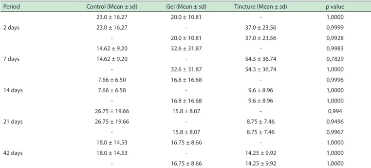
The mean number of mast cells in the superficial region
was greater in animals treated by tincture and gel of Arnica montana at 7th post-treatment days, as shown in Table 1. The
lowest density of mast cells was observed in the control group
Dihydrohelenalin produce anti-inflammatory and analgesic
effects. Sesquiterpene lactones (SL), which are secondary
plant metabolites from the flowerheads of Arnica, exert anti-inflammatory effects mainly by preventing nuclear factor (NF)-kappa B activation because of alkylation of the
p65 subunit.5 Despite its known immunosuppressive action,
Arnica has been classified as a plant with strong potency to
induce allergic contact dermatitis.6
Mast cells are cells of hematopoietic origin that terminally differentiate and become mature in tissues.7 They
contribute to both innate and adaptive immune responses. Mast cells are long-lived secretory cells viewed as sentinels, able to
rapidly respond to modifications in their environment.8-9 Once
activated, mast cells have the ability to secrete a wide array of
inflammatory mediators. Thus, mast cells can modulate the intensity of organ injury depending upon the pathophysiological
context.10 The aim of this study was to investigate the effect of
Arnica montana on mast cells during the wound healing of oral ulcers treated by gel and tincture.
■
MATERIALS AND METHODSEthical Approval
The experimental protocol of the present study was approved by Committee of Ethics in Animal
Experimenta-tion at the Pontifícia Universidade Católica do Paraná.
Animals and drugs
Seventy-five male albino rats (Rattus norvegicus) weighing 200-250 g were used in this study. Animals were
maintained in individual cages and received standard solid food and water ad libitum. An ulcerated lesion was chemically
induced on the surface of the tongue using 40% sodium
hydroxide after general anesthesia induced with thiopental sodium® (Cristália, Brazil, 20 mg/Kg). The ulcerated lesion
was produced by topical application using a small cotton ball soaked in the chemical solution. The chemical agent was left to act on the edge of the tongue for approximately one minute.
Groups
The animals were divided into three groups: i) Control: 25 animals that were treated daily by topic
applications of saline solution during seven days.
ii) Gel: 25 animals received topical applications of a gel of 30% A. montana during seven days.
iii) Tincture: 25 animals received topical applications
of 30% A. montana tincture during seven days.
The animals were treated with an analgesic with
peripheral action (no anti-inflammatory effect) to control
postoperative pain and facilitate the feeding of animals.
The drug used was dipyrone (50 mg/kg) 4 times a day
Table 1 - Mean numbers of mast cells in the supericial region for groups untreated and treated by Arnica montana
Period Control (Mean ± sd) Gel (Mean ± sd) Tincture (Mean ± sd) p value
2 days
23.0 ± 16.27 20.0 ± 10.81 - 1,0000
23.0 ± 16.27 - 37.0 ± 23.56 0,9999
- 20.0 ± 10.81 37.0 ± 23.56 0,9928
7 days
14.62 ± 9.20 32.6 ± 31.87 - 0.9983
14.62 ± 9.20 - 54.3 ± 36.74 0,7829
- 32.6 ± 31.87 54.3 ± 36.74 1,0000
14 days
7.66 ± 6.50 16.8 ± 16.68 - 0,9996
7.66 ± 6.50 - 9.6 ± 8.96 1,0000
- 16.8 ± 16.68 9.6 ± 8.96 1,0000
21 days
26.75 ± 19.66 15.8 ± 8.07 - 0,994
26.75 ± 19.66 - 8.75 ± 7.46 0,9496
- 15.8 ± 8.07 8.75 ± 7.46 0,9967
42 days
18.0 ± 14.53 16.75 ± 8.66 - 1,0000
18.0 ± 14.53 - 14.25 ± 9.92 1,0000
- 16.75 ± 8.66 14.25 ± 9.92 1,0000
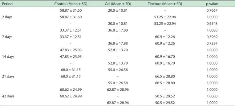
The numbers of mast cells were significantly increased for all groups in the deeper tissues when
compared to the superficial region (p = 0.003), as shows the Figure 1. The mean mast cell number in the
submucosa was greater in the experimental group when compared to control group at 2, and 21 post-treatment days, as shown in Table 2. As previously noted, the lowest density of mast cells was observed in the control group at 14 post-treatment days. However, none of these differences was statistically significant for this
region (p > 0.05)
Figure 1 - Supericial and deep average density of mast cells in the region of an ulcer.
Averages for control and treated animals, which were not diferent between them.
■
DISCUSSIONEvaluation of the biological activities of compounds in Arnica montana and elucidation of the mechanisms of their functions may provide substantial clues for the development of new drug candidates. Some previous studies have demonstrated that Arnica montana exhibits
potential anti-inflammatory activity.11-15 Additionally,
other studies have demonstrated the beneficial effects
of Arnica montana in wound healing,16-18 pain relief,19
antioxidant activity and cytoprotective effect against oxidative damage.20 However, to our knowledge the
mechanism by which Arnica montana acts on the oral wounds is not known.
This study investigated the effect of Arnica montana upon the mast cell population during wound healing of oral ulcers in rats. Mast cells are located in the vicinity microcirculatory vessels involved in vasodilation. They mature under the influence of various cytokines. Human skin and mucosal mast cells play an essential role in various physiological a n d p a t h o l o g i c a l p ro c e s s e s a n d m e d i a t e e a r ly hypersensitive reaction and allergic diseases.21 In the
present experiments, the morphology of mast cells was not altered in the presence of Arnica (gel or tincture).
Furthermore, no statistical difference was observed
Table 2 -Number of mast cells at submucosa for groups untreated and treated by Arnica montana
Period Control (Mean ± SD) Gel (Mean ± SD) Tincture (Mean ± SD) p value
2 days
58.87 ± 31.60 20.0 ± 10.81 - 0,7667
58.87 ± 31.60 - 53.25 ± 22.94 1,0000
- 20.0 ± 10.81 53.25 ± 22.94 0,6548
7 days
33.37 ± 12.51 36.8 ± 17.88 - 1,0000
33.37 ± 12.51 - 60.9 ± 12.26 0,3969
- 36.8 ± 17.88 60.9 ± 12.26 0,7297
14 days
47.83 ± 25.93 52.8 ± 13.70 - 1,0000
47.83 ± 25.93 - 60.9 ± 16.70 1,0000
- 52.8 ± 13.70 60.9 ± 16.70 1,0000
21 days
68.0 ± 31.15 55.0 ± 26.58 - 1,0000
68.0 ± 31.15 - 66.5 ± 28.80 1,0000
- 55.0 ± 26.58 66.5 ± 28.80 1,0000
42 days
60.62 ± 24.99 62.87 ± 28.96 - 1,0000
60.62 ± 24.99 - 50.5 ± 29.52 1,0000
- 62.87 ± 28.96 50.5 ± 29.52 1,0000
The cytoplasm of the resting mast cell is filled with
large granules containing histamine, prostaglandins, and
other proinflammatory mediators.22 The activation and
degranulation of mast cells are major contributors to inflammation.23 This reaction is noticeable when the mean
number of mast cells is analyzed in relation to time. In
our study, the results showed that the lowest number of mast cells present in the surface region of the wound was recorded after 14 days of treatment. This phase of the repair
process coincides with the period of chronic inflammation when collagen fibers are being deposited and angiogenesis
is evident. According to Trautmann et al.24 as the healing
process progresses, mast cells migrate to the site of injury,
dramatically increasing in numbers.
Most cases of oral ulcers observed in general practice are due to recurrent aphthous ulceration, infection or trauma. However, many of the reports in the literature have not been validated in controlled clinical trials. Ulcers related to trauma usually resolve in about seven days after removal of the cause and with the use of pharmacological agents to
minimize the undesirable effects induced by inflammation.1
Ganzera et al.25 demonstrated that sequiterpenes,
flavonoids and phenolic acids are among the biologically active ingredients in the flowerheads of the Arnica Monta -na. Quercetin 3-O-glucuronic acid was the most dominant
flavonoid, whereas 3,5-dicaffeoylquinic acid was the major phenolic acid; the total content of flavonoids and phenolic acids varied in the samples from 0.60 to 1.70%, and 1.03 to 2.24%, respectively.
Although the therapeutic use of A montana is established, some side effects have been described.26 It is
classified as a plant with a strong power to induce allergic
observed in animals during our clinical follow-up and neither in the histological evaluation of mast cells.
This study also showed that the number of mast cells
in the deeper tissues is higher than those in the superficial regions of the ulcer. These findings corroborate the results of Natah et al.27 who observed similar results when
investigating the population of mast cells within traumatic ulcers and recurrent aphthous ulcerations.
The vehicle used for topical applications is a potentially important factor in the pharmacologic effects. Bergamante et al.28 investigated the ability to dissolve,
release, and induce the permeation of helenalin (flavonoid responsible for the anti-inflammatory activity of the Arnica
montana extract) in two types of gels and microemulsions. Their study showed that a microemulsion could be a good vehicle to increase the permeation of helenalin. In our study, the greatest numbers of mast cells were observed the groups treated by the tincture of Arnica Montana. In this scenario, the ethanol used in the tincture may have
influenced the results. Additionally, the mast cell population was significantly increased in the region of submucosa. This result corroborates the findings of Pumpa et al.19 and
demonstrated that both preparations of Arnica Montana used in this study were unable to act on mast cells in the muscle tissue of the tongue.
The present research represents the first study
suggesting that Arnica Montana does not change the mast cell population in oral ulcers during the wound healing.
This fact reinforces the concept that the anti-inflammatory
8. Gurish MF, Austen KF. The diverse roles of mast cells. J Exp Med. 2001;194(1):F1-5.
9. Galli SJ, Nakae S, Tsai M. Mast cells in the development of adaptive immune responses. Nat Immunol. 2005;6(2):135-42.
10. Knol EF, Olszewski M. Basophils and mast cells: Underdog in immune regulation? Immunol Lett. 2011;138(1):28-31.
11. Macêdo SB, Ferreira LR, Perazzo FF, Carvalho JC. Anti-inflammatory
activity of Arnica montana 6cH: preclinical study in animals.
Homeo-pathy. 2004;93(2):84-7.
12. Totonchi A, Guyuron B. A randomized, controlled comparison between
arnica and steroids in the management of postrhinoplasty ecchymosis
and edema. Plast Reconstr Surg. 2007;120(1):271-274.
13. Widrig R, Suter A, Saller R, Melzer J. Choosing between NSAID and
arnica for topical treatment of hand osteoarthritis in a randomised,
double-blind study. Rheumatol Int. 2007;27(6):585-91.
14. Yui F, Linarelli MCB, Zelante PM. Atividade antiinflamatória da Arnica montana/Anti-inflammatory activity of Arnica montana. Rev Ciências
Médicas. 1998;7(1):21-6.
15. Hostanska K, Rostock M, Melzer J, Baumgartner S, Saller R. A home
-opathic remedy from arnica, marigold, St. John’s wort and comfrey accelerates in vitro wound scratch closure of NIH 3T3 fibroblasts. BMC Complement Altern Med. 2012;12:100.
16. Capelari-Oliveira P, Paula CA, Rezende SA, Campos FT, Grabe-Guimarães A, Lombardi JA, Saúde-Guimarães DA. Anti-inflammatory activity of Lychnophora passerina, Asteraceae (Brazilian “Arnica”). J Ethnophar
-macol. 2011;135(2):393-8.
17. Karow JH, Abt HP, Fröhling M, Ackermann H. Efficacy of Arnica mon -tana D4 for healing of wounds after Hallux valgus surgery compared
to diclofenac. J Altern Complement Med. 2008;14(1):17-25. 18. Castro FC, Magre A, Cherpinski R, Zelante PM, Neves LM, Esquisatto MA, et
al. Effects of microcurrent application alone or in combination with topical Hypericum perforatum L. and Arnica montana L. on surgically induced
wound healing in Wistar rats. Homeopathy. 2012;101(3):147-53. 19. Pumpa KL, Fallon KE, Bensoussan A, Papalia S. The effects of topical
Arnica on performance, pain and muscle damage after intense
eccen-tric exercise. Eur J Sport Sci. 2014;14(3):294-300.
20. Craciunescu O, Constantin D, Gaspar A, Toma L, Utoiu E, Moldovan
L. Evaluation of antioxidant and cytoprotective activities of Arnica montana L. and Artemisia absinthium L. ethanolic extracts. Chem Cent
J. 2012;6(1):97.
21. Kritas SK, Saggini A, Varvara G, Murmura G, Caraffa A, Antinolfi P, et al. Impact of mast cells on the skin. Int J Immunopathol Pharmacol. 2013;26(4):855-9.
22. Galli SJ. New concepts about the mast cell. N Engl J Med.
1993;328(4):257-265.
23. Frandsen PM, Krohn IJ, Hoffmann HJ, Schiøtz PO. The Influence of
IgE on Cultured Human Mast Cells. Allergy Asthma Immunol Res.
2013;5(6):409-14.
24. Trautmann A, Toksoy A, Engelhardt E, Bröcker EB, Gillitzer R. Mast cell
involvement in normal human skin wound healing: expression of mo-nocyte chemoattractant protein-1 is correlated with recruitment of mast
cells which synthesize interleukin-4 in vivo. J Pathol. 2000;190(1):100-6. 25. Ganzera M, Egger C, Zidorn C, Stuppner H. Quantitative analysis of fla -vonoids and phenolic acids in Arnica montana L. by micellar
electroki-netic capillary chromatography. Anal Chim Acta. 2008;614(2):196-200. 26. Hörmann HP, Korting HC. Allergie acute contact dermatitis due to
Arnica tincture self-medication. Phytomedicine. 1995;1(4):315-7.
27. Natah SS, Häyrinen-Immonen R, Hietanen J, Malmström M, Konttinen
YT. Quantitative assessment of mast cells in recurrent aphthous ulcers
(RAU). J Oral Pathol Med. 1998;27(3):124-9.
28. Bergamante V, Ceschel GC, Marazzita S, Ronchi C, Fini A. Effect of
vehicles on topical application of aloe vera and arnica montana
com-ponents. Drug Deliv. 2007;14(7):427-32.
ARNICA MONTANA NÃO AFETA POPULAÇÕES DE MASTÓCITOS EM ÚLCERAS ORAIS INDUZIDAS EXPERIMENTALMENTE EM RATOS
OBJETIVO: Sabe-se que a Arnica montana mostra
atividade anti-inflamatória e anti-oxidante e tem sido
usada em medicina tradicional para o tratamento de vários
distúrbios. O objetivo deste estudo foi investigar o efeito da Arnica montana em mastócitos durante a cicatrização de feridas de úlceras orais.
MÉTODO: Uma úlcera foi quimicamente induzida na
língua de 75 ratos albinos machos e, em seguida, tratada topicamente durante sete dias, utilizando solução salina
(controle), gel ou tintura de Arnica montana. Os animais
foram sacrificados após 2, 7, 14, 21 e 42º dia de tratamento. As línguas foram removidas e submetidas a rotina de laboratório (coloração com 0,2% de azul de toluidina). A
densidade de mastócitos foi determinada em duas regiões:
superficial e submucosa.
RESULTADOS: O número de mastócitos aumentou
nitidamente para todos os grupos na região mais profunda
do tecido peri-ulceroso, quando comparada à região
superficial. Nenhuma diferença estatística foi observada no número de mastócitos entre os grupos.
CONCLUSÃO: Este estudo revelou que a tintura
ou o gel de Arnica montana foram incapazes de interferir na população de mastócitos durante a cicatrização da úlcera oral de ratos. De acordo com estes resultados, os efeitos anti-inflamatórios de Arnica montana não foram relacionados à inibição da degranulação dos mastócitos.
UNITERMOS: Arnica montana, mastócitos, mucosa
oral, úlcera oral, da inflamação.
■
REFERENCES1. Scully C, Porter S. Orofacial disease: Update for the dental clinical team: Ulcers, erosions and others causes of sore mouth. Part I. Dental
Update. 1998;25(10):478-84.
2. Oçzelic O, Haytac MC, Akkaya M. Iatrogenic trauma to oral tissues. J Periodontol. 2005;76(10):1793-7.
3. Rawal SY, Claman LJ, Kalmar JR, Tatakis DN. Traumatic lesions of the gingiva: a case series. J Periodontol. 2004;75(5):762-9.
4. Neville B, Allen CM, Damm DD, Bouquot JE. Patologia oral e maxillo
-facial. Rio de Janeiro: Guanabara Koogan; 2004.
5. Lyss G, Schmidt TJ, Pahl HL, Merfort I. Anti-inflammatory activity of Arnica tincture (DAB 1998) using the transcription factor NF-kappaB
as molecular target. Pharm Pharmacol Lett. 1999;9:5-8.
6. Lass C, Vocanson M, Wagner S, Schempp CM, Nicolas JF, Merfort I, et al. Anti-inflammatory and immune-regulatory mechanisms pre -vent contact hypersensitivity to Arnica montana L. Exp Dermatol.
2008;17(10):849-57.
7. Ribatti D, Crivellato E. Mast cell ontogeny: an historical overview.