Received from Hospital Sírio Libanês, São Paulo, Brazil. 1. MD, Anesthesiologist, São Paulo Serviços Médicos de Anestesia. Submitted on July 25, 2011.
Approved on January 19, 2012. Correspondence to: Alexander Alves da Silva, MD R Leoncio de Carvalho, 303 ap 62 04003010 – São Paulo, SP, Brazil. E-mail: alexskin@terra.com.br
SCIENTIFIC ARTICLE
Transesophageal Echocardiography in Anesthesiology:
Characterization of Use Profile in a Tertiary Hospital
Alexander Alves da Silva, TSA
1, Arthur Segurado, TSA
1, Pedro Paulo Kimachi, TSA
1, Enis Donizete Silva, TSA
1,
Fernando Goehler, TSA
1, Fabio Gregory, TSA
1, Claudia Simões, TSA
1Summary: Silva AA, Segurado A, Kimachi PP, Silva ED, Goehler F, Gregory F, Simões C – Transesophageal Echocardiography in Anesthesiol-ogy: Characterization of Use Profile in a Tertiary Hospital.
Background and objective: Since its introduction in the 80s, transesophageal echocardiography (TEE) not only gained popularity but also expe-rienced great advances in technology and currently it is an extremely valuable tool in the intraoperative period. In Brazil, there are no published data on the profile of its use in the intraoperative period by anesthesiologists. The objective of this study was to describe the use of intraoperative TEE in an Anesthesiology Service in a tertiary private hospital.
Patients and methods: Retrospective study from completed medical charts in all cases where the patient was monitored with TEE. Monitoring was applied in patients classified as I-II according to the American Society of Echocardiography and presenting no contraindication to the exami-nation. At the end of procedure, after examination, a note on the chart classified monitoring according to its usefulness in the intraoperative period into three groups: group 1, no interference of TEE in anesthetic or surgical approach; group 2, TEE prompted change in anesthetic approach regarding the administration of volume, introduction and/or modification of vasoactive drugs (here, TEE generated change of anesthetic approach in conjunction with other monitors, but it was the deciding factor); group 3, TEE led to a change in approach or review of surgical procedure per-formed.
Results: From January 2009 to January 2011, 164 intraoperative TEE were performed in our service, with 41 pediatric and 123 adult patients. In all patients, the test was successful and there were no problems regarding the introduction of transesophageal tube. In pediatric sample, group I had 10 patients (24.4%), group II had 27 patients (65.8%), and group III had 4 patients (9.8%). Among adults, group I had 38 patients (30.9%), group II had 81 patients (65.9%), and group III had 4 patients (3.2%).
Conclusion: Despite this small sample size compared to the literature, and the limitations of this study, there was agreement with other reports related to changes in anesthetic-surgical approach based on intraoperative TEE. Our data also strongly suggest that transesophageal echocar-diography is an extremely useful tool for monitoring patients at high cardiovascular risk, even when undergoing noncardiac surgery. Larger studies conducted in our country are needed, as there are no other studies in literature defining the use profile of TEE or even clearly setting out how it has been used in our field.
Keywords: Monitoring, Intraoperative; Echocardiography, Transesophageal; Anesthesia, General; Data Collection; Perioperative Period.
©2012 Elsevier Editora Ltda. All rights reserved.
INTRODUCTION
Since its introduction in the 80s, transesophageal echocar-diography (TEE) has undergone great technological advanc-es and gained popularity among anadvanc-esthadvanc-esiologists. Currently, it is an extremely valuable tool in the intraoperative period and widely used by various anesthesiology services in the USA and Europe 1,2,3.
In cardiac surgery, especially in valvuloplasties, birth de-fect correction, and minimally invasive surgery, TEE has its use well established 4,5. In noncardiac surgery, despite the growing interest, there are still some controversies in literature regarding its routine use 6,7.
According to the type of patient or surgery, TEE can be classified as Class I, no proven benefit of its use; Class II, situ-ations in which the TEE may be useful, but indicsitu-ations need further evidence; and Class III, no current indication for the use of TEE 8. Box 1 shows a summary of the information ac-cording to each class.
The objective of this study was to describe the use of in-traoperative TEE in an Anesthesiology Service in a tertiary private hospital. This retrospective study was conducted with data from medical charts that are completed in all cases of patient monitoring with TEE. In these charts, the patient’s clinical data, indication for TEE, and what was found in the examination are recorded. Moreover, all still images and films are stored on digital media, which allows us to review the cases both for didactic purposes and to clarify questions that may arise.
The apparatus used in all cases was the Sonosite Maxx Micro® with transesophageal probe for adults, also used in the examination of pediatric patients weighing 15 kg or more, considered the minimum weigh compatible with the size of our probe. For all procedures, according to the Infection Control Committee, the probe was coated with a plastic cover suitable for this purpose as seen on Picture 1.
Monitoring with TEE was applied in all patients classified as I-II, according to Box 1, and with no contraindications for the procedure. The contraindication criteria were patients who underwent recent gastric or esophageal surgery (less than six weeks), patients with coagulation disorders with active bleed-ing, esophageal stenosis, tumor involving the esophagus, Ze-nker’s diverticulum, and those with previous history of radio-therapy or esophageal varices.
We define as patients at risk for myocardial ischemia or intraoperative hemodynamic instability those with one of the following conditions: previous myocardial infarction (MI),
his-tory of episodes of angina, myocardial scintigraphy or stress test positive, moderate or severe aortic stenosis, heart failure or pulmonary hypertension (PH).
For TEE examination, we follow the American Society of Echocardiography and American Society of Cardiovascular Anesthesiologists guidelines. In all patients, a baseline exami-nation was performed after induction of general anesthesia and tracheal intubation, and the following evaluations were performed:
1. Left ventricular end-diastolic area (LVEDA) in a trans-gastric transverse plane, considering the areas with values > 5.5 cm2.m-2 and < 11.9 cm2.m-2 as normal. 2. For analysis of segmental systolic function, we adopt
the system suggested by the American Heart Associa-tion (AHA) in conjuncAssocia-tion with the Committees of the American Societies of Echocardiography, Cardiac To-mography, Cardiac Resonance, and Nuclear Medicine. According to the recommendation, the left ventricle is divided into 17 segments (Figure 1), to which a score is assigned according to contractility, being equal to 1 when the contractility is normal (thickening > 30%); 2 when the segment is moderately hypokinetic (thicken-ing between 10% and 30%); 3 to severe hypokinesia (thickening < 10%); 4 to akinesia; and 5 in case of dys-kinesia. Worsening of a segment movement greater than or equal to a score of 2 on the scale and duration over 60 seconds was defined as a new episode sug-gestive of ischemia.
3. Color Doppler was used to evaluate blood flow through the aortic, mitral, tricuspid and pulmonary valves. Systolic pressure in pulmonary artery was assessed whenever tricuspid regurgitation was present, through the modified Bernoulli equation (4V2 + PVC).
4. Cardiac output was calculated using the formula: VTI L-VOT x LVOT Diameter x HR. Where VTILVOT is the ve-locity-time integral (VTI) of the route of left ventricular
Box 1 – Summary of Indications for TEE
Class I Class II Class III
Patients with ventricular hemodynamic instability
Intraoperative monitoring of cardiac patiets
Coronary anatomy and anastomosis patency
Valvuloplasties Valve replacement Patient with uncomplicated endocarditis Correction of
congenital heart defects with CPB*
Aneurysms of the heart muscle
Detection of emboli in orthopedic surgery
Hypertrophic cardiomyopathy
Cardiac tumors Pleuropulmonary diseases Endocarditis with
extension to valve apparatus Intracardiac foreign body Administration of cardioplegia Aortic disease with hemodynamic instability Heart Transplant
Pericardial windows Detection of air during cardiotomy
Picture 1 – Probe Plastic Cover.
outflow tract (LVOT) obtained by continuous wave Dop-pler positioned at LVOT (for best alignment we used the transgastric plane on its long axis with multiplane between 110° and 130° degrees or the deep transgas-tric plane). LVOT diameter was calculated from LVOT radius measured in the long axis of the aorta plane and heart rate (HR) was derived from electrocardiographic monitoring connected to the device and used for other calculations.
5. For ejection fraction (EF), we use Simpson’s method for cases with segmental dysfunction or Teichholz method for cases without dysfunction. The values ob-tained during the examination after intubation were considered as baseline and EF greater than or equal to 55% as normal.
For assessment of volume responsiveness when indicated, we use one of the following methods:
1. Respiratory variation of superior vena cava obtained through mid esophageal bicaval view evaluated by M-mode, using the formula below and considering as re-sponders those with Delta SVC greater than 36% 9:
Maximal expiratory diameter – Minimum inspiratory diameter x 100 Delta svc (%) =
Maximum expiratory diameter + Minimum inspiratory diameter
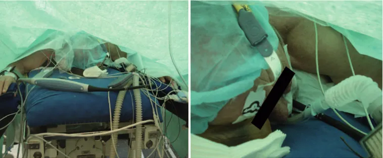
2. Change in peak velocity of blood flow ejected by the left ventricle into the aorta during a respiratory cycle, applying the following formula and considering as re-sponders those with Delta Vpeak greater than 12% 10: Picture 2 – Patient Undergoing Laminectomy and Lumbar Spine Fusion in the Prone Position, Monitored with TEE.
Figure 1 – Model segmentation recommended by the American He-art Association (AHA).
Four Chamber ME
Longitudinal ME AO
TG Transversal MP
TG Transversal Basal
Two Chamber ME
Basal segments 1. Basal anteroseptal
2. Basal anterior
3. Basal lateral
4. Basal posterior
5. Basal inferior
6. Basal septal
Mid segments 7. Mid anteroseptal
8. Mid anterior
9. Mid lateral
10. Mid posterior
11. Mid inferior
12. Mid septal
Apical segments 13. Apical anterior
14. Apical lateral
15. Apical inferior
16. Apical septal
Delta Vpeak (%) = 100 x (Vpeakmax – Vpeakmin) / [ (Vpeakmax+Vpeakmin) /2]
After performing all measures, the transducer was left in the transgastric position showing the transverse plane at the level of papillary muscle to intermittent monitoring of contrac-tility and ventricular volume. A new set of measurements was performed after the identification of ischemia or hemodynamic instability. In all cases, a final examination is performed before the patient leaves the operating room.
At the end of procedure, after examination, a note on the chart classified monitoring according to its usefulness in the intraoperative period into three groups: group 1, no interfer-ence of TEE in anesthetic or surgical approach; group 2, TEE prompted change in anesthetic approach regarding the administration of volume, introduction and/or modification of vasoactive drugs (here, TEE generated change of anesthetic approach in conjunction with other monitors, but it was the deciding factor); group 3, TEE led to a change in approach or review of surgical procedure performed.
RESULTS
From January 2009 to January 2011, 164 intraoperative TEE were performed in our service in 41 pediatric and 123 adult patients. In all patients, examination was successful and there were no problems regarding transesophageal probe insertion. Also, no serious complications arose from monitoring. Table I shows the demographic characteristics of patients included in the study.
All pediatric patients underwent cardiac surgery. Adult pa-tients underwent conventional cardiac surgery (n = 28), percu-taneous aortic valve replacement (n = 10), orthopedic surgery (n = 16), major abdominal surgeries (n = 66), vascular sur-gery via endovascular approach (n = 1), and neurosursur-gery in a seated position (n = 2). Table II shows the conditions found in children.
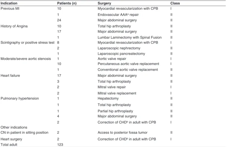
Table III shows the indications for monitoring and types of surgery performed in adults. Taking into account the indica-tion according to each class, 37 patients were classified as Class I and 86 patients as Class II.
Table I – Demographic Characteristics of Patients Pediatric Patients Adult Patients
Sex
Female 22 54
Male 19 69
Age (years) 8.29 ± 4.41 65.68 ± 19.07*
* Mean ± standard deviation
Table IV shows the division of patients according to TEE usefulness on final classification. In pediatric group (Table IV), intraoperative TEE had no interference in the anesthetic or surgical approach in 10 cases, only serving as a monitor; and in 27 cases, it was useful in patient’s hemodynamic manage-ment for adjustmanage-ment or modification of blood volume and ad-ministration of vasoactive drugs. The incidence of second car-diopulmonary bypass (CPB) prompted by TEE was 7.3%.
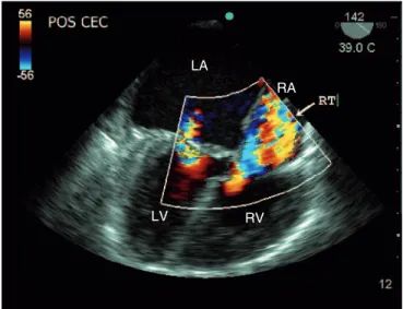
On four occasions, the use of TEE directly interfered in the surgical approach. First, a patient who underwent mitral valve repair has evolved with significant tricuspid insufficiency after weaned fromCPB, having to return to CPB for proper correc-tion (Figure 2). Second, a child who needed a residual atrial
Figure 2 – Four Chamber Cut Inverted with Multiplane angle of 142° Showing Right Chambers on the Right and Left Chambers on the Left of the Screen.
TR: tricuspidregurgitation; RA: right atrium, LA: left atrium, RV: right ventricle, LV: left ventricle.
Table II – Diseases Found in Children
ASD 15
Ostium secundum with PH 1 8
Ostium primum 3
Superior venous sinus 2
Superior venous sinus with ADPV 2 2
VSD 12
Tetralogy of Fallot 4
Isolated mitral cleft 1
Aortic coarctation + SAH 3 1
Tricuspid atresia 2
Mitral regurgitation 3
Aortic insufficiency 3
Total Children 41
1: Pulmonary Hypertension; 2: Anomalous Drainage of Pulmonary Veins; 3: Asymmetric Septal Hypertrophy.
LA
RA
Table III – Indications for Monitoring and Types of Surgery Performed in Adults
Indication Patients (n) Surgery Class
Previous MI 10 Myocardial revascularization with CPB I
1 Endovascular AAAa repair II
24 Major abdominal surgery II
History of Angina 10 Total hip arthroplasty II
17 Major abdominal surgery II
1 Lumbar Laminectomy with Spinal Fusion II Scintigraphy or positive stress test 8 Myocardial revascularization with CPB I
2 Laparoscopic nephrectomy II
1 Laparoscopic pancreatectomy II
Moderate/severe aortic stenosis 1 Aortic valve repair I
10 Percutaneous aortic valve replacement I 1 Conventional aortic valve replacement II
Heart failure 17 Major abdominal surgery II
3 Total hip arthroplasty II
2 Mitral valve repair I
2 Mitral valve replacement I
Pulmonary hypertension 1 Hepatectomy II
1 Total hip arthroplasty II
1 Partial hip arthroplasty II
4 Major abdominal surgery II
2 Correction of CHDb in adult with CPB I Other indications
CN in patient in sitting position 2 Access to posterior fossa tumor II
Heart surgery 2 Correction of CHDb in adult with CPB I
Total adult 123
a: abdominal aortic aneurysm; b: Congenital heart disease.
terfered with surgical procedure. The first case was a patient referred to us for elective repair of a presumed pulmonary valve stenosis previously diagnosed by transthoracic echocardiogra-phy. In this patient, intraoperative examination showed a sub-aortic ventricular septal defect (VSD) with right ventricle out-flow stenosis and normal pulmonary valve. The initial proposed surgical correction of stenosis was changed, and the surgery involved the VSD closure and infundibular widening (Figure 4). In the second case, an adult patient who underwent mitral valve repair returned to CPB immediately after the first attempt of weaning for surgical correction adjustment because the control TEE accused persistence of a moderate insufficiency after he-modynamic parameters normalization.
Considering only the cardiac surgery group in adults, a new information regarding the condition leading to surgery was add-ed in 7.1% of cases, which lead to change in surgical approach in 3.5% of cases. In the last two cases, patients with pulmonary hypertension underwent the initial proposed colon surgery by video-laparoscopy. Immediately after pneumoperitoneum in-sufflation, both patients evolved with significant right ventricular distention and severe hemodynamic deterioration, which lead to conversion to conventional surgery.
In two cases, TEE was fundamental in detecting poten-tially life-threatening situations and allowed us to take quick decisions. In the first case, a patient with PH underwent total
Table IV – Division of Groups According to TEE Intraoperative Usefulness
Group 1 Group 2 Group 3
Pediatric patients (41)
10 (24.4%) 27 (65.8%) 4 (9.8%)
Adult patients (123)
38 (30.9%) 81 (65.9%) 4 (3.2%)
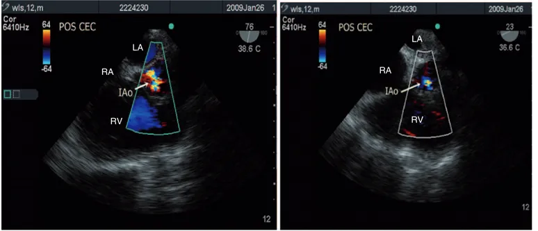
septal defect (ASD) to equalize left-right pressure returned to CPB to increase the ASD diameter. Third, one patient devel-oped significant right ventricular dysfunction after closure of the sternum; so, we opted for maintaining the sternotomy un-til RV function improvement, which occurred on the second postoperative day when he returned to the operating room for closure of his chest. Fourth, a patient who underwent repair of the aortic valve return to CPB for valve reapproachment, since residual aortic insufficiency was quantified as moderate, which was considered unacceptable as a final result (Figure 3).
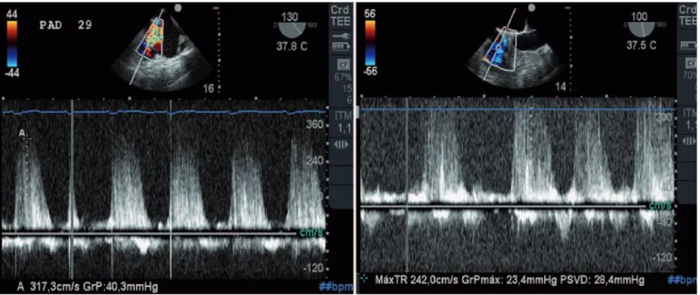
in-hip arthroplasty. At the end of femoral prosthesis component cement, she evolved with progressive increase in pulmonary pressure followed by cardiac arrest (CA). The differential diagnosis between the PH crisis and a possible pulmonary embolism (PE) could only be done because the pulmonary artery systolic pressure (PASP) assessment performed during monitoring showed the gradual increase of pressure, which reached a peak of 69 mm Hg. Resuscitation maneuvers were immediately initiated and, after restoration of spontaneous circulation, we chose the combination of nitric oxide with mil-rinone, which was already being administered from the onset, with excellent response. The patient was transferred to the
in-tensive care unit and extubated 48 hours later, without neuro-logical sequelae and evolving postoperatively without further complications. Below we have the assessment of PASP in two moments: before CA, reaching 69 mm Hg, and after clinical stabilization and installation of nitric oxide, now at lower levels (Figure 5).
The second situation with life-threatening potential oc-curred in a patient with a history of heart failure, coronary ar-tery disease, and restricted to bed for two days due to a femo-ral neck fracture. She underwent a partial hip arthroplasty and after the procedure progressed to cardiac arrest. In this case, diagnostic suspicion arrived from the presence of McConnell’
Figure 3 – Aortic Plan Short Axis with Multiplane Angle in 76° (left) showing the result of control echo after the first attempt of repair, with residual aortic insufficiency quantified as moderate and the same plan (right) showing a mild aortic regurgitation as final result.
Figure 4 – Aortic Plan Long Axis (left) showing the subaorctic VSD and short axis plan (right) showing the right ventricular outflow tract (RVOT). LA
RA
RV
LA
RA
RV
LA
LV
RV LVOT
RV
LA
RA
sign, which is an echocardiographic change characterized by abnormal contraction of the RV free wall, with normal contrac-tion of its apex, and has a 77% sensitivity and 94% specificity for PE diagnosis. Due to the characteristic of CA and patient’s critical situation, a new examination was requested on ICU ar-rival, and transthoracic echocardiography performed by spe-cialist confirmed the TEE finding.
One of the diagnoses made during the intraoperative he-modynamic monitoring of a patient with heart failure, who underwent major abdominal surgery, was not involved in the
proposed surgical procedure. The finding was a functional separation of the right atrium by an elongated Eustachian valve as shown in Figure 6.
The most challenging case in terms of monitoring was that of a patient who underwent lumbar laminectomy with spinal fusion the due to disabling pain. The indication in this case was unstable angina and, because of surgical positioning, TEE was performed throughout the procedure with the patient in the prone position (Figure 2).
Figure 5 – PASP estimation (left) before CA at 69 mmHg and after nitric oxide with lower value. PASP: pulmonaryartery systolic pressure; CA: cardiac arrest.
Figure 6A – Modified Four Chamber Cut Showing Right Atrium Divided by Elongated Eustachian Valve (white arrow).
Figure 6B – Functional Division View after Injection of Micro Bubbles Filling the Lower Portion of the Valve Limited by the Valve. LA
RA
LV
RV
DISCUSSION
The first important aspect identified after the study was how important it is a standardized and organized record of all data for each examination, as it was through these records that we could easily get all data needed to perform the work. In addi-tion, the digital storage of still images and films allowed us to resolve any doubts, either through review by our staff anes-thetist or the opinion of an echocardiographer, if necessary.
Although our sample is small compared to those published outside Brazil, we found similarities between the results, which suggest great utility of TEE.
In pediatric patients undergoing heart surgery, our inci-dence of second CPB prompted by TEE was 7.3%, which we consider not so far from the 8.5% reported by Bettex et al. 11 in a retrospective of 10 years experience specifically using TEE in pediatric heart surgery. Other case series 12,13 have reported up to 9.6% return to CPB prompted by TEE, which is also close to our results. The most important fact of our small case series is that after returning to CPB, in all three cases, significant surgical adjustment was required, which probably would need further surgery if they were not re-approached. In the patient whose chest was left open, in a subsequent analy-sis, the team thought this attitude was the most appropriate allowing the good outcome.
Regarding its use in adult heart surgery, the numbers are quite variable, and some published works have shown that in-traoperative TEE may add new information regarding disease that prompted operation in 10% to 40% of cases and that this new information may result in change of surgical procedure in 4% to 15% of cases 14,15,16. In our series, a new information regarding disease that prompted surgery was added in 7.1% of cases, which led to change in surgical procedure in 3.5% of cases. This difference may be explained by the small sample size of our study (28 patients), a number much lower than those reported in other studies 17,18.
Similar to the study by Schulmeyer et al. 19, we also classify the usefulness of intraoperative TEE according to change in the information obtained from the echo, but we included only three groups in our study. Perhaps that was the reason why 65.9% of our patients were allocated to group II in which ETE moti-vated change in anesthetic approach versus 48% in the above mentioned study in which four groups were considered.
The use of TEE in noncardiac surgery is still controversial in literature and there are no large randomized, controlled, multicenter or meta-analysis studies confirming or completely denying its usefulness in this type of surgery.
Denault et al. 20 published a paper showing that TEE was able to change the anesthetic-surgical procedure in up to 60% of cases in Class I patients, 31% in Class II, and in patients currently classified as Class III (i.e., those not eligible for in-traoperative TEE), 21% had benefits and changes in the man-agement of their cases. Similar results were obtained by other European studies that also associated the use of TEE with the same classification 21,22. Among our patients, most were clas-sified as Class II and almost all patients clasclas-sified as Class I underwent heart surgery, which did not allow us to make comparisons with these data.
Another fact observed, also reported elsewhere, is that TEE is more likely to produce decisive changes in case man-agement of patients with severe cardiovascular diseases undergoing noncardiac surgery. That became clear in cases of anesthesia in patients with pulmonary hypertension. In all cases, intraoperative TEE was essential regarding changes in management procedures.
In the patient undergoing total hip replacement, TEE has enabled us to gain a few precious seconds, recognizing the crisis of pulmonary hypertension, and eliminated the differ-ential diagnosis of pulmonary embolism. This occurred in another case, also during an orthopedic surgery, and was recognized by the McConnell sign. In other patients, the change in the indication of video laparoscopy to convention-al surgicconvention-al technique was only possible because TEE convention-also allowed the immediate diagnosis of acute right ventricular failure.
From this experience, we can also state that, although some information obtained depend on interpretation, most data that are important to the anesthesiologist comes from quantitative measurements performed on the basis of stan-dardized procedures that can be easily reproduced by another examiner in case of uncertainty.
Similar to other technologies that have been incorporated into the operating room routine over time, we believe that very soon TEE will also be part of the hemodynamic monitoring arsenal of Brazilian hospitals that perform surgeries of high complexity.
REFERENCES
1. Denault A, Couture P, McKenty S, et al. – Perioperative use of tran-sesophageal echocardiography by anesthesiologists: Impact in non-cardiac surgery and in the intensive care unit. Can J Anasth, 2002;49:287-294.
Anaesthesia 53:767-773, 1998.
3. Morewood GH, Gallagher ME, Gaughan JP et al. – Current practice patterns for adult perioperative transesophageal echocardiography in the United States. Anesthesiology, 2001;95:1507-1512.
4. Ramamoorthy C, Lynn AM, Stevenson JG – Pro: Transesophageal echocardiography should be routinely used during pediatric open car-diac surgery. J Cardiothorac Vasc Anesth, 1999;13:629-631. 5. Qaddoura FE, Abel MD, Mecklenburg KL et al. – Role of
intraopera-tive transesophageal echocardiography in patients having coronary artery bypass graft surgery. Ann Thorac Surg, 2004;78:1586-1590. 6. Hofer C, Zollinger A, Rak M et al. – Therapeutic impact of
intraope-rative transesophageal echocardiography during noncardiac surgery. Anaesthesia, 2004;59:3-9.
7. Suriani RJ, Neustein S, Shore-Lesserson L et al. – Intraoperative tran-sesophageal echocardiography during noncardiac surgery. J Cardio-thorac Vasc Anesth, 1998;12:274-280.
8. Shanewise J, Cheung A, Aronson S et al. – Practice guidelines for perioperative transesophageal echocardiography. Anesthesiology, 1996;84:986-1006.
9. Vieillard-Baron A, Chergui K, Rabiller A et al. – Superior vena caval collapsibility as a gauge of volume status in ventilated septic patients. Intensive Care Med, 2004;30(9):1734-9.
10. Feissel M, Michard F, Mangin I , Ruyer O, Faller JP, Teboul JL – Respiratory changes in aortic blood velocity as an indicator of fluid responsiveness in ventilated patients with septic shock. Chest, 2001;119:867-873.
11. Bettex DA, Prêtre R, Jenni R, Schmid ER – Cost-Effectiveness of Routine Intraoperative Transesophageal Echocardiography in Pediatric Cardiac Surgery: A 10-Year Experience. Anesth Analg, 2005;100:1271-1275.
12. Stevenson JG – Adherence to physician training guidelines for pediat-ric transesophageal echocardiography affects the outcome of patients undergoing repair of congenital cardiac defects. J Am Soc Echocar-diogr, 1999;12:165-172.
13. Roberson DA, Muhiudeen IA, Cahalan MK et al. – Intraoperative tran-sesophageal echocardiography of ventricular septal defect. Echocar-diography, 1991;8:687-697.
14. Jneid H, Bolli R – Inotrope use at separation from cardiopulmonary bypass and the role of pre bypass TEE. J Cardiothorac Vasc Anesth, 2004;8:401-403.
15. Hillel Z – Refining intraoperative echocardiography. J Cardiothorac Vasc Anesth, 2003;17:419-421.
16. Thys DM – Echocardiography and anesthesiology successes and challenges. Anesthesiology, 2001;95:1313-1314.
17. Fanshawe M, Ellis C, Habib S, Konstadt SN, Reich DL – A Retros-pective Analysis of the Costs and Benefits Related to Alterations in Cardiac Surgery from Routine Intraoperative Transesophageal Echo-cardiography. Anesth Analg, 2002;95:824-827.
18. Qaddoura FE, Abel MD, Mecklenburg KL et al. – Role of Intraoperati-ve Transesophageal Echocardiography in Patients Having Coronary Artery Bypass Graft Surgery. Ann Thorac Surg, 2004;78:1586-1590. 19. Schulmeyer MCC, Santelices E, Vega R, Schmied S – Impact of
In-traoperative Transesophageal Echocardiography During Noncardiac Surgery. J Cardioth Vasc Anest, 2006;20(6):768-771.
20. Denault A, Couture P, McKenty S et al. – Perioperative use of transesophageal echocardiography by anesthesiologists: Impact in noncardiac surgery and in the intensive care unit. Can J Anesth, 2002;49:287-294.
21. Patteril M, Swaminathan M – Pro: Intraoperative transesophageal echocardiography is of utility in patients at high risk of adverse cardiac events undergoing noncardiac surgery. J Cardiothorac Vasc Anesth, 2004;18:107-109.
