Multidetector-row computed tomography angiography
for the diagnosis of anomalous pulmonary venous
drainage: an initial experiment*
Angiotomografia com múltiplos detectores no diagnóstico de drenagem venosa pulmonar anômala: experiência inicial
Letícia Yurie Kimura1, Gustavo Spadaccia dos Santos Fernandes1, Karina Tavares de Melo Nóbrega1, Luiz Augusto Gadia Gabure1, César Augusto Mastrofrancisco Cattani2, José Pedro da Silva3, Beatriz Helena Sanches Furlanetto4
OBJECTIVE: We aimed to determine if multidetector-row computed tomography angiography is an effective and non-invasive method for diagnosing anomalous pulmonary venous drainage. MATERIALS AND METHODS: We performed a retrospective review of 2905 cases from July 2003 to November 2007 in which cardiac multidetector-row computed tomography was used. Of the cases, 393 evaluated congenital cardiopathy, with the others (2512) evaluating the coronary arteries. RESULTS: Anomalous pulmonary venous drainage was found in 21 cases, with 7 (33.3%) from total anomalous pulmonary venous drainage (3 supracardiac, 3 infracardiac and 1 cardiac), and 14 (66.7%) from partial anomalous pulmonary venous drainage. CONCLUSION: Multidetector-row computed tomography angiography can be useful for diagnosing the described anomalies because of its non-invasiveness, and its ability to evaluate intra- and extra-cardiac structures. The technique allows a thorough study of the thoracic anatomy and contributes to surgical conduct, consequently improving patient prognosis, in particular by allowing the diagnosis of clinically unsuspected malformations.
Keywords: Anomalous drainage; Pulmonary veins, Congenital abnormalities; Multidetector-row computed tomography.
OBJETIVO: Demonstrar que a angiotomografia computadorizada com múltiplos detectores é um método efetivo e não invasivo para o diagnóstico de drenagem venosa pulmonar anômala. MATERIAIS E MÉTODOS: Rea-lizou-se estudo retrospectivo de 2905 angiotomografias computadorizadas com múltiplos detectores cardía-cas realizadas em nosso serviço no período de julho de 2003 a novembro de 2007. Destas, 393 foram des-tinadas para avaliar cardiopatias congênitas e as 2.512 restantes, para analisar as artérias coronárias. RE-SULTADOS: Encontraram-se 21 casos de drenagem venosa pulmonar anômala, sendo 7 (33,3%) do tipo total (3 do tipo supracardíaco, 3 do tipo infracardíaco, 1 do tipo cardíaco) e 14 (66,7%) do tipo parcial. CONCLUSÃO: A angiotomografia computadorizada com múltiplos detectores tem demonstrado fundamen-tal importância no diagnóstico destas anomalias, principalmente por tratar-se de um método não invasivo capaz de analisar estruturas intra e extracardíacas e por permitir um estudo completo da anatomia torácica, contribuindo sobremaneira na conduta cirúrgica e, consequentemente, no prognóstico destes pacientes, es-pecialmente por diagnosticar malformações não suspeitadas clinicamente.
Unitermos: Drenagem anômala; Veias pulmonares; Anomalias congênitas; Tomografia computadorizada multidetectores.
Abstract
Resumo
* Study developed at the Unit of Cardiac Radiology of Med Imagem, Real e Benemérita Associação Portuguesa de Benefi-cência de São Paulo, São Paulo, SP, Brazil.
1. MDs, Unit of Cardiac Radiology of Med Imagem, Real e Benemérita Associação Portuguesa de Beneficência de São Paulo, São Paulo, SP, Brazil.
2. PhD, MD, Unit of Cardiac Radiology of Med Imagem, Real e Benemérita Associação Portuguesa de Beneficência de São Paulo, São Paulo, SP, Brazil.
3. Surgeon-in-Chief, Unit of Cardiac Surgery, Real e Benemé-rita Associação Portuguesa de Beneficência de São Paulo, São Paulo, SP, Brazil.
4. Physician Assistant at Unit of Cardiac Surgery, Real e
Be-known etiology that manifests as a devel-opment failure in the pulmonary veins. It represents less than 1% of all congenital heart defects. APVD is defined as the drain-age of one or more pulmonary veins from the left atrium, with direct or indirect pul-monary venous return into the right atrium(1–4).
APVD is classified as either total APVD, in which all four pulmonary veins
Kimura LY, Fernandes GSS, Nóbrega KTM, Gabure LAG, Cattani CAM, Silva JP, Furlanetto BHS. Multidetector-row com-puted tomography angiography for the diagnosis of anomalous pulmonary venous drainage: an initial experiment. Radiol Bras. 2010;43(6):347–353.
nemérita Associação Portuguesa de Beneficência de São Paulo, São Paulo, SP, Brazil.
Mailing address: Dra. Letícia Yurie Kimura. Rua Maestro Car-dim, 476, ap. 83, Bela Vista. São Paulo, SP, 01323-000, Bra-zil. E-mail: [email protected]
Received February 10, 2010. Accepted after revision May 12, 2010.
INTRODUCTION
un-drain out of the left atrium, or partial APVD, in which one to three pulmonary veins drain either into a systemic vein or into the right atrium.
The anomalous pulmonary venous con-nection in the systemic venous circuit has been known in its partial form since 1739, and in its total form, since 1798. However, variations in pulmonary venous connection are rarely observed, occurring in 0.4% of autopsy specimens(5).
In the general population, total APVD is a rare condition, but it is frequently found in patients at institutions that specialize in congenital heart defects(4–7).
According to Darling, Rothney and Craig, total APVD is anatomically classi-fied into four types. In the supracardiac type, drainage is through a vertical vein, generally into the left innominate vein. This is the most frequent type, accounting for 40% to 50% of cases. In the cardiac type, drainage is directly into the right atrium or into the coronary sinus (20%). In the infracardiac type, drainage is through a vertical vein into the portal vein or into the inferior vena cava. This occurs in 10%-20% of cases. The mixed type represents about 10% of cases and is a combination of two or more of the above mentioned types. Association with atrial septal defect is always observed in all types of total APVD, since the absence of this defect with total APVD is incompatible with sur-vival(4,6,7).
Until recently, APVD diagnosis was made using echocardiography and conven-tional angiography. The main disadvan-tages of echocardiography are operator dependency, dependence on the patient’s acoustic window, and difficulty in evalu-ating extracardiac structures, particularly pulmonary venous drainage into the left atrium because of posterior positioning. The presence of a real communication be-tween the pulmonary veins and the left atrium is often in doubt. The main disad-vantages of conventional angiography is its invasiveness and higher risks for compli-cations.
This study aimed to determine if multi-detector-row computed tomography an-giography (MCTA) is an effective, non-invasive method for the diagnosis of APVD.
MATERIALS AND METHODS
All cardiac MCTA studies were per-formed with a 16-row Aquilion (Toshiba Medical Systems; Tokyo, Japan) or a 64-row Light Speed VCT (GE Healthcare; Milwaukee, WI, USA), from July 2003 to November 2007 were retrospectively re-viewed, and a database was constructed. Our database showed 2905 cardiac MCTA studies performed from July 2003 to No-vember 2007. These were divided accord-ing to their original objective into 393 stud-ies intended to evaluate congenital cardio-pathies, and 2512 intended to evaluate coronary arteries.
Almost all studies that evaluated con-genital cardiopathies were performed un-der anesthesia, except for those where the patient could cooperate with apnea. Stud-ies performed under anesthesia required prior fasting for 6 hours for patients solely fed by breastfeeding, or 8 hours for patients on other diets. For patients who did not require anesthesia, a 4-hour fast was re-quested.
All studies were retrospectively syn-chronized to electrocardiograms with two acquisitions, one arterial phase, and one venous phase, with an iodinated contrast (Optiray 320, Mallinckrodt; St. Louis, MO, USA) injection at a dose of 2 ml/kg, fol-lowed by a dose of 2 ml/kg of 0.9% saline solution, both in bolus. Beta-blocker drugs were not required in cases of suspected congenital cardiopathy.
In children weighing < 10 kg, peripheral venous access was obtained with a Jelco 24 or 22 catheter with the entire contrast me-dium volume manually injected with a 10 ml syringe, followed by saline solution. The first-phase acquisition was initiated when half the contrast medium volume was injected, followed by the second-phase acquisition with no waiting period, always under apnea supported by the anesthetist. In patients weighting > 10 kg, an injection pump was used, with peripheral injection at 2 ml/s into a Jelco 22 catheter, with the first scan initiated as half the volume of contrast medium was injected, also under apnea. For adult patients who could coop-erate with inspiratory apnea, an injection pump with 4 ml/s injection was used to achieve a total dose of 1.2 ml/kg of contrast
medium injected through peripheral venous access with a Jelco 20 or 18 cath-eter, followed by 40 ml of saline solution injected at the same rate, monitoring con-trast uptake in the left atrium, with the two scans initiated with a six-second respiratory interval.
All MCTA reports on congenital cardio-pathies were evaluated, and all cases of APVD were recorded. Reports were pre-pared by professionals with four-, two- and one-year of experience in MCTA, and all diagnoses were surgically confirmed and had clinical follow-up to monitor patient survival.
RESULTS
Of the 393 MCTA studies that evaluated congenital cardiopathies, 21 cases of APVD were observed, with 7 (33.3%) cases of total APVD, and 14 (66.7%) of partial APVD. No cases of APVD were found in the cardiac studies performed to evaluate coronary arteries.
Fifteen patients were men (71%) and six were women (29%), with ages ranging from 7 days to 35 years (mean 3.3 years, standard deviation 8.9 years). Anomalous pulmonary venous drainage was found in only two adult patients (24 and 35 years). Of the total APVD cases, three were supracardiac, three infracardiac, and one cardiac. All are in Table 1 with cases of partial APVD.
Supracardiac-type total APVD was seen in two cases in which four pulmonary veins drained into a vertical vein that drained into the left brachiocephalic vein. In another case, the left superior pulmonary vein drained directly into the left persistent su-perior vena cava, while the other pulmo-nary veins drained into a collector tube which also drained into the left persistent superior vena cava.
Infracardiac-type total APVD was found in patients in whom four pulmonary veins drained into a vertical vein that drained into the portal vein.
Cardiac-type APVD was characterized by four pulmonary veins draining directly into the right atrium.
cases presented with abnormality on both sides (28.6%).
The partial APVD cases presented with a high variation in drainage types (Table 1). Nine deaths (42.8%) were recorded, eight (88.9%) in the first six months of life. The other death occurred in a two-year-old child. Of the patients who progressed to death, three had total APVD (33.3%), and the other had partial APVD.
Figures 1 to 5 demonstrate the main types of APVD found at the authors’ insti-tution.
DISCUSSION
Although APVD is a rare congenital cardiac anomaly, with a prevalence of <1% reported in the literature, we observed a prevalence of 5.34%, probably because the study was conducted at a center of refer-ence for congenital cardiopathies. In agree-ment with other studies, we found the prevalence of APVD to be higher at spe-cialized institutions(1–6).
Most patients with partial APVD are asymptomatic, and the condition is inciden-tally diagnosed after chest radiography or computed tomography(5,7–10). The method of choice for diagnosis is transthoracic Table 1 Distribution of anomalous pulmonary venous drainage types diagnosed by multidetector-row
computed tomography angiography for evaluation of congenital cardiopathies.
Patient
T.O.S. T.H.A. A.B.B. V.H.R.V.S.
E.S.B. A.S.S.
T.H.F.S. K.L.R.V. A.C.C. S.B.S.P. E.C.A.A. M.M.S. A.A.B.S.B.
N.C.S. L.R.S. P.H.R.S.
N.M.C. D.A.S.C.
J.J.A.
G.M.R. P.L.G.O.
Sex
Female Male Male Male Male Male
Male Male Male Female Female Male Female
Male Male Male Female Female Male
Male Male
Age in months
24 10 2 2 5 48
1 3 1 0.3333 0.2333 288
5
420 0.1333
12 6 0.0666
24
0.2333 3
Anomalous pulmonary venous drainage
RIPV–RA LSPV–coronary sinus Total APVD: vertical vein–portal vein
LIPV–RA; RPVs–RA
Total APVD: vertical vein–left brachiocephalic vein RSPV–SVC; RIPV–RA/LA; LSPV–left vertical vein
RPVs–IVC
Total APVD: LSPV–LSVC; others, vertical vein–LSVC LSPV–left vertical vein
RSPV–SVC RPVs–RA
LPVs–left brachiocephalic vein RPVs–SVC
Total APVD: vertical vein–left brachiocephalic vein Total APVD: vertical vein–portal vein
RSPV–RA; RIPV–RA/LA RIPV–IVC; LIPV–left hepatic vein
RPVs–SVC; LSPV–LSVC Total APVD: RPVs and LPVs–RA
Total APVD: vertical vein–portal vein RSPV–SVC
RIPV, right inferior pulmonary vein; RA, right atrium; LSPV, left superior pulmonary vein; APVD, anomalous nary venous drainage; LIPV, left inferior pulmonary vein; RPVs, right pulmonary veins; RSPV, right superior pulmo-nary vein; SVC, superior vena cava; RA/LA, right atrium/left atrium; IVC, inferior vena cava; LSVC, persistent left superior vena cava; LPVs, left pulmonary veins.
Figure 1. Multiplanar reconstruction of MCTA (A) A male, one-month-old patient, demonstrating partial anomalous pulmonary venous drainage with right pulmonary veins draining into the inferior vena cava in the shape of a curved “Turkish sword” (scimitar), confirmed by conventional angiography (B).
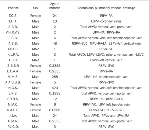
Figure 2. Multiplanar (A) and three-dimensional (3D) (B) re-construction of MCTA in a male, 35-year-old patient sub-mitted to examination because of suspicion of a mediastinal tumor, demonstrating a total anomalous pulmonary venous connection represented by all the four veins draining into the vertical vein that drains into the left brachiocephalic vein, to-wards the superior vena cava.
A B
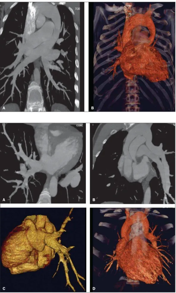
Figure 3. Multiplanar (A,B) and 3D (C,D) reformation of MCTA in a male, 24-year-old patient with partial anomalous pulmonary venous drainage in which the right pulmonary veins drain into the left atrium and the left pulmonary veins drain into the vertical vein towards the left brachiocephalic vein.
D C
Figure 4. Multiplanar reconstruction of MCTA in a female, 6-month-old patient with partial anomalous pulmonary venous drainage, demonstrating that the right inferior pulmonary vein drains into the inferior vena cava (A) and the left inferior pulmonary vein drains into the left hepatic vein (B,C).
A B C
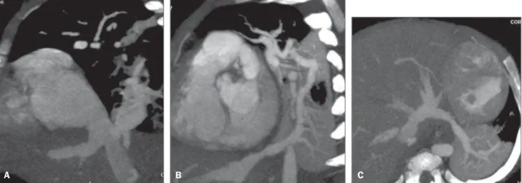
Figure 5. Multiplanar (A,C) and 3D (B,D) reformatation of the MCTA in a male, 7-day-old patient, demonstrating total anomalous pulmonary venous drainage, with all pulmonary veins draining into a vertical vein that drains into the portal vein.
A
B
echocardiography or by conventional an-giography, but these have some disadvan-tages. These include operator dependency, dependence on the patient’s acoustic win-dow, and difficulty in evaluating extracar-diac structures, particularly the pulmonary venous drainage with its posterior position-ing. Another disadvantage is invasiveness, with higher risks for death, especially in patients with obstructed APVD(10–12). Mag-netic resonance imaging has also been used to diagnose APVD(10,13–15), but has not been adopted at the authors’ institution because of the difficulty in monitoring in the exami-nation room. This method also has lower spatial resolution for patients weighing less than 10 kg.
Computed tomography angiography is a valuable method for evaluating several vascular alterations in adults, particularly in cases of aortic conditions such as aneu-rysms and dissections(16–18). The main dis-advantages of this method are the necessity for apnea, requiring general anesthesia of pediatric patients, and the use of iodinated contrast media and ionizing radiation, which is similar to conventional angiogra-phy. In 2000, Kim et al. reported the appli-cation of helical computed tomography angiography in neonates and children with total APVD, which had not been studied before. However, those examinations were performed without apnea(12).
We found no studies that used MCTA for diagnosing APVD, and only one study using magnetic resonance imaging. Other studies used magnetic resonance imaging to evaluate congenital abnormalities in general(10,13–15).
In this study, the majority of cases were partial APVD, which is consistent with reports in the literature, and the consider-ation that most patients with total APVD die in their first years of life. The patients in our study were not asymptomatic, and multidetector-row CT angiography had been requested with a precise indication for preoperative evaluation, since most pa-tients presented with the disease in asso-ciation with severe congenital cardiopa-thy(5,7–9,10).
Patients with total APVD rarely survive to adulthood, and this abnormality almost always requires emergency neonatal
sur-gery, as soon as the diagnosis is made, to avoid irreversible pathological alterations in the pulmonary vascular bed. In adult-hood, patients may present with fatigue or dyspnea with stress because of the blood volume overload produced by the left-to-right shunt(5,7–10,19–23). The two adult pa-tients were found in this study to be asymp-tomatic. One had total APVD and the other had partial APVD. The latter underwent tomography because of suspicion of a me-diastinal tumor from enlargement of the mediastinum observed on a chest radio-graph.
In cases of partial APVD, the right side is usually more involved than the left side with a ratio of 2:1 to 10:1. In cases of left-side APVD, the pulmonary veins generally demonstrate venous return to a vertical vein that drains into the left brachiocephalic vein, with the typical “snowman” sign on radiography and/or computed tomogra-phy(8–10). Our data corroborate the previous findings. Seven (50%) of the partial APVD patients presented with involvement of only the right side, and four demonstrated partial APVD in both sides (28.6%). Only three (21.4%) presented with partial APVD isolatedly involving the left side. Partial APVD may lead to hemodynamic changes such as blood volume overload in the left ventricle and pulmonary circulation, result-ing in congestive heart failure. Addition-ally, systemic and pulmonary venous blood mixing occurs in the right atrium, leading to arterial desaturation(5,7,8,19–23).
Total APVD is incompatible with life in the absence of a coexisting interatrial com-munication that allows the passage of oxy-genated blood into the left heart chambers. More than 75% of children with total APVD die in the first year of life (7,8,19–23). In this study, nine patients died, eight in the first months of life (88.89%), consistent with reports in the literature. Of the patients who died, three presented with total APVD (33.33%).
The first successful surgery for repair of total APVD was performed in 1951. Since then, surgical mortality rates have de-creased, but remain high, particularly in patients younger than three years of age who have severe pulmonary hypertension or pulmonary venous obstruction that
de-termines preoperative hemodynamic insta-bility(5,7).
Early diagnosis of APVD and its imme-diate repair are of paramount importance for the prognosis of these children, consid-ering that clinical and hemodynamic dete-rioration worsens the surgical outcomes in these patients(5). Of the total APVD cases in this series, four occurred in patients who were younger than five months of age, one was in a four-month-old patient, and an-other was in a 35-year-old patient. The lat-ter represents a rare case report in the lit-erature.
Surgical treatment consists of the rein-sertion of the four pulmonary veins into the left atrium, and is indicated as soon as the diagnosis is made, to prevent hemody-namic changes and lesions on the pulmo-nary vascular bed, which generally occur in the first years of life(1–7,9,11,12,19–23).
Occlusion of a catheterized or central vein is a frequent complication, occurring in approximately 23% of patients submit-ted to central venous access procedures. Symptomatic occlusion occurs in 1% to 4% of cases, presenting with sudoresis, pain, and superior vena cava syndrome that can be managed with a combination of throm-bolysis angioplasty and stent implanta-tions(8–10). As previously described, this study neither analyzed postoperative com-plications nor causes of death. This topic deserves further investigation in future studies.
In spite of requiring anesthesia, the use of iodinated contrast medium and ionizing radiation with MCTA is useful for diagnos-ing APVD because of its non-invasiveness, and the ability to evaluate intra- and extra-cardiac structures. This allows a thorough study of the thoracic anatomy and makes a positive contribution to the surgery, and improves patient prognosis. In particular, it allows the diagnosis of clinically unsus-pected malformations.
CONCLUSION
In spite of being rare, anomalous pul-monary venous drainage can be lethal un-less it is diagnosed early. MCTA is a highly accurate and non-invasive method for di-agnosing this condition. As an alternative to other, more invasive diagnostic methods, it can reduce costs and the morbimortality of APVD patients.
Acknowledgements
We thank Valdomiro Ferreira França for the support in the editing and finishing of the images.
REFERENCES
1. Geva T, Van Praagh S. Anomalies of the pulmo-nary veins. In: Allen H, Gutgesell Clark E, Driscoll D, editors. Moss and Adams’ heart dis-ease in infants, children, and adolescent. Phila-delphia, PA: Lippincott Williams & Wilkins; 2001. p. 736–72.
2. Lupinetti FM, Kulik TJ, Beekman RH 3rd, et al. Correction of total anomalous pulmonary venous connection in infancy. J Thorac Cardiovasc Surg. 1993;106:880–5.
3. Reitz BA, Yuh DD. Cyanotic defects. In: Reitz BA, Yuh DD, editors. Congenital cardiac surgery. New York, NY: McGraw-Hill; 2002. p. 148–52.
4. Miller SW. Congenital heart disease. In: Miller SW, editor. Cardiac imaging: the requisites. Phila-delphia, PA: Elsevier Mosby; 2005. p. 316–23.
5. Albert D, GironaJ, BonjochC, et al. Retorno
ve-noso pulmonar total anómalo en pediatría: impor-tancia del diagnóstico ecocardiográfico y de la cirugía precoz. Rev Esp Cardiol. 2000;53:810–4. 6. Raisher BD, Grant JW, Martin TC, et al. Complete repair of total anomalous pulmonary venous con-nection in infancy. J Thorac Cardiovasc Surg. 1992;104:443–8.
7. Behrendt DM, Aberdeen E, Waterson DJ, et al. Total anomalous pulmonary venous drainage in infants. I. Clinical and hemodynamic findings, methods, and results of operation in 37 cases. Circulation. 1972;46:347–56.
8. Levy JM, Smyth SH. Partial anomalous pulmo-nary venous return: iatrogenic occlusion of the innominate vein producing right-to-left shunt. J Vasc Interv Radiol. 2002;13:423–5.
9. Cooley DA, Collins HA. Anomalous drainage of entire pulmonary venous system into left innomi-nate vein: clinical and surgical considerations. Circulation. 1959;19:486–95.
10. Masui T, Seelos KC, Kersting-Sommerhoff BA, et al. Abnormalities of the pulmonary veins: evaluation with MR imaging and comparison with cardiac angiography and echocardiography. Radiology. 1991;181:645–9.
11. Vicente WVA, Dias-da-Silva PS, Vicente LM, et al. Correção cirúrgica de drenagem venosa pulmo-nar anômala total em adulto. Arq Bras Cardiol. 2006;87:e172–5.
12. Kim TH, Kim YM, Suh CH, et al. Helical CT angiography and three-dimensional reconstruc-tion of total anomalous pulmonary venous con-nections in neonates and infants. AJR Am J Roentgenol. 2000;175:1381–6.
13. Didier D, Higgins CB, Fisher MR, et al. Congeni-tal heart disease: gated MR imaging in 72 pa-tients. Radiology. 1986;158:227–35.
14. Choe YH, Lee HJ, Kim HS, et al. MRI of total
anomalous pulmonary venous connections. J Comput Assist Tomogr. 1994;18:243–9.
15. White CS, Baffa JM, Haney PJ, et al. MR imag-ing of congenital anomalies of the thoracic veins. Radiographics. 1997;17:595–608.
16. Chung JW, Park JH, Im JG, et al. Spiral CT an-giography of the thoracic aorta. Radiographics. 1996;16:811–24.
17. Murayama S, Hashiguchi N, Murakami J, et al. Helical CT imaging of bronchial arteries with curved reformation technique in comparison with selective bronchial arteriography: preliminary report. J Comput Assist Tomogr. 1996;20:749– 55.
18. Kopecky KK, Gokhale HS, Hawes DR. Spiral CT angiography of the aorta. Semin Ultrasound CT MR. 1996;17:304–15.
19. Atik FA, Irun PE, Barbero-Marcial M, et al. [To-tal anomalous drainage of the pulmonary veins – surgical therapy for the infradiaphragmatic and mixed anatomical types]. Arq Bras Cardiol. 2004; 82:259–63.
20. Serraf A, Belli E, Roux D, et al. Modified supe-rior approach for repair of supracardiac and mixed total anomalous pulmonary venous drainage. Ann Thorac Surg. 1998;65:1391–3.
21. Cope JT, Banks D, McDaniel NL, et al. Is verti-cal vein ligation necessary in repair of total anomalous pulmonary venous connection? Ann Thorac Surg. 1997;64:23–9.
22. Shah MJ, Shah S, Shankargowda S, et al. L–R shunt: a serious consequence of TAPVC repair without ligation of vertical vein. Ann Thorac Surg. 2000;70:971–3.