Peripartum Cardiomyopathy Treatment with
Dopamine Agonist and Subsequent Pregnancy
with a Satisfactory Outcome
Tratamento da miocardiopatia periparto com agonista
dopaminérgico e subsequente gestação com resultado
satisfatório
Maria Adélia Medeiros e Melo
1Jordão Sousa Carvalho
1Francisco Edson de Lucena Feitosa
1Edward Araujo Júnior
2Alberto Borges Peixoto
2Francisco Herlânio Costa Carvalho
1Regina Coeli Marques Carvalho
11Fetal Medicine Service, Maternidade-Escola Assis Chateaubriand, Universidade Federal do Ceará, Fortaleza, CE, Brazil
2Department of Obstetrics, Escola Paulista de Medicina, Universidade Federal de São Paulo, São Paulo, SP, Brazil
Rev Bras Ginecol Obstet 2016;38:308–313.
Address for correspondence Edward Araujo Júnior, PhD, Escola Paulista de Medicina, Universidade Federal de São Paulo (EPM-Unifesp), Rua Napoleão de Barros, 875, Vila Clementino, 04024-002, São Paulo, SP, Brazil (e-mail: [email protected]).
Keywords
►
peripartum
cardiomyopathy
►
congestive heart
failure
►
dopamine agonists
►
pregnancy outcome
Abstract
Pathophysiological mechanisms of peripartum cardiomyopathy are not yet completely
de
fi
ned, although there is a strong association with various factors that are already
known, including pre-eclampsia. Peripartum cardiomyopathy treatment follows the
same recommendations as heart failure with systolic dysfunction. Clinical and
experi-mental studies suggest that products of prolactin degradation can induce this
cardiomyopathy. The pharmacological suppression of prolactin production by D2
dopamine receptor agonists bromocriptine and cabergoline has demonstrated
satis-factory results in the therapeutic response to the treatment. Here we present a case of
an adolescent patient in her
fi
rst gestation with peripartum cardiomyopathy that
evolved to the normalized left ventricular function after cabergoline administration,
which was used as an adjuvant in cardiac dysfunction treatment. Subsequently, despite
a short interval between pregnancies, the patient exhibited satisfactory progress
throughout the entire gestation or puerperium in a new pregnancy without any cardiac
alterations. Dopamine agonists that are orally used and are affordable in most tertiary
centers, particularly in developing countries, should be considered when treating
peripartum cardiomyopathy cases.
received
January 16, 2016
accepted
May 3, 2016
DOI http://dx.doi.org/ 10.1055/s-0036-1584567.
ISSN 0100-7203.
Introduction
Peripartum cardiomyopathy (PPCM) is a dilated cardiomy-opathy that clinically manifests with systolic dysfunction of the left ventricle (LV) and ejection fraction (EF) of<45%. In the absence of other causes of heart failure, PPCM most often occurs at the end of pregnancy or in the months following childbirth, and it is, therefore a diagnosis of exclusion.1The incidence of PPCM varies depending on the geographical area. The highest incidences of PPCM are found in studies from South Africa (1 in 1000) and Haiti (1 in 400).2,3In the United States, it is estimated that PPCM incidence is 1 case for every 3200 live births,4,5 and the disease more frequently occurs in African–American women.6,7
Pathophysiological mechanisms of PPCM remain unclear, although there is a strong association with several well-known factors, such as malnutrition, poor selenium intake, multiparity, multiple pregnancies, advanced maternal age (> 35 years), viral myocarditis, autoimmune response, genetic susceptibility, and, more recently, association with pre-eclampsia.8–11
PPCM treatment follows the same recommendations as heart failure with systolic dysfunction; the treatment in-cludes angiotensin-converting enzyme (ACE) inhibitors,β -blockers, systemic vasodilators, and diuretics, which im-prove symptoms. In some cases, prognosis remains uncer-tain, with mortality rates varying from 4 to 30%.8,12,13 Recovery of myocardial function occurs in only 23–32% of PPCM cases, and 25% of cases evolve into the progressive deterioration of the disease, in which only cardiac transplant remains as the last recourse for patient survival.5,14,15
Clinical and experimental studies suggest that the
products of prolactin degradation can induce
PPCM.15–20The increase in oxidative stress during preg-nancy may trigger the activation of proteinase cathepsin D, which cleaves the 23-kDa molecular weight prolactin into smaller fragments, generating a 16-kDa prolactin
subfragment. This prolactin subfragment causes an angio-genic imbalance with the breaking down of capillaries and endothelial damage, along with inhibiting vasodilatory factors, such as nitric oxide, culminating in damaging cardiomyocytes, dilating cardiac chambers, and conse-quent myocardial dysfunction. The pharmacological sup-pression of prolactin production by agonist of D2 dopamine receptor, bromocriptine or cabergoline has demonstrated satisfactory results in the therapeutic re-sponse to PPCM treatment.17,21–23
Here we present a case of a pregnant woman with PPCM who progressed into the normalization of the left ventricular function after being administered cabergoline, and who subsequently presented a satisfactory progress throughout the entire gestation or puerperium in a new pregnancy without exhibiting any cardiac alterations.
Case Report
A 16-year old mixed-race female, who was in her first pregnancy with a gestational age of 36 weeks and 4 days that was estimated by the date of her last menstrual period, was admitted to the emergency ward of the Maternidade-Escola Assis Chateaubriand. She reported that 24 hours prior to that, she sought care at an emergency obstetrics clinic with complaints of dyspnea, cough with hemoptoic sputum and fever. She had no history of heart disease or any similar clinical condition. According to the referral report, at the time of examination, her blood pressure (BP) was 160/ 120 mm Hg; thus, hydralazine was intravenously adminis-tered (a total of 20 mg) along with magnesium sulfate to treat the clinical suspicion of severe pre-eclampsia.
At the time of admission, the patient exhibited tachypnea with a respiratory rate of 28 breaths/min, temperature of 37.2°C, and BP of 130/80 mm Hg. Pulmonary auscultation revealed reduced breath sounds at the bases of the lungs, particularly on the right side, with the presence of crackling
Resumo
Os mecanismos
fi
siopatológicos da miocardiopatia periparto ainda não são totalmente
de
fi
nidos, apesar de haver forte associação com vários fatores já conhecidos, incluindo
a pré-eclâmpsia. O tratamento segue as mesmas recomendações para a insu
fi
ciência
cardíaca com disfunção sistólica. Estudos clínicos e experimentais recentes sugerem
que os produtos de degradação da prolactina podem induzir a miocardiopatia. A
supressão farmacológica da produção de prolactina por agonista do receptor D2 da
dopamina, bromocriptina ou cabergolina, vem demonstrando resultados satisfatórios
na resposta terapêutica do tratamento. Apresentamos o relato de uma primigesta,
adolescente, com miocardiopatia periparto que evoluiu para a normalização da função
ventricular esquerda após a administração da cabergolina, utilizada como adjuvante na
terapêutica da disfunção cardíaca. Subsequentemente, apesar do intervalo entre as
gestações ser considerado curto, apresentou evolução satisfatória em uma nova
gestação sem qualquer alteração cardíaca durante todo o período gestacional ou
puerpério. Os agonistas dopaminérgicos, drogas de uso oral e de preço acessível para a
maioria dos centros terciários, em particular em países subdesenvolvidos, não podem
ser esquecidos frente a casos de miocardiopatia periparto.
Palavras-chave
►
miocardiopatia
periparto
►
falência cardíaca
congestiva
►
agonistas
dopaminérgicos
►
resultado da
gravidez
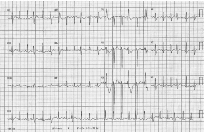
rales. Cardiac auscultation revealed regular heart rhythm without murmurs and a heart rate (HR) of 132 bpm. Obstetric examination revealed a gravid uterus that was compatible with the gestational age, and fetal heartbeat was 154 bpm. Electrocardiogram (ECG) revealed sinus tachycardia (►Fig. 1). Laboratory test results were as follows: hemoglo-bin (Hb), 9.5 g/dL; hematocrit (Ht), 30.3%; platelet count, 314,000/mm3; urea, 35 mg/dL; creatinine, 1.4 mg/dL; sodi-um, 132 mmol/L; potassisodi-um, 2.9 mmol/L; magnesisodi-um, 3.7 mg/L; chloride, 101 mmol/L; alanine aminotransferase, 2.0 U/L; aspartate aminotransferase, 12 U/L; total bilirubin, 2.37 mg/dL; direct bilirubin, 1.97 mg/dL; indirect bilirubin, 0.4 mg/dL; and uric acid, 6.2 mg/dL. Test results of arterial blood gases were pH, 7.2; pCO2, 44.1 mm Hg; pO2, 411.21 mm Hg; BE, 7.3; anion gap, 10.3 mmol/L; glutamate, 130 mg/dL; and lactate, 1.37.
With respect to the clinical data and laboratory tests, a diagnosis of severe pre-eclampsia with acute pulmonary edema was made, and the interruption of gestation via abdominal delivery under general anesthesia was indicated along with posterior investigation of possible maternal cardiopathy.
In the immediate postoperative period, the patient was transferred to the intensive care unit (ICU). When admitted to the ICU, the patient was still on mechanical ventilation (MV), with 50% fraction of inspired oxygen with a sedation of 03 on the Ramsey scale. Overall, the patient’s condition was compromised; she was afebrile, with HR of 128 bpm, BP of 94/56 mm Hg, and mean arterial pressure of 68 mm Hg.
Heartbeat was regular over three periods, with the third heart sound present.
The patient remained sedated and was administered a maintenance dose for sulfate therapy. A chest X-ray was taken, demonstrating lung congestion and increased cardio-thoracic ratio (►Fig. 2). ECG revealed sinus tachycardia, HR of 100 bpm, and 600° axis, with a slow R-wave progression from Fig. 1 Electrocardiogram taken on admission showing sinus tachycardia.
V1 to V4; transthoracic Doppler echocardiogram revealed the presence of a diffuse hypokinesia, systolic dysfunction, and left ventricular ejection fraction (LVEF) of 40%. With this additional data, continuous infusion of furosemide, 5 mcg/ kg/min dobutamine, and 10 mcg/min nitroglycerin was initiated.
On the 3rd day of hospitalization, the patient remained on MV and exhibited fever spikes (39.5°C) that were accompanied with thick yellow secretions from the air-ways; subsequently, antibiotic therapy was initiated with piperacillin sodium and tazobactam sodium. After thera-py, laboratory test results were as follows: Hb, 7.3 g/dL; Ht, 23.7%; platelet count, 372,000/mm3; urea, 39 mg/dL; creatinine, 1.0 mg/dL; sodium, 156 mmol/L; potassium, 4.0 mmol/L; and magnesium, 1.9 mg/L. The patient sud-denly exhibited severe ventricular arrhythmia with several episodes of sustained ventricular tachycardia (SVT), which satisfactorily responded to an intravenous administration of 300 mg amiodarone (►Fig. 3). Despite continuing 5 mcg/kg/min amiodarone, ventricular ar-rhythmia persisted with several repeated episodes of non-SVT, which only returned to a sinus rhythm after an intravenous administration of 2% (1 mg/kg) lidocaine that was maintained at 4 mg/kg/min for 72 hours.
Another transthoracic Doppler echocardiogram was per-formed on the 11th day of hospitalization and revealed similarfindings to the previous examination (LVEF of 40%); however, the chest X-ray demonstrated improvement in lung congestion. On the 13th day of hospitalization, the patient’s clinical condition had sufficiently improved to indicate re-moval from MV. However, this was unsuccessful because the
patient presented laryngeal stridor and required re-intuba-tion followed by tracheostomy. On the 25th day, the patient was successfully weaned off tracheostomy and was trans-ferred to the ward with the following medications: 0.125 mg/ day digoxin, 40 mg/day furosemide, 12.5 mg/day carvedilol, 75 mg/day captopril, and 25 mg/day spironolactone.
In the ward, the patient reported generalized weakness, dyspnea at minimum exertion, and restriction to bed (New York Heart Association functional class III-IV). The patient again presented a productive cough and fever, and during a physical therapy session, she complained of malaise, dizziness, hypotension (BP, 67/32 mm Hg) and irregular pulse (122 bpm), and was again transferred to the ICU. Upon re-admission to the ICU, the patient was conscious, febrile, cooperative, eupneic, 15 on the Glasgow scale, spontaneously breathing without oxygen support, had an oxygen saturation of 99%, a breathing rate of 28 breaths/min, BP of 88/60 mm Hg, and capillary glucose level of 139 mg/dL. ECG revealed sinus rhythm with the presence of supraventricular extrasystoles. The patient re-mained in the ICU for another 56 hours, and on the 33rd day of hospitalization she was transferred back to the ward, and antibiotics Tazocin™ and teicoplanin were added to her medication. On the 37th day, the patient underwent another transthoracic Doppler echocardiogram that revealed worsen-ing of LVEF (37%) and diffuse hypokinesia, without a significant increase in intracavity diameters, moderate contractile defi -cits, moderate systolic dysfunctions, and mild diastolic dys-functions of LV (Stage I) (►Fig. 4). On the 39th day, the patient continued in the functional class III-IV without clinical im-provement, despite optimized therapy for systolic heart fail-ure. For studies report improved peripartum cardiomyopathy Fig. 3 Electrocardiogram showing sustained ventricular tachycardia.
with inhibitor of prolactin secretion was decided in a joint evaluation with an obstetrician and cardiologist prescribe cabergoline 1mg once a week, there were two doses. After the third day of thefirst dose of drug, the patient presented significant clinical improvement, and as symptoms decreased and functional capacity improved, digoxin and furosemide were discontinued. On the 9th day after thefirst cabergoline administration, a thoracic X-ray was performed, which re-vealed a reduction in the cardiac area. A new transthoracic Doppler echocardiogram revealed the right ventricle with preserved contractility and improvement in the systolic dys-function with an EF of 43%, but with continuing diffuse hypokinesia and moderate systolic dysfunction and left ven-tricular relaxation deficit. Despite clinical improvements, the third and last weekly dose of cabergoline was administered.
The patient was discharged without symptoms, and had progressive improvement in functional capacity by taking 6.25 mg carvedilol every 12 hours and 25 mg/day spirono-lactone. Ninety days after hospital discharge, the currently completely asymptomatic patient reported delayed men-struation and had positiveβ-HCG results. Transvaginal ul-trasound confirmed a single pregnancy at 9 weeks and 6 days. Transthoracic Doppler echocardiogram revealed an EF of 73%, cavities with normal dimensions and wall thick-ness, normal heart valves, preserved contractility, normal systolic function in both ventricles, normal pericardial leaf-lets, and an ECG in sinus rhythm without changes. The use of all medications was suspended.
The second pregnancy progressed without any clinical complications to the fetus or mother. During examinations at the end of pregnancy, the patient continued to have a normal ECG and a LVEF of 70%. The baby was abdominally delivered
without complications, and the patient was discharged after 48 hours.
Discussion
PPCM remains a diagnosis of exclusion, with no specific treatment and uncertain prognosis. National and interna-tional records, whether systematic or collected from clinical cases, are required to better understand this disease. Multi-center studies are also required to improve the understand-ing of the pathogenic mechanisms of PPCM, includunderstand-ing its genetic potential, sociocultural aspects involved in its devel-opment, and its association with cardiovascular comorbid-ities and, more recently, with pre-eclampsia.9
Currently, PPCM treatment follows the same guidelines provided for dilated cardiomyopathy; the treatment includes drug therapy with ACE inhibitors,β-blockers, vasodilators and diuretics. However, there is no specific therapy for this disease.12,17Haghikia et al24established a prospective reg-istry with confirmed PPCM present in 115 patients (mean baseline LVEF: 279%). Follow-up data (63 months) showed LVEF improvement in 85% and full recovery in 47%, while 15% failed to recover, with death in 2% of patients. A high recovery rate (96%) was observed in patients obtain-ing combination therapy withβ-blockers, angiotensin-con-verting enzyme (ACE) inhibitors/angiotensin-receptor-blockers (ARBs) and bromocriptine.
heart failure, and heart transplant is the only option for patient survival.1,10–13
Recent clinical and experimental findings describe a common mechanism in the development of PPCM: the increase in gestational period-related oxidative stress may be involved in PPCM pathogenesis. The activation of en-zymes, such as metalloproteinase and cathepsin resulting from oxidative stress, in cardiomyocytes cleaves the 23-kDa molecular weight prolactin into smaller 16-kDa fragments. These 16-kDa fragments are angiostatic and pro-infl amma-tory factors, and are involved in the apoptosis of cardiomyo-cytes and, subsequently, the dilation of the heart chambers and myocardial dysfunction.15–20
The suppression of prolactin production by D2 dopamine receptor agonists bromocriptine and cabergoline represents a new therapeutic approach in PPCM and could change the course of PPCM and decrease maternal morbidity and mor-tality associated with this disease. Few cases have been reported that describe the use of dopamine agonists in patients with PPCM; however, they report a more rapid improvement in ventricular function with the use of both drugs.17,21–23Experimental studies, particularly randomized clinical trials, in the coming years are likely to yield more data regarding the real advantages of the use of dopaminer-gic agonists as adjuvants in the treatment of similar cases. They also may indicate whether there are clinical differences between bromocriptine and cabergoline.
We sought to describe another case of satisfactory prog-ress with the use of a dopaminergic agonist, which is a drug orally used and affordable in most reference centers, partic-ularly in developing countries. It is easy to use; thus, it should be considered as an adjuvant to the other therapeutic options when treating PPCM cases for faster recovery of ventricular function.
In particular, this case was the first to demonstrate satisfactory progress in a subsequent gestation. Despite the short interval between pregnancies, this case may also suggest more satisfactory long-term progress.
References
1 Sliwa K, Hilfiker-Kleiner D, Petrie MC, et al; Heart Failure Associa-tion of the European Society of Cardiology Working Group on Peripartum Cardiomyopathy. Current state of knowledge on aeti-ology, diagnosis, management, and therapy of peripartum cardio-myopathy: a position statement from the Heart Failure Association of the European Society of Cardiology Working Group on peri-partum cardiomyopathy. Eur J Heart Fail 2010;12(8):767–778 2 Hilfiker-Kleiner D, Sliwa K. Pathophysiology and epidemiology of
peripartum cardiomyopathy. Nat Rev Cardiol 2014;11(6):364–370 3 Fett JD, Carraway RD, Dowell DL, King ME, Pierre R. Peripartum cardiomyopathy in the Hospital Albert Schweitzer District of Haiti. Am J Obstet Gynecol 2002;186(5):1005–1010
4 Elkayam U. Clinical characteristics of peripartum cardiomyopathy in the United States: diagnosis, prognosis, and management. J Am Coll Cardiol 2011;58(7):659–670
5 McNamara DM, Elkayam U, Alharethi R, et al; IPAC Investigators. Clinical outcomes for peripartum cardiomyopathy in North
America: results of the IPAC Study (Investigations of Pregnan-cy-Associated Cardiomyopathy). J Am Coll Cardiol 2015;66(8): 905–914
6 Gentry MB, Dias JK, Luis A, Patel R, Thornton J, Reed GL. African-American women have a higher risk for developing peripartum cardiomyopathy. J Am Coll Cardiol 2010;55(7):654–659 7 Goland S, Modi K, Hatamizadeh P, Elkayam U. Differences in
clinical profile of African-American women with peripartum cardiomyopathy in the United States. J Card Fail 2013;19(4): 214–218
8 Pearson GD, Veille JC, Rahimtoola S, et al. Peripartum cardiomy-opathy: National Heart, Lung, and Blood Institute and Office of Rare Diseases (National Institutes of Health) workshop recom-mendations and review. JAMA 2000;283(9):1183–1188 9 Bello N, Rendon IS, Arany Z. The relationship between
peclampsia and peripartum cardiomyopathy: a systematic re-view and meta-analysis. J Am Coll Cardiol 2013;62(18): 1715–1723
10 Johnson-Coyle L, Jensen L, Sobey A; American College of Cardiol-ogy Foundation; American Heart Association. Peripartum cardio-myopathy: review and practice guidelines. Am J Crit Care 2012; 21(2):89–98
11 Blauwet LA, Cooper LT. Diagnosis and management of peripartum cardiomyopathy. Heart 2011;97(23):1970–1981
12 Sliwa K, Fett J, Elkayam U. Peripartum cardiomyopathy. Lancet 2006;368(9536):687–693
13 Ramaraj R, Sorrell VL. Peripartum cardiomyopathy: Causes, diag-nosis, and treatment. Cleve Clin J Med 2009;76(5):289–296 14 Rasmusson K, Brunisholz K, Budge D, et al. Peripartum
cardiomy-opathy: post-transplant outcomes from the United Network for Organ Sharing Database. J Heart Lung Transplant 2012;31(2): 180–186
15 Biteker M, Kayatas K, Duman D, Turkmen M, Bozkurt B. Peripar-tum cardiomyopathy: current state of knowledge, new develop-ments and future directions. Curr Cardiol Rev 2014;10(4): 317–326
16 Hilfiker-Kleiner D, Sliwa K, Drexler H. Peripartum cardiomyopa-thy: recent insights in its pathophysiology. Trends Cardiovasc Med 2008;18(5):173–179
17 Yamac H, Bultmann I, Sliwa K, Hilfiker-Kleiner D. Prolactin: a new therapeutic target in peripartum cardiomyopathy. Heart 2010; 96(17):1352–1357
18 Gonzalez C, Corbacho AM, Eiserich JP, et al. 16K-prolactin inhibits activation of endothelial nitric oxide synthase, intracellular calci-um mobilization, and endothelicalci-um-dependent vasorelaxation. Endocrinology 2004;145(12):5714–5722
19 Hilfiker-Kleiner D, Kaminski K, Podewski E, et al. A cathepsin D-cleaved 16 kDa form of prolactin mediates postpartum cardio-myopathy. Cell 2007;128(3):589–600
20 Patten IS, Rana S, Shahul S, et al. Cardiac angiogenic imbalance leads to peripartum cardiomyopathy. Nature 2012;485(7398): 333–338
21 Jahns BG, Stein W, Hilfiker-Kleiner D, Pieske B, Emons G. Peri-partum cardiomyopathy—a new treatment option by inhibition of prolactin secretion. Am J Obstet Gynecol 2008;199(4):e5–e6 22 Sliwa K, Blauwet L, Tibazarwa K, et al. Evaluation of bromocriptine in
the treatment of acute severe peripartum cardiomyopathy: a proof-of-concept pilot study. Circulation 2010;121(13):1465–1473 23 de Jong JS, Rietveld K, van Lochem LT, Bouma BJ. Rapid left
ventricular recovery after cabergoline treatment in a patient with peripartum cardiomyopathy. Eur J Heart Fail 2009;11(2):220–222 24 Haghikia A, Podewski E, Libhaber E, et al. Phenotyping and outcome on contemporary management in a German cohort of patients with peripartum cardiomyopathy. Basic Res Cardiol 2013;108(4):366