Arq Bras Cardiol 2002; 78: 239-41.
Barberato et al Aneurysm of the right atrial appendage
2 3 9 2 3 9
CEPEC - Centro Paranaense de Ecocardiografia, Hospital Universitário Cajuru da PUC-PR and Hospital Universitário Evangélico de Curitiba
Mailing address: Silvio Henrique Barberato – Rua Manoel Eufrásio, 403/4 – 80030-440 – Curitiba, PR, Brazil – E-mail: [email protected]
English version by Stela Maris C. e Gandour
Silvio Henrique Barberato, Márcia Ferreira Alves Barberato, Bianca Milanese Ávila, Sonia Perretto, Liliam do Rocio Gavazzoni Blume, Miguel Chamma Neto
Curitiba, PR - Brazil
Aneurysm of the Right Atrial Appendage
Case Report
Atrial aneurysms involving the free wall or atrial ap-pendage are rare entities in cardiology practice and may be associated with atrial arrhythmias or embolic pheno-mena. We review the literature and report a case of aneu-rysm of the right atrial appendage in a young adult, whose diagnosis was established with echocardiography after an episode of paroxysmal atrial flutter.
Atrial aneurysms are extremely rare entities in cardiolo-gy practice. In the literature, we found 49 reported cases in the left atrium and only 7 in the right atrium 1-2.
Left atrial aneurysms may be congenital and intraperi-cardial3 or secondary to the partial absence of the
pericardi-um 4 (due to herniation of the left atrial auricula). They most
frequently manifest as recurring or incessant atrial arrhyth-mias 2, which may be refractory to medicamentous treatment
and require surgical resection of the aneurysm. In addition, systemic embolization may occur as a severe complication 5;
therefore, long-term anticoagulation is indicated.
Right atrial aneurysms may be congenital and intraperi-cardial involving the free wall or they may result from trauma
6. No report of aneurysm of the right atrial appendage exists in
the literature. Patients may be asymptomatic 7 or have atrial
arrhythmias 8,9 and repetitive pulmonary embolism 10.
Case report
The patient is a 23-year-old male who sought emergen-cy treatment complaining of rhythmic tachycardic palpita-tions of sudden onset. He reported a similar episode 4 years before, which was treated in the emergency department with intravenous medication. The physical examination was wi-thin the normal range, except for a heart rate of 150 bpm and deviation of the ictus cordis to the left. The chest X-ray showed global enlargement of the cardiac area. The elec-trocardiogram diagnosed regular atrial flutter (type I), which
was successfully treated with chemical cardioversion with amiodarone. The following complementary examinations (thoracic echocardiogram, transesophageal echocardio-gram and chest tomography) revealed a giant intrapericar-dial aneurysm of the right atrial appendage. On thoracic echocardiogram, the aneurysm measured 15x8 cm, caused compression of the middle and basal regions of the right ventricle and deviation of the cardiac structures to the left (fig. 1). Both ventricles were of normal size and function. The left atrium was normal. On Doppler, significant diastolic restriction to the filling flows of both ventricles was not found. During transesophageal echocardiography, intense spontaneous contrast (stasis) was detected inside the aneurysm, but with no thrombi (figs. 2, 3, and 4). No thrombi could be seen in the left atrium. Patent oval foramen was al-so diagnosed, with no hemodynamic repercussions.
The patient refused to undergo surgery, therefore, being kept on clinical medicamentous treatment with amio-darone and oral anticoagulant. Currently, the patient is asymptomatic and free from morbid events in the 9th month
of ambulatory follow-up.
Discussion
The case we report differs from those in the consulted literature in regard to anatomical features and clinical evolu-tion. In regard to the location of the aneurysm, all those re-ported aneurysms were located in the trabecular portion of the right atrial free wall, anterior to the right ventricle. Our patient is the first reported with an aneurysm located speci-fically in the right atrial appendage, similar to the cases re-ported for the left atrial appendage (this location is more common for aneurysms of the left side). In regard to clinical evolution, even though the patient may be asymptomatic and the diagnosis established as a surgical or complemen-tary examination finding, the most common occurrence was the arrhythmic manifestation. The resulting atrial arrhyth-mias evolve in an incessant or recurring way, and the poten-tial risk of systemic or pulmonary embolic phenomena oc-curs. In the literature, we found reports of 2 asymptomatic patients. One was only diagnosed in the surgical suite 7
during myocardial revascularization, when the aneurysm was resected and the patient evolved uneventfully; the other
2 4 0 2 4 0
Barberato et al
Aneurysm of the right atrial appendage
Arq Bras Cardiol 2002; 78: 239-41.
patient, whose diagnosis was an examination finding 11,
chose clinical management. One patient had a recurring pul-monary embolism 10, was treated with oral anticoagulation,
and remained asymptomatic for more than 4 years, when the case was published. One patient was diagnosed in the
prenatal period, and gestation was interrupted 12. The
remai-ning 3 cases had incessant atrial arrhythmias (1 fibrillation, 1 flutter and 1 atrial tachycardia). Due to refractoriness to clinical treatment, the surgical treatment was indicated and resolved the arrhythmias found in 2 patients 8-9. The
unsuc-cessful case 1, a patient with atrial fibrillation, also had 1
com-plication on the 4th postoperative day, embolism to the anterior
descending artery, which had not been recanalized by angio-plasty, and new surgery was required. This event showed the need for surgical exploration of the left atrium in surgical resection of right atrial aneurysms, even when complementary examinations do not reveal thrombi. No death related to right atrial aneurysm has been reported in the literature. Our patient had only 2 episodes of arrhythmia during his 23 years of life; he evolved asymptomatically and not medicated until the 2nd
epi-sode. No manifestation of embolic phenomena could be de-tected through current or previous histories or on physical or complementary examinations. Because of the good clinical evolution, absence of incessant arrhythmia, and the patient’s desire not to undergo surgery on that occasion, we chose medicamentous treatment with oral anticoagulants and antiarrhythmic agents, with follow-up with a complementary imaging examination every 6 months.
Despite the tendency found in the literature reports to-wards surgical indication, especially in symptomatic cases, the rarity of the affliction does not allow conclusions about the efficacy of surgery in curing arrhythmia and improving the prognosis of embolic events. We believe that, when fa-cing such a rare affliction, individualization of the treatment according to clinical features (valuing the presence of inces-sant atrial arrhythmia and embolism) and complementary exa-minations (valuing the presence of thrombi, compression of adjacent structures, and associated diseases) is required.
In conclusion, aneurysm of the right atrium or atrial auricula is a rare malformation, which may evolve with high morbidity, and, therefore, should be remembered as a poten-tial anatomic cause of atrial arrhythmias or embolic pheno-mena, or both. The diagnosis may be easily established through noninvasive complementary techniques, such as echocardiography.
Fig. 1 - Transthoracic echocardiogram: aneurysm of the right atrial appendage produ-cing compression of the right ventricle. RA- right atrium; AN- aneurysm; LV- left ventricle; LA- left atrium.
RV
LV
RA LA AN
Fig. 3 – Transesophageal echocardiogram: intense auto-contrast (arrow tip) inside the aneurysm.
AUTOCONTRAST ANEU
Fig. 2 – Transesophageal echocardiogram. RV- right ventricle; RA- right atrium; ANEU- right atrial appendage aneurysm; LV- left ventricle; LA - left atrium.
ANEU
LA
RA
LV
RV
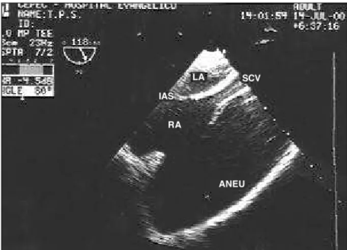
Fig. 4 - Transesophageal echocardiogram: longitudinal section showing relation of the aneurysm and atrial structures. LA- left atrium; IAS- interatrial septum; RA- right atrium; SCV- superior vena cava; ANEU- aneurysm of the right atrial appendage.
ANEU
LA SCV
IAS
Arq Bras Cardiol 2002; 78: 239-41.
Barberato et al Aneurysm of the right atrial appendage
2 4 1 2 4 1
1. Suedkamp M, Horst M, Mehlhorn U, Hoppe U, Arnold G, Dalichau H. Surgical repair of right atrial aneurysm. Thorac Cardiov Surg 2000; 48: 35–7. 2. Gold JP, Afifi HY, Ko W, Horner N, Hahn R. Congenital giant aneurysm of the left
atrial appendage: diagnosis and treatment. J Card Surg 1996: 11: 147–50. 3. Zimand S, Frand M, Hegesh J. Congenital giant left atrial aneurysm in infant. Eur
Heart J 1997; 18: 1034-5.
4. Ruys F, Paulus W, Stevens C, Brutsaert D. Expansion of the left atrial appendage is a distinctive cross-sectional echocardiographic feature of congenital defect of pericardium. Eur heart J 1983; 4: 738.
5. Gullestad L, Flogstad T, Nordstrand K, et al. Intrapericardial left atrial aneurysm diagnosed by transesophageal echocardiography and nuclear magnetic resonance imaging. Eur Heart J 1991; 12: 277–9.
6. Von der Emde J, Cesnjevar RA, Kretschmer S, Janssen GH, Wittekind C. Posttraumatic aneurysm of the right atrium. Ann Thorac Surg 1996; 62: 1507–9.
References
7. Zeebregts CJ, Hensens AG, Lacquet LK. Asymptomatic right atrial aneurysm: fortuitous finding and resection. Eur J Cardiothorac Sug 1997; 11: 591–3. 8. Scalia GM, Stafford WJ, Burstow DJ, Carruthers T, Tesar PJ. Successful treatment
of incessant atrial flutter with excision of congenital giant right aneurysm diagnosed by transesophageal echocardigraphy. Am Heart J 1995; 129: 834-5. 9. Miyamura H, Nakagomi M, Eguchi S, Aizama Y. Successful surgical treatment of incessant automatic atrial tachycardia with atrial aneurysm. Ann Thorac Surg 1990; 50: 476–8.
10. Staubach P. Large right atrial aneurysm: rare cause of recurrent pulmonary embolism. Z Kardiol 1998; 87: 894–9.
11. Kozlj M, Angelski R, Pavcnik D, Zorman D. Idiopatic enlargement of the right atrium. Pediatr Cardiol 1998; 19: 420-1.