Arq Bras Cardiol 2002; 78: 589-91.
Frota Fo et al
Cardiac angiosarcoma
5 8 9
Hospital São Francisco - Santa Casa de Porto Alegre
Mailing address: José Dario Frota Filho – Rua Prof. Freitas Cabral, 305/602 – 90690-130 – Porto Alegre, RS - E-mail: dariofrota@cardiol.br
English version by Stela Maris C. e Gandour
Arq Bras Cardiol, volume 78 (nº 6), 589-91, 2002
José Dario Frota Filho, Fernando A. Lucchese, Paulo Leães, Luís Antônio Valente, Mariana S. Vieira, Celso Blacher
Porto Alegre, RS - Brazil
Primary Cardiac Angiosarcoma. A Therapeutical Dilemma
Case Report
Cardiac angiosarcomas are malignant tumors that almost invariably have a short and fatal evolution. The therapeutic approach includes surgery, chemotherapy, and radiation therapy, alone or in combination. Heart transplantation is an attractive option in nonresectable tumors, even though the current experience is still limited. However, in most patients, the diagnosis is still establi-shed late, and survival is only slightly altered by the pro-posed treatments, mainly due to previously existing and undetected metastases. We report a case that illustrates the therapeutic dilemma faced with this neoplasia, and we discuss the case based on a literature review.
Approximately 25% of all primary cardiac tumors have malignant characteristics and an invasive behavior. Sarcomas account for almost all these malignant tumors, which rank second in the general incidence of cardiac tu-mors, right after myxomas. Sarcomas derive from the mesen-chymal tissue and have a variable morphology, which makes possible their classification as angiosarcomas, rhabdomyo-sarcomas, fibrorhabdomyo-sarcomas, and others. From the clinical point of view, they have a rapid and fatal evolution, and the surgical results remain unsatisfactory, regardless of the techniques used. Surgery, however, allows the differential diagnosis between benign and malignant neoplasia, which is not always possible through invasive and noninvasive diagnostic examinations. Even the therapeutic combina-tions involving surgery and radiation therapy or chemothe-rapy, or both, have not proved to be effective in managing cardiac sarcomas.
Case Report
The patient was a 74-year-old female who presented to the emergency service with acute pulmonary edema. The
patient complained of progressive dyspnea on exertion over the past few months, prior to which she reported being heal-thy. The patient denied rheumatic and cardiovascular disea-ses. The physical examination was compatible with modera-te mitral and aortic smodera-tenosis, and functional class IV. Radio-graphy of the chest, heart, and great vessels showed an elongated aorta with parietal calcifications, in addition to small left pleural effusion. The coronary arteries were normal on coronary angiography, and atrial fibrillation was evidenced on electrocardiography.
On transthoracic and transesophageal echocardio-graphy, a lobulated mass of large dimensions, attached to the interatrial septum was evidenced. This mass slid through the mitral valve to the left ventricular cavity also filling the left atrial auricle. Maximum and medium mitral gradients were 44 and 22mmHg, respectively. Global systo-lic function was preserved with an ejection fraction above 70%. The aortic valve had calcifications and a systolic flow with maximum and medium gradients of 67 and 40mmHg, respectively. The tricuspid valve had a mild regurgitation jet and a systolic pressure in the pulmonary artery of 64mmHg. Fibrinous pleural and small pericardial effusions were ob-served. These findings were suggestive of left atrial myxo-ma with functional mitral stenosis.
5 9 0 Frota Fo et al
Cardiac angiosarcoma
Arq Bras Cardiol 2002; 78: 589-91.
valvoplasty (annuloplasty), aortic valvoplasty (decalcifica-tion of the 3 leaflets), and exclusion of the left atrial auricle with inner ligation were also performed.
The analysis of the pericardial effusion was negative for bacteria and tumor cells, revealing only erythrocytes (90,000/mm3) and other cells (100/mm3).
The anatomicopathological examination revealed a high-grade malignant epithelioid neoplasia with a high
mitotic index and nuclear pleomorphism. The immuno-histochemistry profile showed focal expression of CD31 and intense positivity of vimentin and type IV collagen in the neoplastic cells, indicating a high-grade sarcoma, angiosarcoma (fig. 4).
The postoperative transesophageal echocardiogra-phy showed the mitral valve with a normal opening, maxi-mum and medium gradients of 10 and 3mmHg, respectively, with no regurgitation. The aortic valve maximum and medium gradients were 20 and 9mmHg, respectively. The left atrium was slightly enlarged (44mm), but no masses were present inside. The ejection fraction was 80%. No examina-tions to detect cerebral and pulmonary metastases were performed. The early evolution was uneventful, and the pa-tient was discharged from the hospital in good condition and using oral anticoagulants. The patient evolved asymptomatically, and, 6 months afterwards, she died suddenly at home, probably due to ventricular arrhythmia.
Discussion
Primary cardiac tumors are not frequent. The incidence of primary cardiac neoplasias ranges from 0.001 to 0.03% in autopsy reports 1. In adults, approximately 75% of the
tumors are benign, the most common being myxoma, and the remaining 25% are malignant. Angiosarcoma accounts for at least one third of the latter, is the most frequent ma-lignant primary cardiac tumor, being reported in patients aged from 9 to 80 years, in an approximate proportion of 2 males to every female 2,3. Angiosarcomas manifest mainly as
a pediculate intracavitary formation in the right atrial cham-ber, but they may also originate in the left atrial wall 3. They
are characteristically lobulated masses, with necrotic foci, ranging from 2 to 30cm in size, and may extend beyond the epicardium, reaching the pericardial sac. Microscopically, they comprise anastomotic vascular channels formed by malignant cells, solid areas of spindle cells, and other areas of primarily anaplastic cells 4.
The natural history of cardiac angiosarcomas is cha-racterized by a short clinical course and fatal evolution,
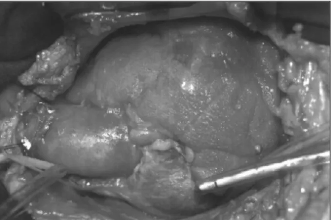
Fig. 1 - View of the pericardium recovered by a fibrin net resulting from sanguineous effusion.
Fig. 2 - Posterior portion of the angiosarcoma attached to the mitral ring and mitral posterior leaflet, evidencing a large amount of atrial thrombi.
Fig. 3 – Gross aspect of the anterior portion of the angiosarcoma, in which its pedicle of implantation in the mitral posterior ring can be seen.
Arq Bras Cardiol 2002; 78: 589-91.
Frota Fo et al
Cardiac angiosarcoma
5 9 1 even after surgery, chemotherapy, and radiation therapy 1. A
review of the literature shows that this is an aggressive tu-mor with 80% of the patients with metastases at the time of diagnosis and 90% of them surviving less than 9 months after the diagnosis (mean ranging from 6 to 11 months 1). In
the past, the diagnosis was established only on autopsy in 55% to 88% of the cases 4. The clinical presentation is
uns-pecific and relates to the location of the primary tumor, de-gree of local involvement, and presence or absence of me-tastases, which are mainly pulmonary. All these features lead to late diagnosis. Initial signs and symptoms may sug-gest pericarditis, with pleuritic chest pain, pericardial fric-tion, and nonspecific electrocardiographic alterations 4.
Cy-tology of the thoracocentesis and pericardiocentesis fluids or of the biopsy may provide the diagnosis of tumor malig-nancy 5. The signs and symptoms may include fever,
dysp-nea, cardiac murmurs, weight loss, orthopdysp-nea, fatigue, ano-rexia, vomiting, peripheral edema, and right heart failure 3.
Extrinsic tumoral compressions in the venae cava and obs-tructions of the valvular orifices lead to hepatomegaly and jugular venous distention. Hemoptysis and cardiac tampo-nade usually represent catastrophic events in the course of disease. Electrocardiographic alterations, such as elevation of the ST segment, inversion of the T wave, low-voltage QRS, left axial deviation, right bundle-branch block, and atrial fibrillation, are frequent findings. From the radiological point of view, we may observe cardiomegaly, mediastinal distortions secondary to invasion of the pericardial sac by tumor or hemorrhage 4, and abnormalities in the pulmonary
fields, a consequence of metastases. The differential diagnosis is necessary, not only between primary and me-tastatic, malignant and benign cardiac tumors, but also between neoplastic or nonneoplastic masses 6. Imaging
techniques, such as echography, tomography, magnetic re-sonance imaging, and angiography allow the rapid recogni-tion of these neoplasias. The advanced stage and the di-mensions of this tumor are limiting factors of successful surgical treatment. In case of tumors confined to the atrial free wall, the interatrial septum, or to a small portion of the ventricle or of a cardiac valve, complete tumor resection should be performed, in an attempt to attenuate symptoms and increase postoperative survival 7,3. Often, the tumor is
1. Kakizaki S, Takagi H, Hosaka Y. Cardiac angiosarcoma responding to multidisciplinary treatment. Int J Cardiol 1997; 62: 273-5.
2. Klima U, Wimmer-Greinecker G, Harringer W, Mair R, Grob CH, Brucke P. Car-diac angiosarcoma- a diagnostic dilemma. Cardiovasc Surg 1993; 1: 674-6. 3. Putnam JB, Sweeney MS, Colon R, Lanza LA, Frazier OH, Cooley DA. Primary
cardiac sarcomas. Ann Thorac Surg 1991; 51: 906-10.
4. Herrmann MA, Shankerman RA, Edwards WD, Shub C, Schaff HV. Primary car-diac angiosarcoma: a clinicopathologic study of six cases. J Thorac Cardiovasc Surg 1992; 103: 655-64.
5. Rudoff J, Slavin RE. Cardiac angiosarcoma arising in a coronary artery: angiogra-phic and pathologic findings. Cathet Cardiovasc Diag 1995; 34: 215-8.
References
6. Basso C, Valente M, Poletti A, Casarotto D, Thiene G. Surgical pathology of pri-mary cardiac and pericardial tumors. Eur J Cardiothorac Surg 1997; 12: 730-8. 7. McFadden PM, Ochsner JL. Atrial replacement and tricuspid valve
reconstruc-tion after angiosarcoma resecreconstruc-tion. Ann Thorac Surg 1997; 64: 1164-6. 8. Crespo MG, Pulpón LA, Pradas G, et al. Heart transplantation for cardiac
angio-sarcoma: should its indication be questioned? J Heart Lung Transplant 1993; 12: 527-30.
9. Baay P, Karwande SV, Kushner JP, Olsen S, Renlund DG. Successful treatment of a cardiac angiosarcoma with combined modality therapy. J Heart Lung Trans-plant 1994; 13: 923-5.
nonresectable due to the extension of myocardial invasion 1.
In these cases, adjuvant chemotherapy and radiation the-rapy have been frequently used. More recently, cardiac transplantation has become an alternative; most of the time, however, survival does not differ from that in which transplantation is not performed 1. A hypothesis to explain
the unaltered mortality after transplantation is the pos-sibility that immunosuppressive treatment causes disse-mination of preexisting micrometastases not detected at the time of diagnosis 8. On the other hand, several
studies emphasize cardiac transplantation as the treat-ment of choice for patients with nonresectable tumors, locally aggressive but with no metastases. Chemothe-rapy and radiation theChemothe-rapy neither alleviate symptoms as surgery does nor seem to prolong patients’ survival 3.
The possibility of a treatment combining these therapeu-tic modalities has already been reported by Baay et al 9.
In that case, the patient underwent initial chemotherapy with doxorubicin, dacarbazine, ifosfamide, and mesna, which was complemented with a total dose of radiation of 2600cGy, being then transplanted. Two months later, the patient received 2 additional courses of chemothe-rapy with the same drugs and was maintained on cy-closporine and prednisone. The clinical outcome was fa-vorable, and metastases were not detected up to 33 months after surgery.
The aggressiveness of angiosarcoma continues to challenge the current methods of treatment, and the ideal therapy is yet to be discovered. In addition, postoperative survival seems not to significantly differ in patients treated with or without surgery 1, independent of the extension of
the surgical resection, or even of the transplant replace-ment of the organ.