2 1 0
Castro et al
Reduction in QTc interval dispersion after pyridostigmine
Arq Bras Cardiol volume 75, (nº 3), 2000
Universidade Federal Fluminense - Niterói e Hospital Pró-Cardíaco - Rio de Janeiro Mailing address: Antonio Claudio Lucas da Nóbrega – Depto de Fisiologia -Instituto Biomédico-UFF – Rua Cinco de Julho, 318/1001 – 24220-111 – Niterói, RJ, Brazil
English version by Syela Maris C. Gandour
Objective – Parasympathetic dysfunction is an
inde-pendent risk factor in individuals with coronary artery di-sease, and cholinergic stimulation is a potential therapeu-tical option. We determined the effects of pyridostigmine bromide, a reversible anticholinesterase agent, on electro-cardiographic variables of healthy individuals.
Methods – We carried out a cross-sectional, double
blind, randomized, placebo-controlled study. We obtained electrocardiographic tracings in 12 simultaneous leads of 10 healthy young individuals at rest before and after oral administration of 45 mg of pyridostigmine or placebo.
Results – Pyridostigmine increased RR intervals
(be-fore: 886±27 ms vs after: 1054±37 ms) and decreased QTc dispersion (before: 72±9ms vs after: 45±3ms), without changing other electrocardiographic variables (PR seg-ment, QT interval, QTc, and QT dispersion).
Conclusion – Bradycardia and the reduction in QTc
dispersion induced by pyridostigmine may effectively present a protective mechanism if these results can be re-produced in individuals with cardiovascular diseases.
Key words - autonomic nervous system, QTc dispersion,
pyridostigmine
Arq Bras Cardiol, volume 75 (nº 3), 210-213, 2000
Renata Rodrigues Teixeira de Castro, Salvador Manoel Serra, Antonio Claudio Lucas da N óbrega
Niterói, RJ - Brazil
Reduction of Qtc interval dispersion. Patential
mechanism of cardiac protection of pyridostigmine
bromide
Original Article
Cardiovascular diseases are one of the major health problems in the industrialized world 1, including Brazil 2. In
the United States of America, 1.5 million people experience annually acute myocardial infarction, i.e., every 20s one per-son experiences acute myocardial infarction 1. Therefore, it
is increasingly necessary to identify and control not only the risk factors for developing the atherosclerotic process, but also the conditions that increase mortality in patients with heart disease, and acute myocardial infarction.
Hypoactivity of the parasympathetic branch of the au-tonomic nervous system (ANS), identified by a reduction in the heart rate variability, is an independent risk factor in pa-tients after acute myocardial infarction 3,4. Odemuyiwa et al5
reported that the low heart rate variability, when compared with the reduced left ventricular ejection fraction, has a si-milar predictive power for overall mortality. On the other hand, the low heart rate variability proved to be a better pre-dictor of sudden death and arrhythmic events than the ejec-tion fracejec-tion in the first 6 months after acute myocardial in-farction 5.
Even though recognizing the clinical importance of sympathetic hyperactivity and its treatment in cardiovas-cular diseases 6, studies evaluating therapeutical
alterna-tives for low vagal activity are scarce. Pyridostigmine bro-mide is an anticholinesterase agent that causes a parasym-pathomimetic effect by increasing the concentration of endogenous acetylcholine. This drug is widely used in pa-tients with myasthenia gravis to increase the concentration of acetylcholine in the motor plate and, therefore, reduce the deficit in muscular strength.
Arq Bras Cardiol volume 75, (nº 3), 2000
Castro et al Reduction in QTc interval dispersion after pyridostigmine
2 1 1
Methods
Ten healthy volunteers (3 males and 7 females) with a mean age (mean ± standard deviation) of 28±6 years, wei-ghing 67.4±16.5kg, and measuring 170±6 cm of height un-derwent a cross-sectional, double-blind, randomized pro-tocol on two different mornings. After resting in the dorsal decubitus position, a surface electrocardiogram with 12 si-multaneous leads (software ErgoPC®, Micromed, Brazil) was performed on each volunteer, before and 2 hours after oral administration of 45mg of pyridostigmine bromide (Mestinon®, Roche, Brazil) or placebo. All individuals were nonsmokers in a fasting period and were not on any medica-tion. They were instructed not to ingest any substance con-taining alcohol or caffeine and to avoid strenuous physical exercise during the 2 days preceding the examination. The-se individuals were considered healthy baThe-sed on normal re-sults of clinical and electrocardiographic examinations, two-dimensional echocardiography with Doppler, and cardio-pulmonary exercise testing.
All volunteers gave written consent to take part in the study after being informed about the procedures they wo-uld undergo and risks they wowo-uld be exposed to. The study was approved by the Institutional Committee on Ethics.
The same observer measured the following variables in the 12 electrocardiographic leads: PR segment, RR and QT intervals. In addition, QTc interval, and QT and QTc disper-sions were calculated. The software used recorded the 12 electrocardiographic leads simultaneously in a digital form, storing the signals for later analysis. During the analysis, tracings and time recording could be widened, allowing measurements with greater resolution. To compare the va-lues calculated, we used the arithmetic mean of the results obtained in the 12 leads. The simultaneous 12-lead recor-ding is an indispensable tool for calculating spatial disper-sion of QT and QTc intervals at the same time.
The QT interval was measured from the first deflection of the QRS complex until the return point of the T wave to the base line or the lowest point between the T and U waves7.
Correction of the QT interval for the heart rate (QTc) was ob-tained through Bazett’s formula (QT/√RR) 8-10. Dispersions
of the QT and QTc intervals were calculated, respectively, as the subtraction between the greatest and the smallest QT and QTc intervals in the 12 electrocardiographic leads 11-13.
Statistical assessment of data was based on a repeated measurements analysis of variance (ANOVA). When the F value was significant, the ANOVA was followed by the Student-Newman-Keuls test for post-hoc paired compa-risons. For the statistical analysis of the symptoms repor-ted by the patients while using pyridostig–mine and pla-cebo, we used the chi-square test. The results were conside-red statistically significant when p<0.05.
Results
Even though undesired effects occurred with a greater frequency with the use of pyridostigmine (p=0.041), the
symptoms were mild, self-limited, and were reported by only 4 individuals as follows: sialorrhea (n=3), abdominal colic and diarrhea (n=1), and epigastric discomfort (n=1).
Two hours after administration of pyridostigmine, we observed significant bradycardia (p=0.01) (table I) shown as an increase in the duration of the RR intervals and reduction in QTc dispersion. No significant differences were observed in the other electrocardiographic variables studied (QT and QTc intervals, QT dispersion, and PR segment).
Discussion
Autonomic nervous system dysfunction due to sym-pathetic hyperactivity or parasymsym-pathetic hypoactivity is associated with an increase in the risk of arrhythmias and other cardiac events in patients with heart disease 4,14.
Therefore, dysautonomia increases cardiovascular morbi-dity and mortality. Adrenergic hyperactivity after acute myocardial infarction has been known for decades, and its clinical relevance has been soundly characterized by a re-duction in mortality in patients treated with beta-blockers 15,
a management currently considered standard in the posta-cute myocardial infarction treatment 16. More recently,
ana-lysis of neuroendocrine features of congestive heart failure has culminated with the indication for use of beta-blockers also in this disease 17. Even though a reduction in mortality
in congestive heart failure has been observed specifically with the use of carvedilol 18, the general concept that a
re-duction in the noxious effects of the sympathetic hyperac-tivity on the heart has been characterized as an efficacious way of reducing cardiac events.
On the other hand, parasympathetic dysfunction gained importance from studies showing its role as an independent risk factor in patients after acute myocardial infarction 3,5, and,
more recently, also in patients with congestive heart failure 19.
Paradoxically, studies aiming to investigate possible speci-fic therapeutical measures for vagal hypoactivity are sce. Aerobic training may promote an increase in vagal car-diac activity as shown by the greater variability heart rate of patients undergoing this type of training 20. In regard to
possible pharmacological alternatives, in the beginning of the ‘90s, four independent groups 21-24 published studies
about the cardiovascular effects of scopolamine after acute
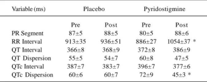
Table I – Electrocardiographic variables of healthy individuals at rest before (pre) and 2h after (post) oral administration of placebo
or 45mg of pyridostigmine in different days
Variable (ms) Placebo Pyridostigmine
Pre Post Pre Post
PR Segment 87±5 88±5 80±5 88±6
RR Interval 913±35 936±51 886±27 1054±37 * QT Interval 366±8 368±9 372±8 386±9 QT Dispersion 55±5 54±7 60±8 47±5 QTc Interval 387±7 383±7 396±7 377±6 QTc Dispersion 60±6 60±7 72±9 45±3 *
2 1 2
Castro et al
Reduction in QTc interval dispersion after pyridostigmine
Arq Bras Cardiol volume 75, (nº 3), 2000
myocardial infarction. Scopolamine is a muscarinic choliner-gic antagonist that may have a parodoxical effect when admi-nistered at low doses (0.4 to 0.6mg) 25. All those studies 21-24
showed a reduction in heart rate and an increase heart rate variability, therefore, suggesting that scopolamine might have a protective effect against cardiac events after acute myocardial infarction. However, the only study that specifi-cally investigated this hypothesis has not confirmed the speculation of the above cited authors 26. Hull Jr et al 26
found that, even though the drug increases heart rate varia-bility in chronically instrumented, dogs it was not able to prevent ventricular fibrillation during physical exertion and artificially induced coronary artery occlusion. Therefore, no experimental or clinical studies exist indicating a pharma-cological therapeutical alternative for parasympathetic hy-poactivity that may protect against arrhythmogenic sudden death and cardiac mortality in general. Thus, the search for drugs that may cause cholinergic cardiac stimulation and reactivate the possibility of treating vagal hypoactivity pre-sent in diverse cardiovascular diseases still continues.
Pyridostigmine bromide is a reversible anticholineste-rase agent that does not cross the blood-brain barrier at usu-al doses and causes a cholinomimetic action by decelera-ting hydrolysis of endogenous acetylcholine and, conse-quently, increasing its concentration in the synaptic cleft. Its mostly known clinical indication is for the treatment of the skeletal muscular paralysis of myasthenia gravis 27, in
which the daily dosage may reach 720mg. Pyridostigmine may also be used in multiple sclerosis, amyotrophic lateral sclerosis, spinal myotrophies, and paresis consecutive to poliomyelitis. Other less common indications of pyridostig-mine include prevention of disorders after lumbar puncture and meningism after electroencephalography, and treat-ment of migraine and headache. Another recently reported use 28 of pyridostigmine was the intake of 30mg every 8
hours by the American soldiers who took part in the Gulf War. The purpose was to prevent intoxication by gases with irreversible anticholinesterase action in case of an attack with chemical weapons.
The cardiovascular action of pyridostigmine is usually considered a side effect, because in the treatment of
myasthenia gravis, the objective is to cause a cholinomi-metic effect on the skeletal muscle. This is why systematic studies of the hemodynamic effects of pyridostigmine are not found.
Our team has been studying the cardiovascular action of pyridostigmine. The single dose of 30mg caused brady-cardia at rest 29 and during dynamic exercise in healthy
indi-viduals without, however, impairing tolerance to exertion 30.
In addition, pyridostigmine maintained bradycardia for 24 hours with the use of 30 mg every 8 hours, and it increased in heart rate variability analyzed in the time domain 31. These
effects observed in healthy volunteers occurred with no change in systolic and diastolic cardiac function 32. When
used at the oral dosage of 45mg, pyridostigmine limited the elevation of the double product of healthy individuals un-der mental stress in a cross-sectional, double blind, rando-mized, placebo-controlled study 33.
Parasympathomimetic drugs are known to lower the conduction of the electric cardiac impulse, inducing brady-cardia. Drugs that increase duration of the RR intervals on the electrocardiogram would also potentially increase other electrocardiographic variables, such as the PR segment and the QT interval. Depending on the magnitude of these in-creases, they could mean, respectively, pathological de-crease in atrioventricular conduction (atrioventricular blo-cks of different degrees) or increase in duration of ventricu-lar repoventricu-larization. Therefore, when parasympathomimetic drugs are studied, evaluation of their potential effects on clinically relevant electrocardiographic variables are manda-tory independently of their effect on heart rate.
Even though some authors consider it unnecessary to calculate the QTc interval, i.e., to correct the QT interval ac-cording to heart rate, in our case, this correction is very im-portant. As the drug we are studying is known to cause bradycardia, the calculation of the QTc interval allows a bet-ter analysis of cardiac repolarization independent of the du-ration of the RR interval. That is why an analysis of the de-gree of spatial dispersion of the refractory periods between different areas of the cardiac surface is obtained eliminating the influence that heart rate exerts on such a variable throu-gh the analysis of QTc interval dispersion.
QTc dispersion has a clinical importance similar to that of the QT and QTc intervals. Prolongation of QTc dispersion indicates an increase in the degree of temporal dispersion of the refractory periods between different areas of the cardiac surface, potentiating the occurrence of electric stimulus reentrant phenomena and, consequently, an increase in the risk of ventricular arrhythmias and sudden death 34. In this
way, QTc dispersion proved to be an independent predictor of cardiac and cerebrovascular mortality 35-37. On the other
hand, a smaller QTc dispersion is associated with a smaller mortality rate after acute myocardial infarction 38, in addition
to improving prognosis in individuals with congestive heart failure 39. In our study, administration of pyridostigmine
bromide increased the duration of the RR interval and decreased QTc dispersion without altering the other electro-cardiographic variables studied. Thus, in addition to
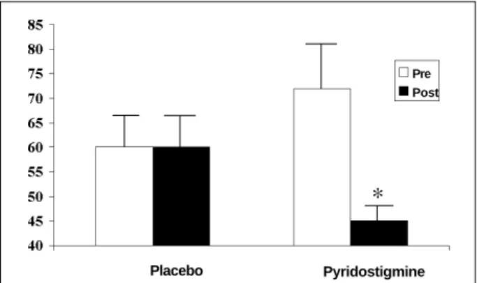
Fig. 1 – Corrected QT interval (QTc) of healthy individuals at rest before (pre) and 2h after (post) oral administration of placebo or 45mg of pyridostigmine in different days * P = 0.04 vs pre pyridostigmine.
Pre
Post
Pyridostigmine Placebo
Arq Bras Cardiol volume 75, (nº 3), 2000
Castro et al Reduction in QTc interval dispersion after pyridostigmine
2 1 3
inducing bradycardia, pyridostigmine bromide at the dose used showed a potential cardioprotective effect in regard to the occurrence of ventricular arrhythmias and sudden death. The study here described was carried out in healthy young individuals and, therefore, may not be necessarily re-producible in individuals with cardiovascular diseases. In addition, all studies performed so far by our team have involved the use of a single dose 29,30,32,33, or a maximum 3
doses 31 of pyridostigmine bromide. Studies with patients
may analyze the effect of pyridostigmine bromide adminis-tration in fractionated doses at long-term.
1. American Heart Association: Heart and Stroke Facts: 1996 Statistical Supplement. Dallas, American Heart Association, 1996, pp. 1-23.
2. DATASUS. Morbidade Hospitalar do SUS. Ministério da Saúde. http:// www.datasus.gov.br
3. Kleiger RE, Miller JP, Bigger JTJ and The Multicenter Post-Infarction Research Group. Decreased heart rate variability and its association with increased mortality after acute myocardial infarction. Am J Cardiol 1987; 59: 256-62. 4. La Rovere MT, Bigger Jr JT, Marcus FI, Mortara A, Schwartz PJ. Baroreflex
sensitivity and heart-rate variability in prediction of total cardiac mortality after myocardial infarction. Lancet 1998; 351: 478-84.
5. Odemuyiwa O, Malik M, Farrel TG, et al. A comparision of the predictive carach-teristics of heart rate variability and left ventricular ejection fraction for all-cause mortality, arrhythmic events and sudden death after acute myocardial infarction. Am J Cardiol 1991; 64: 434-9.
6. Corr PB, Gillis RA. Autonomic neural influences on the dysrhythmias resulting from myocardial infarction. Circulation Res 1978; 43: 1.
7. Lepeschkin E, Surawicz B. The measurement of the Q-T interval of the electrocar-diogram. Circulation 1952; 6; 378-88.
8. Bazett HC. An anallysis of the time-relations of eletrocardiograms. A.N.E 1997; 02(02): 177-94.
9. Ashman R. The normal duration of the Q-T interval. Am Heart J 1962; 23: 522-34. 10. Simonson E, Cady L, Woodbury M. The normal Q-T interval. Am Heart J 1962;
63: 747-53.
11. Higham PD, Campbell RWF. QT dispersion. Br Heart J 1994; 71: 508-10. 12. Hnatkova K, Malik M, Kautzner J, Gang Y, Camm AJ. Adjustment of QT
dispersion assessed from 12 lead eletrocardiograms for different numbers of analysed eletrocardiographic leads: comparision of stability of different methods. Br Heart J 1994; 72: 390-6.
13. Pye M, Quinn AC, Cobbe SM. QT interval dispersion: a non-invasive marker of susceptibility to arrhythmia in patients with sustained ventricular arrhythmias? Br Heart J 1994; 71: 511-14.
14. Schwartz PJ, La Rovere MT, Vanoli E. Autonomic nervous system and sudden death. Experimental basis and clinical observations for post-myocardial infarc-tion risk stratificainfarc-tion. Circulainfarc-tion 1992; 85(suppl I): I 77-99.
15. Bigger JT, Coromilas J. How do beta-blockers protect after myocardial infarcti-on? An Intern Med 1984; 101: 256-8.
16. Antman EM, Braunwald E. Acute myocardial infarction. In: Braunwald E. Heart Disease. A Textbook of Cardiovascular Medicine. Philadelphia: WB Saunders, 1997; 1184-288.
17. Barnett DB. Beta-blockers in heart failure: a therapeutic paradox. Lancet 1994; 343: 557-8.
18. Packer M, Bristow MR, Cohn JM, et al. The effect of carvedilol on morbidity and mortality in patients with chronic heart failure. N Engl J Med 1996; 334: 1349-55.
19. Nolan J, Batin PD, Andrews R, et al. Prospective study of heart rate variability and mortality in chronic heart failure: results of the United Kingdom heart failure evaluation and assessment of risk trial (UK-heart). Circulation 1998; 98: 1510-6.
20. La Rovere MT, Mortara A, Sandrome G, Lombardi F. Autonomic nervous system adaptation to short-term exercise training. Chest 1992; 101: 299s-303s. 21. Casadei B, Pipilis A, Sessa F, Conway J, Sleight P. Low doses of scopolamine
in-References
crease cardiac vagal tone in the acute phase of myocardial infarction. Circulation 1993; 88: 353-7.
22. De Ferrari GM, Mantica M, Vanoli E, Hull SS Jr, Schwartz PJ. Scopolamine increa-ses vagal tone and vagal reflexes in patients after myocardial infarction. J Am Coll Cardiol 1993; 22: 1327-34.
23. Pedretti RF, Colombo E, Braga SS, Carú B. Influence of scopolamine on cardiac sympathovagal interaction after acute myocardial infarction. Am J Cardiol 1993; 72: 384-92.
24. Vybiral T, Glaeser DH, Morris G, et al. Effects of low-dose transdermal scopola-mine on heart rate variability in acute myocardial infarction. J Am Coll Cardiol 1993; 22: 1320-36.
25. Brown JH, Taylor P. Muscarinic receptor agonists and antagonists. In: Goodman & Gilman’s. The Pharmacological Basis of Therapeutics. Tennessee: McGraw-Hill, 1996: 149-151.
26. Hull Jr SH, Vanoli E, Adamsom PB, et al. Do increases in markers of vagal activity imply protection from sudden death? The case of scopolamine. Circulation 1995; 91: 2516-9.
27. Taylor P. Anticholinesterase agents. In: Goodman & Gilman’s. The Pharmacolo-gical Basis of Therapeutics. Tennessee: McGraw-Hill, 1996: 161-76. 28. Volans AP. Sarin: guidelines on the management of victims of a nerve gas attack.
J Accid Emerg Med 1996; 13: 202-6.
29. Nóbrega ACL, Carvalho ACG, Bastos BG. Resting and reflex heart rate res-ponses during cholinergic stimulation with pyridostigmine in humans. Braz J Med Biol Res 1996; 29: 1461-5.
30. Serra SM, Vivacqua R, Ramalho SHR, Santos KB, Bastos BG, Nóbrega ACL. Cardiopulmonary exercise testing during cholinergic stimulation with pyridos-tigmine in healthy subjects. J Am Coll Cadiol 1998; 31: 407C.
31. Reis AF, Moraes RS, Bastos BG, Ferlin EL, Ribeiro JP, Nóbrega ACL. Heart rate variability during cholinergic stimulation with piridostigmine in healthy subjects. J Am Coll Cardiol 1998; 31: 407C.
32. Pontes PV, Nóbrega ACL, Mesquita ET, Bastos BG, Carvalho ACG, Romêo LJM. Estudo das variáveis hemodinâmicas e da função cardíaca sistólica e dias-tólica com emprego de piridostigmina. Arq Bras Cardiol, 1999 (no prelo). 33. Nóbrega ACL, Carvalho ACG, Santos KB, Soares PPS. Cholinergic
stimula-tion with pyridostigmine blunts the cardiac responses to mental stress. Clin Auton Res 1999; 9: 1-6.
34. Ashikaga T. Increased QTc dispersion predicts lethal ventricular arrhythmias complicating coronary angioplasty. Am J Cardiol 1998; 15: 814-6
35. de Bruyne MC, Hoes AW, Kors JA, Hofman A, van Bemmel JH, Grobbee DE. QTc dispersion predicts cardiac mortality in the elderly: the Rotterdam Study. Circu-lation 1998; 97: 467-72.
36. Sawicki PT, Kiwitt S, Bender R, Berger M. The value of QT interval dispersion for identification of total mortality risk in non-insulin-dependent diabetes melli-tus. J Intern Med 1998; 243: 49-56.
37. Batur MK, Aksoyek S, Oto A, Yildirir A, Ozer N, Atalar E, et al. Circadian varia-tions of QTc dispersion: is it a clue to morning increase in sudden cardiac death? Clin Cardiol 1999; 22: 103-6.
38. Glancy JM, Garratt CJ, Woods KL, de Bono DP. Qt dispersion and mortality after myocardial infarction. Lancet 1995; 345: 945-8.
39. Barr CS, Naas AA, Fenwick M, Struthers AD. Enalapril reduces QTc dispersion in mild congestive heart failure secondary to coronary artery disease. Am J Car-diol 1997; 79: 328-33.
If the effects demonstrated so far in healthy indi-viduals may be reproduced in these patients, the per-formance of controlled studies aiming to evaluate a po-tential protective effect of pyridostigmine may be jus-tifiable.