Ten-Year Experience with the Ross Operation
Francisco Diniz Affonso da Costa, Elaine Welk Lopes Pereira, Luiz Eduardo Barboza, Hermínio Haggi Filho, Claudinei
Collatusso, Carlos Henrique Gori Gomes, Sérgio Augusto Veiga Lopes, Evandro Antônio Sardetto, Andréa Dumsch de
Aragon Ferreira, Marise Brenner Affonso da Costa, Iseu Affonso da Costa
Aliança Saúde Santa Casa – PUCPR, Curitiba, PR, Brazil
objective: To evaluate the 10-year outcomes of the Ross Operation, analyzing survival rate, incidence of reoperations, and late performance of pulmonary autografts and homografts in the reconstruction of the right ventricular outflow tract.
Methods: Two hundred and twenty seven patients with a mean age of 29.1±11 years underwent Ross operation from May 1995 to February 2005. The most prevalent etiology was rheumatic disease in 61% of the cases. Autografts were implanted using the total root replacement technique in 202 cases, with intraluminal cylinder in 20, and in the subcoronary position in 5. The right ventricular outflow tract was conventionally reconstructed with cryopreserved homografts (n = 160), with proximal extension of the homograft with pericardium (n = 41), and with decellularized homografts (n = 26). The postoperative follow-up ranged from 1 to 118 months (mean = 45.5 months).
Results: Hospital mortality was 3.5%, and long-term survival was 96.9% at ten years. No episodes of thromboembolism and only two cases of endocarditis occurred. Eleven patients underwent reoperation because of problems related to the auto and/or homograft, progression of rheumatic mitral valve disease, and iatrogenic coronary insufficiency. After 10 years, 96.4% and 96.2% of the patients were free from reoperation in the autograft and homograft groups, respectively. No late autograft dilatation was observed. Reconstruction of the left ventricular outflow tract with decellularized homografts significantly reduced the incidence of gradients on late follow-up.
Conclusion: Late outcomes with the Ross Operation were associated with an excellent long-term survival and a low incidence of reoperations and late morbidity. We consider this procedure the best option for the surgical treatment of aortic valve disease in children and young adults.
palavras-chave: Autografts, Ross operation, aortic valve replacement.
Mailing Address: francisco diniz Affonso da Costa •
Rua Henrique Coelho Neto, 55 – 82220-120 – Curitiba, PR, Brazil
The optimal prosthesis for aortic valve replacement remains highly controversial. However, many consider the Ross Operation the best option, especially in children and young adults1,2. Among the advantages of the pulmonary autograft are: the durability, the physiologic hemodynamic performance with preservation of the normal valve opening and closing mechanism, the absence of thromboembolic complications, and the low incidence of infectious complications, in addition to the growth potential when implanted in children. These advantages have been confirmed by mid and long-term clinical results in several series3,4.
On the other hand, the operation is technically more complex and subject to criticism for its potential of inducing disease in two valves in patients with primary disease in only one. Additionally, variations in the surgical procedure may result in different types of complications with an important impact on results5.
Long-term outcomes of pioneer Ross cases reflect, in their majority, the experience with pulmonary autograft implantation in the subcoronary position6. However, currently,
the most frequently used technique has been the total aortic root replacement because a geometrically aligned and competent graft is easier to be consistently obtained3.
Despite the ability of the valve and pulmonary arterial wall to adapt to the pressure regimen of the systemic circulation, there is a growing concern that pulmonary autografts are subject to progressive dilatation with formation of neoaortic aneurysm, and concurrent valve prolapse and regurgitation7,8. Additionally, the incidence of degeneration and dysfunction of the valve autograft implanted in the right ventricular outflow tract can also increase late morbidity, and the patients become thus subject to the need for reoperations9,10.
portion of the aortic arc. In no case was the distal anastomosis reinforced with Teflon or bovine pericardium strips.
The pulmonary autograft was always placed so that its thinner sinus lacking a pericardium lining was positioned toward the left coronary sinus, thus being at least partially supported by the cardiac structures posterior to the ascending aorta.
For the reconstruction of the right ventricular outflow tract, the patients were divided into three groups according to the surgical procedure or method of graft preservation used. In group 1 (n = 160), outflow tract reconstruction was performed with fresh or cryopreserved valve homografts sutured proximally and distally, without the interposition of any type of prosthetic material. In group 2 (n = 41), the reconstruction of the right ventricular outflow tract was performed with cryopreserved homografts. However, the proximal anastomosis was performed with the interposition of a bovine pericardium or autologous pericardium patch immersed in glutaraldehyde, so as to elongate the homograft and avoid any tension in the anastomoses or in the body of the homograft. Group 3 (n = 26) was comprised of patients whose right ventricular outflow tract was reconstructed with
Methods
Two hundred and twenty seven patients underwent aortic valve replacement with pulmonary autograft at the Cardiac Surgery Service of Aliança Saúde Santa Casa – PUCPR, from May 1995 to February 2005. One hundred and seventy one patients (71%) were male and the age ranged from 5 to 56 years (mean = 29.1±11 years). Thirty five patients were younger than 18 years of age. The most frequent etiology of the heart valve disease was rheumatic disease in 140 cases (61%). Fifteen patients had severe associated mitral valve dysfunction. Ten patients had bacterial endocarditis in the native valve or in the valve prosthesis, and four had ascending aortic aneurysm. Thirty four patients had already undergone one or more previous surgeries in the aortic valve, namely aortic valvuloplasty in 14, valvuloplasty plus subvalvar membrane resection in 7, and biological prosthesis implantationin 13. Some clinical and laboratory test data are listed in Table 1.
All surgeries were performed by the same surgeon and the surgical procedure had beenthoroughly described in previous publications11. However, some relevant technical details for the correlation with occasional valve autograft and/or homograft dysfunctions are reviewed here.
The surgeries were performed with extracorporeal circulation, moderate hypothermia of 30-32ºC, and myocardial protection with intermittent cold blood cardioplegia into the coronary ostia. The aortic clamping time was 104±22 min (min= 69, max= 175) and that of extracorporeal circulation was 137±26 min (min= 92, max= 230).
The most frequently used technique was the total aortic root replacement in 202 cases, whereas the intraluminal cylinder technique (inclusion) was used in 20 patients and the subcoronary implantation in five.
During the autograft preparation, a 2-3mm muscular border was left below the valvar level. For the proximal anastomosis separate sutures were used, and care was taken to pass the sutures next to the base of the valve cusps. Regardless of the technique used, the proximal anastomosis was always intra-annular, so that the native aortic ring could support the pulmonary autograft.
When the total aortic root replacement was used, the proximal anastomosis was always reinforced all around its circumference with a Teflon or bovine-pericardium strip, in an attempt to prevent any further dilatation of the aortic ring. Likewise, any proximal or distal diameter discrepancy was always adjusted to make the pulmonary autograft dimensions compatible with those of the aortic ring and of the ascending aorta. In patients with mild annular dilatation a plication of the intercommissural triangle was performed (n = 8) and, in the cases with a more intense annuloectasia, external reduction of the aortic ring was performed with a Teflon strip (n = 14). Enlargement of the aortic arc usingMannougian(n = 6) or Ross-Konno procedures (n = 5) was performed in patients with aortic ring hypoplasia12,13. The ascending aorta was enlarged in five cases of hypoplasia and, more frequently, it was reduced using a wedge resection of part of its wall in 18 patients with dilatation. In the cases with aneurysm of the ascending aorta, the autograft was expanded with a Dacron tube up to the initial
data n %
heart valve lesion
AoS 56 (24.6)
AoR 108 (47.5)
DAoL 63 (27.9)
Etiology
Rheumatic 140 (61.6)
Congenital 53 (23.3)
Degenerative 11 (4.8)
Prosthesis dysfunction 13 (5.7)
Endocarditis 10 (4.6)
functional class
I 22 (9.7)
II 150 (66.1)
III 48 (21.1)
IV 7 (3.0)
operation
Primary 193 (85)
Reoperation 34 (15)
Ejection fraction
> 50% 198 (87.2)
35-50% 24 (10.6)
< 35% 5 (2.2)
AoS - aortic stenosis; AoR - aortic regurgitation; DAoL - double aortic lesion; n - number; % - percentage.
decellularized valve homografts. The implantation procedure in this group was similar to that of group 1.
Associated procedures were necessary in 28 patients: mitral valvuloplasty in 15, resection of aneurysm of the ascending aorta in 4, VSD correction in 3, and coronary artery bypass grafting with venous graft for the right coronary artery in cases with right ventricular dysfunction due to reduced coronary flow in 6 cases.
No patient used anticoagulation drugs, and the prescription of cardiotonic or heart failure drugs was left to the discretion of the patient’s cardiologist. The observation of postoperative complications was made according to well-established guidelines14.
All patients had M-mode and two-dimensional transthoracic echocardiograms with Doppler performed prior to hospital discharge and were advised to repeat this test at the 6th and 12th postoperative months and yearly thereafter. Left ventricular systolic and diastolic dimensions were recorded, as well as septal and posterior wall thickness, and the calculation of the left ventricular mass was estimated using the equation LVM= 0.80 X 1.05 X [(Septal+Posterior Wall+LV Systolic Dimension)3 – LV Systolic Dimension2].
Transvalvular gradients in the pulmonary autograft and in the right ventricular outflow tract homograft were calculated using Bernoulli’s modified equation based on flow velocities through the valves. The severity of the heart valve regurgitation was estimated by the regurgitant jet width at the left ventricular outflow tract as described by Perry, and graded as absent, trivial, mild, moderate or severe15. The dimensions of the pulmonary autograft at the annulus, of the sinuses of Valsalva, and of the sinotubular junction were measured using Roman et al’s method16. In the late echocardiographic assessment of the pulmonary gradients and of the pulmonary autograft dimensions only the tests performed by our institution’s echocardiographist according to standardized procedures performed by a single operator were considered.
Three physicians obtained the late postoperative clinical data and performed the control echocardiogram in a guided manner at our institution from July 2004 to February 2005. One hundred and ten patients (50.2%) came to our institution to be interviewed and undergo clinical examination and control echocardiogram. In 61 patients (27.8%) we were able to obtain clinical information and information on the up-to-date echocardiogram by interviewing the patients and/or their cardiologists, whereas in other 31 cases (14.1%) only the clinical information could be obtained. Seventeen patients(7.7%) could not be contacted and were considered lost to follow-up. The total follow-up time ranged from 1 to 118 months (mean = 45.5 months).
The Kruskal-Wallis non-parametric test was used for the comparison of late pulmonary gradients between the three groups analyzed, given the data skewness and heterogeneity of variances. The Mann-Whitney non-parametric test was used in the analysis of risk factors for the development of late pulmonary gradients. Actuarial survival and event-free curves were performed using the Kaplan-Meier method.
Results
Hospital mortality was 3.5%(8/227) The causes were low output syndrome in 2 cases, uncontrollable intraoperative hemorrhage in 2, irreversible ventricular fibrillation in the immediate postoperative period, injury to the anomalous circumflex artery, injury to the first septal branch, and angulation of the left coronary in 1 case each.
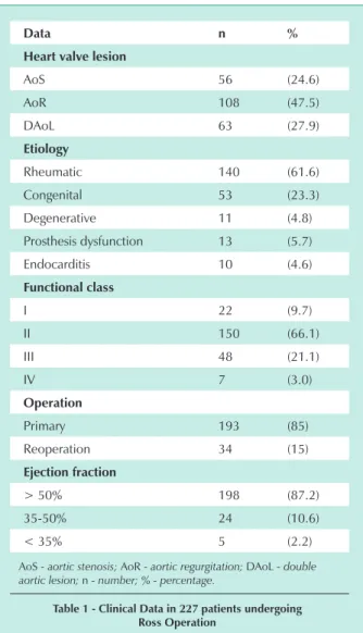
Five late deaths occurred. Four of them were sudden, in the 1st, 2nd, 4th and 36th months, whereas the fifth death resulted from endocarditis in the right homograft in the 4th year of follow-up. In the first three patients who had a sudden death, the clinical course was quite satisfactory and there were no clinical conditions indicating the occurrence of unfavorable events. The patient who had a sudden death in the 36th month had congenital aortic and subaortic stenosis with 4 previous surgeries, complete atrioventricular block, and was pacemaker dependent. He died in the 3rd postoperative year of a Ross-Kono operation. The late actuarial survival was 96.9% (95%CI = 94.2% - 99.6%) at 10 years, when only late deaths were considered, and 93.5% (95%CI = 90.1% - 97.0%) when hospital mortality was included (Fig. 1).
Of the 197 patients with known clinical course, 181 are in functional class I, and 15 in functional class II. One patient undergoing surgery with AoR and dilated cardiomyopathy remains with severe left ventricular dysfunction and is currently in functional class III.
No case of thromboembolism was detected in the late follow-up. Bacterial endocarditis occurred twice: one in the right ventricular outflow tract homograft causing one of the late deaths and another in the pulmonary autograft which despite having been eradicated with the use of antibiotics resulted in severe heart valve regurgitation becauseof perforation of one the valve cusps; thus reoperation was required. No recurrences were observed among the 10 patients undergoing surgery because of infeccious endocarditis.
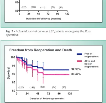
In the follow-up 11 reoperations were performed. They are listed in Table 2. The interval between the primary operation and the reoperation ranged from 6 to 60 months. Two patients underwent reoperation with simultaneous dysfunction of the valve auto and homograft; three patients had pulmonary autograft dysfunction; two of the right valve homograft; two underwent surgery because of progression of rheumatic mitral valve disease; one patient required coronary artery bypass grafting because of ostial lesion of the left coronary artery; and one patient who had tricuspid endocarditis that progressed to the ring of the pulmonary autograft required aortic and tricuspid replacement. No deaths occurred in the reoperations. The probability of freedom from reoperation was 92.3% (95%CI – 87.8% - 96.8%) after 10 years of follow-up. The probability of survival and freedom from reoperation at 10 years was 89.4% (95%CI = 84.3% - 94.5%) (Fig. 2).
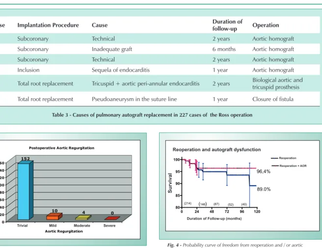
Pulmonary autograft replacement was required in six patients, as shown in Table 3.
and inadequate autograft in the other. All had their pulmonary autografts replaced by cryopreserved aortic homografts.
No patients undergoing surgery with the total aortic root replacement procedure required reoperation because of primary dysfunction of the pulmonary autograft.
One patient developed bacterial endocarditis in the tricuspid heart valve, which progressed to the Teflon strip around the pulmonary autograft with formation of a significant paravalvar abscess. Although the pulmonary autograft had not been affected by the infeccious process and had a normal function, it was injured during debridement of the infected tissues, and the patient had to undergo placement of biologic prostheses in the aortic and tricuspid positions.
One patient presented pseudoaneurysm in the proximal suture line of the pulmonary autograft with the aortic ring, which could be corrected directly without valve replacement.
In the last echocardiographic assessment, 152 pulmonary autografts were competent or showed trivial regurgitation, 10 presented mild regurgitation, whereas four patients had moderate heart valve regurgitation (Fig. 3).
Three of them underwent surgery in 1995. They were rheumatic and the autografts were implanted with the total root replacement procedure. In the first case, the heart valve regurgitation had already been graded as moderate in the 1st year of follow-up and remained unchanged up to the 9th year of follow-up. In the second patient, there was a
strong suspicion of new rheumatic attacks because despite the development of moderate regurgitation in the pulmonary autograft the progression of the mitral valve disease was even more significant. In the third patient, the pulmonary autograft presented mild regurgitation in the 3rd year of follow-up, progressing to moderate in the 9th year of late follow-up.
Another patient with aneurysm of the ascending aorta, bicuspid aortic valve and marked dilation of the aortic valve ring presented moderate heart valve regurgitation of the autograft as from the 6th postoperative month, but remained stable up to the second year of follow-up.
The probability of freedom from reoperation with the pulmonary autograft was 96.4% (95%CI = 93.6% - 99.2%) at 10 years of follow-up, whereas 89% (95%CI = 79.6% - 93.6%) were free from reoperation and with a normofunctional autograft (Fig. 4).
Pulmonary autograft gradients were consistently low both in the immediate phase and later. In the last echocardiographic assessment, mean peak instantaneous gradients was 8 ± 3.2mmHg (min = 2, max= 32). In only 2 cases the gradients were higher than 20 mmHg. In the first, the 24-mmHg residual gradient is found in the left ventricular outflow tract in a patient with congenital aortic and subaortic stenosis despite an enlargement using the Ross-Konno procedure. The second case results from recurrent rheumatic attack, and the patient has been currently diagnosed with moderate DAoL (gradient = 32 mmHg) and DMiL after 9 years of follow-up. As a result of the good hemodynamic performance of the autografts, the left ventricular mass decreased from 288± 45g in the preoperative period to 197±39g later.
The dimensions of the autograft at the annulus, of the sinuses of Valsalva, and of the sinotubular junction in the 110 patients with late control echocardiogram performed in our institution are listed in Table 4. These tests were performed after a mean 41-month follow-up (min=2, max=114). No aneurysmatic dilatations were observed in the autograft, and the largest dimension observed was 4.2 and 4.3 cm in the sinuses of Valsalva and in the sinotubular junction, respectively.
Four patients underwent reoperation to replace the valvar homograft in the right ventricular outflow tract. In all, the valvar cusps were morphologically normal; however, a significant perivascular fibrotic reaction with marked retraction and extrinsic compression of the arterial wall of the conduit causing
Cause of reoperation Cases
Auto + homograft dysfunction 2
Primary autograft dysfunction 2
Autograft pseudoaneurysm 1
Homograft dysfunction 2
Progression of mitral heart valve disease 2
Ostial lesion of the left coronary artery 1
Tricuspid and aortic endocarditis 1
table 2 - Causes of Reoperation in 227 patients undergoing the Ross operation
Fig. 1 - Actuarial survival curve in 227 patients undergoing the Ross operation.
Curva atuarial de Sobrevida
0 24 48 72 96 120
80 85 90 95 100
Incluindo a mortalidade imediata
Somente —bitos tardios
(227) (153) (111) (71) (40)
96,5%
93,5%
tempo de Seguimento (meses)
Late deaths
early mortality and late deaths
actuarial survival curve
Duration of Follow-up (months)
Su
rv
iv
a
l
Fig. 2 - Actuarial curve showing the probability of freedom from reoperation and survival free from reoperation.
Free of reoperations alive and free of reoperations
Freedom from reoperation and Death
Duration of Follow-up (months)
Su
rv
iv
a
l
0 24 48 72 96 120
80 85 90 95 100
(227) (146) (105) (64) (36)
92.38%
diffuse tubular stenosis was observed.
In addition, ten patients although asymptomatic had peak instantaneous gradients higher than 40 mmHg in the late echocardiographic assessment. In 8 of them, these gradients had already been detected in the first two years of postoperative follow-up and remained unchanged or had a slight progression in subsequent years. The probability of freedom from reoperation in the right heart valve homograft was 96.2% (95% CI = 94.1% - 98.3%) at 10 years of follow-up, and 85.5% (95% CI = 82.9% - 88.3%) are free from reoperation and with peak instantaneous gradients ≤ 40 mmHg (Fig. 5).
The reconstruction procedure and/or method of homograft preservation had a significant influence on the late gradients found in the right ventricular outflow tract. In group 1 (conventional homografts, without pericardial extension) mean peak instantaneous gradients was 24.2±17.7mmHg, and 9 patients (13.4%) had a gradient higher than 40 mmHg. In group 2 (conventional homografts with proximal extension performed with pericardium) the peak instantaneous gradient was 17.2±11.8 mmHg , and only 1 patient (4.1%) had a gradient higher than 40mmHg. In group 3 (decellularized homografts), the peak instantaneous gradient was 10.7±4.4mmHg, and the highest gradient observed in this group was 22mmHg.
The univariate analysis of risk factors for the development of high gradients in the right ventricular outflow tract
demonstrated that, in addition to the method ofhomograft preservation, only a patient’s age - under 20 years - was associated with late stenoses (Tab. 5).
Discussion
This study corroborates the thesis that the Ross operation is a safe option in the surgical treatment of children and young adults with aortic heart valve disease. The hospital mortality of 3.5% seems quite acceptable especially when the complexity of our cases which include mitral-aortic patients, reoperations, aneurysms of the ascending aorta and bacterial endocarditis in native heart valves and prosthetic valves is considered. The increased risks of the Ross operation in these circumstances have already been extensively discussed5. In addition, with a larger experience we were able to improve immediate results even further so that only one hospital death occurred in the past eighty cases operated.
The advantages of the use of a physiological valve substitute were evidenced by the excellent late survival with absence of thromboembolic events even when no anticogulation therapy was used, as well as the low prevalence of infectious complications, and the significant functional recovery. Although this study is not comparative, the longer survival and improved quality of life in patients undergoing Ross operation when compared to other valvar substitutes have already been well documented by other authors2,17.
Case implantation procedure Cause duration of
follow-up operation
1 Subcoronary Technical 2 years Aortic homograft
2 Subcoronary Inadequate graft 6 months Aortic homograft
3 Subcoronary Technical 2 years Aortic homograft
4 Inclusion Sequela of endocarditis 1 year Aortic homograft
5 Total root replacement Tricuspid + aortic peri-annular endocarditis 2 years Biological aortic and
tricuspid prosthesis
6 Total root replacement Pseudoaneurysm in the suture line 1 year Closure of fistula
table 3 - Causes of pulmonary autograft replacement in 227 cases of the Ross operation
Fig. 3 - Late aortic regurgitation.
152
10 4
0
0 20 40 60 80 100 120 140 160
N
Trivial Leve Moderada Severa
Aortic Regurgitation Postoperative Aortic Regurgitation
trivial Mild Moderate Severe
Free of reoperations Free of reoperations and without autograft dysfunction
Fig. 4 - Probability curve of freedom from reoperation and / or aortic regurgitation.
Reopera‹o e disfun‹o do autoenxerto
0 24 48 72 96 120 80
85 90 95
100 Reopera›es
Reopera›es +Iao
(214) (146) (87) (52) (40)
96,4%
89.0%
tempo de Seguimento (meses)
reoperation and autograft dysfunction
Su
rv
iv
a
l
Duration of Follow-up (months)
reoperation
Nevertheless, the occasional need of late reoperations in the valve auto and/or homograft remains the major concern following the procedure.
More recently, some authors have reported progressive dilation and aortic valve regurgitation as the major cause of late reoperation in patients undergoing the Ross operation with the total root replacement procedure. David et al analyzed 118 patients with a mean follow-up duration of 44 months and found 7 cases of moderate or severe aortic regurgitation, which represented 92% of freedom from this complication after 6 years of follow-up. According to the authors, patients with bicuspid aortic valve and aortic ring dilation comprise a subset of patients more prone to late dilation, because they have more marked degenerative alterations in the wall of the ascending aorta and pulmonary truncus7. In Elkins et al’s experience, only 86% of the patients were free from reoperation in pulmonary autograft after 8 years of follow-up. In 206 patients with a mean 28-month follow-up 11 reoperations were necessary because of valvar regurgitation, and annular dilation was the most frequent cause. Using multiple regression analysis, they found that preoperative diagnosis of aortic stenosis, previous sternotomy, and the total root replacement procedure were factors that decreased the risk of development of late valve regurgitation5. On the other hand, in David et al’s experience the inclusion procedure was associated with less dilation and valve regurgitation than the total aortic root replacement procedure7. Kouchoukos et al reoperated 11 pulmonary autografts among 119 patients with a mean 51-month follow-up. The majority of the reoperations occurred after the fifth year of follow-up, and progressive autograft dilation was the most common cause. The probability of freedom from reoperation in the pulmonary autograft was
only 75% at ten years. As an addendum to their publication, the authors informed that seven additional patients were reoperated because of aortic regurgitation between 5.5 and 11.5 years of follow-up8.
Our results do not corroborate these observations; they show significant differences not only in the prevalence but also in the way and temporal relation of this complication. In the 227 patients of this series, only 6 underwent reoperation because of autograft dysfunction, which resulted in 96% free from this event at 10 years of follow-up. All reoperations were necessary before the fifth postoperative year, and technical failure and bacterial endocarditis were the most frequent causes of dysfunction. It is important to point out that none of the patients undergoing surgery with the total aortic root replacement was reoperated in the first ten years. Of the four patients with late moderate AoR none had dilation of the annulus and of the sinuses of Valsalva. In two of our cases, there is a strong suspicion of new rheumatic attacks as the cause. Data similar to ours were reported by Carr-White et al who did not demonstrate dilations greater than 20% in the autografts of 49 patients with up to 4 years of follow-up18.
Several factors may influence the dilation of the pulmonary autograft. Details in the surgical procedure such as intra-annular implantation, space orientation of the graft, reinforcement of the proximal suture with Teflon or pericardium strips, and correction of discrepancies between the diameters of the autograft and the proximal and distal portions of the aorta are not performed evenly among the surgical groups18-20. The adequate control of blood pressure, especially in the first few postoperative months, may also be significant because in this phase the pulmonary arterial wall is not yet adequately remodeled to support systemic pressures18.
Some studies demonstrated that the pulmonary arterial wall of patients undergoing the Ross operation had varying degrees of defficiency and fragmentation of its elastic fibers preoperatively, especially in those with bicuspid aortic valve7,18. In most of the American series with the Ross operation, bicuspid aortic valve is the most frequent etiology, which can explain the high incidence of late dilation8. Our series is comprised of 60% of rheumatic patients and this can, at
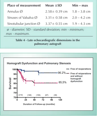
place of measurement Mean ±Sd Min – max
Annulus Ø 2.58± 0.39 cm 1.8 – 3.8 cm
Sinuses of Valsalva Ø 3.31± 0.58 cm 2.0 – 4.2 cm
Sinotubular junction Ø 3.37± 0.55 cm 1.9 – 4.3 cm
ø - diameter; SD - standard deviation; min - minimum; max - maximum.
table 4 - late echocardiografic dimensions in the pulmonary autograft
Disfun‹o do Homoenxerto e Estenose Pulmonar
0 24 48 72 96 120 70
75 80 85 90 95 100
Livres de Reopera‹o
e de Gradientes > 40 mmHg
Livres de Reopera‹o
96.2%
85,5%
(214) (146) (106) (67) (38)
tempo de Seguimento (meses)
Fig. 5 - Probability curve of freedom from reoperation and from right heart valve homograft stenosis.
Free of reoperations Free of reoperations and without homograft dysfunction
Homograft Dysfunction and Pulmonary Stenosis
Su
rv
iv
a
l
Duration of Follow-up (months)
parameter p Significance
Decellularized X cryopreserved 0.0002 S
Decellularized X enlargement 0.07 NS*
Enlargement X cryopreserved 0.06 NS*
Recipient age < 20y 0.03 S
Homograft diameter 0.41 NS
Homograft diameter / Body
surface 0.81 NS
ABO compatibility 0.09 NS
Donor age 0.74 NS
Donor age – recipient age 0.84 NS
* Borderline statistical significance.
least in part, justify the difference in the results. In addition, our mean 44-month follow-up may also be insufficient for more definitive conclusions, which emphasizes the need of a continued observation of these patients. The late dimensions observed here suggest that the pulmonary autograft geometry is different from that of the native aortic root, and sinotubular junction diameters are significantly larger than those at the annulus.Our measurements are similar to those presented by Carr-White et al, and the fact that the duration of our follow-up is much longer reinforces the stability and maintenance of autograft diameters throughout time18.
Another relevant aspect in the Ross operation is related to the growth potential when implanted in children21. Elkins et al analyzed 86 children undergoing this surgery and demonstrated that the growth of the pulmonary autograft was proportional to the somatic development. Unfortunately, in our case series data regarding dimensions of the ring, of the sinuses of Valsalva and of the sinotubular junction were only collected in the late assessment and could not be systematically compared to the immediate postoperative dimensions, which prevents us from documenting the growth of the pulmonary autograft in our pediatric patients.
The involvement of the pulmonary autograft in new attacks of rheumatic disease has already been recorded by other authors, and this is probably the cause of moderate AoR in two of our cases. For this reason, the adequate prophylaxis against this disease should be strictly followed22.
Although valve homografts last longer in the right side of the circulation because of lower pressure levels, they are also subject to dysfunction and the need of reoperations. The possibility of stenosis due to retraction of the arterial wall of the conduit or located in the area of the distal anastomosis is a well-established complication following the Ross Operation9,10.
Our experience corroborates other authors’ observation, demonstrating that although homografts show a normal hemodynamic performance immediately after surgery, a mild/moderate increase in gradients frequently occurs as a result of fibrotic retraction of the conduits. This is an early process occurring in the first two postoperative years and which tends to stabilize thereafter. In most of the patients, peak instantaneous gradients did not exceed 20 mmHg and probably have a limited clinical impact. However, in some cases the inflammatory reaction is more intense, causing more severe stenosis and reoperation may be required. As already demonstrated in other studies, children and adolescents have a higher risk of developing this complication and this also occurred in our series9,10,23.
In 114 patients undergoing the Ross performed by Ward et al, 20% showed peak instantaneous gradients between 25-40 mmHg in the pulmonary homograft and in 4% of the cases this gradient was higher than 50 mmHg. In patients with severe stenosis, the high gradients were detected early, between the 4th and 12th postoperative months. Using echocardiography the authors concluded that most of the homografts show an approximately 15% retraction or more in some cases. As a result, they recommend the routine use of oversized homografts10. However, our experience corroborates Moidl et al’s observations that the use of oversized homografts by itself
was not enough to prevent this complication24.
Carr-White et al conducted a detailed study on the performance of homografts in the Ross Operation and detected pulmonary gradients higher than 30 mmHg in 17% of the cases, and higher than 50 mmHg in approximately one third. In patients who developed late gradients, the magnetic resonance imaging demonstrated not only a circumferential retraction in the conduits, but also an approximately 40% reduction in their length. Among the possible cause for this retraction, the authors suggest that the tension on the conduit wall may cause the release of tissue factors that stimulate a fibrotic healing reaction9. These findings encouraged us to modify the procedure of the reconstruction of the right ventricular outflow tract, by elongating the proximal portion of the homograftswith pericardium patches and by relieving the tension in the graft body and in the anastomoses (group 2). As a result, a reduction in late gradients occurred (group 1 = 24.2 mmHg versus group 2 = 17.2 mmHg), and only one patient undergoing this procedure had a gradient higher than 40 mmHg. The statistical difference between the groups was borderline (p = 0.06), but we believe that this was due to the relatively small number of cases studied. The efficiency of this maneuver in reducing late pulmonary gradients was corroborated by Betchet et al25.
Although the mechanisms causing stenosis in right homografts are not completely understood, the occurrence of postoperative fever and the presence of a chronic inflammatory reaction in the adventitia with perivascular lymphocytic infiltrate in explanted homografts suggest that immunological phenomena are involved9,10. In addition, several studies have demonstrated that the use of valve homografts results in elevation of circulating HLA class I and II antibodies. However, the correlation between the degree of immune rejection and late graft dysfunction is still controversial26,27.
The decellularization process eliminates endotelial and interstitial cells from the cusps and arterial wall of homografts, significantly reducing their immunogenic potential which could, at least in theory, reduce or postpone the occurence of stenosis in homografts implanted in the right ventricular outflow tract28,29. In Betchel et al’s experience, the use of decellularized homografts with Synergraft technology was efficient in reducing the immune reaction but did not prevent the development of late gradients25.
Our experience with decellularized homografts is not in line with that of Betchel et al’s, possibly because of different decellularization techniques25,30. We recently published our results with decellularized homografts which confirm a significant reduction in antigenicity and in late gradients31. The patients in the present study (group 3) corroborate the thesis that up to 3 years of follow-up decellularized homografts have a normal hemodynamic performance with no elevation in late gradients. This fact is quite encouraging, given that in our experience the complications of right homografts were the most prevalent following the Ross Operation. Obviously, long-term results will be required to confirm the advantages of this new technology.
functional recovery. The pulmonary autograft had a low incidence of dysfunction and no late dilations were observed during the follow-up. No patients undergoing surgery with the total aortic root replacement procedure were reoperated because of primary dysfunction. Modifications in the surgical and/or graft tissue preservation procedures minimized the problems related to the right ventricular outflow tract.
Based on these results, we consider the Ross operation to be the best alternative in the surgical treatment of aortic valve disease in children and young patients.
potential Conflict of interest
No potential conflict of interest relevant to this article was reported.
1. Elkins RC, Lane MM, McCue C. Ross operation in children: late results. J Heart Valve Dis. 2001; 10(6): 736-41.
2. Concha M, Aranda PJ, Casares J, Merino C, Alados P, Munoz I, et al. Prospective evaluation of aortic valve replacement in young adults and middle-aged patients: mechanical prosthesis versus pulmonary autograft. J Heart Valve Dis. 2005; 14(1): 40-6.
3. Elkins RC, Knott-Craig CJ, Ward KE, Lane MM. The Ross operation in children:10-year experience.Ann Thorac Surg. 1998; 65(2): 496-502.
4. Bohm JO, Botha CA, Hemmer W, Starck C, Blumenstock G, Roser D, et al. Older patients fare better with the Ross operation. Ann Thorac Surg. 2003; 75(3): 796-801; discussion 802.
5. Elkins RC, Lane MM, McCue C. Pulmonary autograft reoperation: incidence and management. Ann Thorac Surg. 1996; 62(2): 450-5.
6. Chambers JC, Somerville J, Stone S, Ross DN. Pulmonary autograft procedure for aortic valve disease: long-term results of the pioneer series. Circulation. 1997; 96(7): 2206-14.
7. David TE, Omran A, Ivanov J, Armstrong S, de Sa MP, Sonnenberg B, et al. Dilation of the pulmonary autograft after the Ross procedure. J Thorac Cardiovasc Surg. 2000; 119(2): 210-20.
8. Kouchoukos NT, Masetti P, Nickerson NJ, Castner CF, Shannon WD, Davila-Roman VG. The Ross procedure: long-term clinical and echocardiographic follow-up. Ann Thorac Surg. 2004; 78(3): 773-81; discussion.
9. Carr-White GS, Kilner PJ, Hon JK, Rutledge T, Edwards S, Burman ED, et al. Incidence, location, pathology, and significance of pulmonary homograft stenosis after the Ross operation. Circulation. 2001; 104(12 Suppl 1): I16-20.
10. Ward KE, Elkins RC, Overholt ED, Knott-Craig CJ, Razook JD, Lane MM, et al. Evaluation of cryopreserved homografts in the right ventricular outflow tract after the Ross procedure: intermediate-term follow up. J Heart Valve Dis. 1997; 6(2): 130-3.
11. Costa FDA, Poffo R, Matte E, Sardeto EA, Schneider RA, Ada EP, et al. Cinco anos de experiência com a operação de Ross. O que aprendemos? Rev Bras Cir Cardiovasc. 2000;15(2):109-28.
12. Manouguian S. Patch enlargement of the aortic valve ring. Description of a new operation method. An experimental and clinical study. Fortschr Med. 1983; 101(1-2): 31-3.
13. Hraska V, Krajci M, Haun Ch, Ntalakoura K, Razek V, Lacour-Gayet F, et al. Ross and Ross-Konno procedure in children and adolescents: mid-term results. Eur J Cardiothorac Surg. 2004; 25(5): 742-7.
14. Edmunds LH Jr, Clark RE, Cohn LH, Grunkemeier GL, Miller DC, Weisel RD. Guidelines for reporting morbidity and mortality after cardiac valvular operations. Eur J Cardiothorac Surg. 1996; 10(9): 812-6.
15. Perry GJ, Helmcke F, Nanda NC, Byard C, Soto B. Evaluation of aortic insufficiency by Doppler color flow mapping. J Am Coll Cardiol. 1987; 9(4): 952-9.
16. Roman MJ, Devereux RB, Kramer-Fox R, O’Loughlin J. Two-dimensional echocardiographic aortic root dimensions in normal children and adults. Am J Cardiol. 1989; 64(8): 507-12.
17. Carr-White GS, Glennan S, Edwards S, Ferdinand FD, Desouza AC, Pepper
JR, et al. Pulmonary autograft versus aortic homograft for rereplacement of the aortic valve: results from a subset of a prospective randomized trial. Circulation. 1999; 100(19 Suppl.): II103-6.
18. Carr-White GS, Afoke A, Birks EJ, Hughes S, O’Halloran A, Glennen S, et al. Aortic root characteristics of human pulmonary autografts. Circulation. 2000; 102(19 Suppl 3): III15-21.
19. Hokken RB, Bogers AJ, Taams MA, Schiks-Berghourt MB, van Herwerden LA, Roelandt JR, et al. Does the pulmonary autograft in the aortic position in adults increase in diameter? An echocardiographic study. J Thorac Cardiovasc Surg. 1997; 113(4): 667-74.
20. Sievers HH, Leyh R, Loose R, Guha M, Petry A, Bernhard A. Time course of dimension and function of the autologous pulmonary root in the aortic position. J Thorac Cardiovasc Surg. 1993; 105(5): 775-80.
21. Elkins RC, Knott-Craig CJ, Ward KE, McCue C, Lane MM. Pulmonary autograft in children: realized growth potential. Ann Thorac Surg. 1994; 57(6): 1387-93.
22. Pieters FA, Al-Halees Z, Hatle L, Shahid MS, Al-Amri M. Results of the Ross operation in rheumatic versus non-rheumatic aortic valve disease. J Heart Valve Dis. 2000; 9(1): 38-44.
23. Xie GY, Bhakta D, Smith MD. Echocardiographic follow-up study of the Ross procedure in older versus younger patients. Am Heart J. 2001; 142(2): 331-5.
24. Moidl R, Simon P, Kupilik N, Chevtchik O, Heinrich N, Moritz A, et al. Increased pulmonary flow velocities in oversized homografts in patients after the Ross procedure. Eur J Cardiothorac Surg. 1997; 12(4): 569-72; discussion 573.
25. Bechtel JFM, Steinhardt MM, Schnidtke C, Brunswik A, Stierle U, Sievers HH. Evaluations of the Decellularized Valve Homograft (SynerGraftTM). J Heart Valve Dis. 2003;12:734-40 – in discussion.
26. Welters MJP, Oei FBS, Witvliet MD, Vaessen LMB, Dijkhuis AHC, Borges JJC, et al. A Broad and Strong Humoral Immune Response to Donor HLA after Implantation of Cryopreserverd Human Heart Valve Allografts. Human Immunology. 2002;63:1019-1025.
27. Dignan R, O´Brien M, Hogan P, Thornton A, Fowler K, Byrne D, et al. Aortic valve allograft structural deterioration is associated with a subset of antibodies to human leukocyte antigens. J Heart Valve Dis. 2003;12(3):382-90; discussion 390-1.
28. O´Brien MF, Goldstein S, Walsh S, Black, Elkins R, Clarke D. The synergraft valve: A new acellular (nonglutaraldehyde-fixed) tissue heart valve for autologous recellularization First Experimental Studies Before Clinical Implantion. Semin Thorac Cardiovasc Surg. 1999; 11:194-200.
29. Elkins RC, Lane MM, Capps SB, McCue C, Dawson PE. Hunoral Humoral immune response to allograft valve tissue pretreated with an antigen reduction process. Semin Thorac Cardiovasc Surg. 2001;13(suppl I):82-6.
30. Kasimir MT, Rieder E, Seebacher G, Silberhumer G, Wolner G, Weigel G, et al. Comparision Comparison of different decellularization procedures of porcine heart valves. Int J Artif Organs. 2003;26:421-7.
31. Costa FDA, Dohmen PM, Duarte D, Glenn CV, Lopes SV, Haggi H, et al. Immunological and echocardiographic evaluation of decellularized versus cryopreserved allografts during the Ross operation. Eur J Cardiothorac Surg. 2005 27: 572-8.