Prognostic Value of Left Atrial Volume Index in Hemodialysis
Patients
Silvio H. Barberato and Roberto Pecoits Filho
Pontiical Catholic University of Paraná – Curitiba, PR - Brazil
Summary
Objective: To evaluate the prognostic value of left atrial volume index (LAVi) in the clinical course of hemodialysis (HD) patients, compared with previously established echocardiographic and clinical parameters.
Methods: Echocardiograms were obtained from 118 hemodialysis patients, who were then followed for 19 ± 8 months. Study endpoint was a composite of all-cause mortality and nonfatal cardiovascular events. Cox multivariate analysis was used do assess the independent prognostic value of LAVi.
Results: On univariate analysis, LAVi and other clinical and echocardiographic parameters were predictive of prognosis. Multivariate analyses showed that LAVi was an independent predictor of prognosis (hazard ratio 1.03 per ml/m2, 95%
conidence interval: 1.01 to 1.05, p=0.014), and added incremental information to the model containing traditional predictors of cardiovascular risk, such as left ventricular mass, ejection fraction, and clinical variables (p=0.02).
Conclusion: LAVi is an independent predictor of prognosis in HD patients, providing incremental information to traditional clinical and Doppler echocardiographic data. (Arq Bras Cardiol 2007;88(6):568-575)
Key words: Hypertrophy, left ventricular; renal dialysis; risk assessment; echocardiography, Doppler.
Mailing address: Silvio H. Barberato •
Rua Saint Hilare, 122/203 - 80240-140 – Curitiba, PR - Brazil E-mail: [email protected]
Manuscript received November 7, 2006; revised manuscript received December 18, 2006; accepted December 18, 2006.
Introduction
Cardiovascular disease is the leading cause of death in chronic renal failure patients on renal replacement therapy with hemodialysis (HD)1,2. The excess cardiovascular risk in
this group is caused by the interaction between traditional risk factors for cardiovascular disease and those related to chronic kidney disease (CKD)1. Even though these patients have
accelerated atherosclerosis3, structural cardiac changes such as
hypertrophy and left ventricular dilation (resulting in diastolic and systolic dysfunction) are associated with the incidence of congestive heart failure and high morbidity and mortality rates4,5. For many decades now Doppler echocardiography has
been widely used to evaluate cardiac structure and function, and thus has played a key role in characterizing individuals at higher cardiovascular risk. Former studies using the method to predict cardiovascular risk in hemodialysis population focused primarily on the role of hypertrophy4,6 and left
ventricular (LV) systolic dysfunction7,8. It has been recently
postulated that left atrial (LA) dilation, best represented by planimetric determination of LA volume index (LAVi) on two-dimensional echocardiography9, is related to the duration of
left ventricular diastolic dysfunction10,11, and is a powerful
marker of cardiovascular risk in the general population11,12
and in some clinical populations as well13-16. As LV diastolic
function seems to be impaired in most HD patients, even in
those asymptomatic5, we speculate that LAVi may be useful
in stratifying cardiovascular risk in this group. This study was designed to assess the prognostic value of LAVi in HD patients, compared with previously established clinical and Doppler echocardiographic parameters.
Methods
Survival analysis - Demographics, the presence of comorbidities (history of diabetes mellitus, hypertension, acute myocardial infarction, angina documented by coronary angiography, stroke, and clinical diagnosis of heart failure), drugs in use, and routine laboratory tests (hemoglobin, albumin, calcium, and phosphorus) were determined based on a thorough analysis of the medical chart combined with an interview with the patient or the attending physician. Study patients were prospectively followed from June 2003 to July 2006 or until an endpoint occurred. The study’s composite endpoint was all-cause mortality and nonfatal cardiovascular events. The following events were observed: 1) death; 2) new coronary event, deined as nonfatal acute myocardial infarction or angina pectoris with coronary stenosis > 50% on coronary angiography; 3) ischemic or hemorrhagic stroke, transient ischemic attack excluded; 4) clinical diagnosis of congestive heart failure requiring hospitalization. These events were researched through periodic review of medical documentation, including clinical records and death certiicates, in addition to contact with the doctor and patient’s relatives. In case of multiple events in a single patient, only the irst event was considered. Patients who underwent renal transplantation were censored in the analysis.
Statistical analysis - Data are presented as mean and standard deviation (parametrically distributed continuous variables), median (nonparametrically distributed continuous variables), and percentage (categorical variables). Diferences between groups were determined using the unpaired Student’s t test, the Mann-Whitney test, or the chi-square test. LAVi independent prognostic value was tested using Cox multivariate survival analysis. Initially, groups with and without the study endpoint were compared, in order to select variables for proportional risk analysis. The E/E’ ratio was specifically included in the model because an earlier study showed that this index was associated with increased mortality in candidates for renal transplantation31.
Significant univariate predictors were added to the Cox model (entry and retention set at a signiicance level of 0.1 and 0.05, respectively) in several steps, simulating the clinical reasoning). Thus, the first step used clinical and biochemical variables as baseline risk factors. Subsequently, traditional Doppler echocardiographic variables were added. The last steps consisted of the sequential introduction of E/E’ ratio and LAVi as continuous variables. The increase in predictive power at each step was assessed by change in chi-square value. Kaplan-Meier curves were plotted for LAVi as categorical variable, using a partition value of 32 ml/m2. The
statistical signiicance level was set at 5% (p < 0.05). Data were processed by using “SPSS 13.0 for Windows” (SPSS INC, Chicago, Illinois).
Results
One hundred and thirty-one subjects were assessed. Nine patients were excluded (three because of arrhythmias, three because of major mitral regurgitation, one because of aortic stenosis, and one because of pericardial efusion), and four patients moved to another city and were lost to follow-up. Table 1 summarizes demographic, clinical, biochemical, height and weight. Body surface area was calculated according
to DuBois & DuBois’s simpliied formula (0.20247 x weight0.425
x height0.725)18. Body mass index was calculated by dividing
weight (kg) by squared height (m). Those with BMI > 30 were considered obese.
Doppler echocardiogram - All examinations were performed by a single examiner (S.H.B.) on interdialytic days (Tuesday or Thursday only) between noon and 6 PM, as previously recommended19. M-mode, two-dimensional and
Doppler echocardiography (pulsed, continuous, color, and tissue imaging) were performed using an HDI 3000 system (ATL-Philips Ultrasound Systems, Bothell, Washington, USA) equipped with a 2,5-4 MHz transducer capable of operating with fundamental and second-harmonic imaging. According to the Penn convention20, the following linear measurements
were obtained: interventricular septal thickness, posterior wall thickness, LV end-diastolic and end-systolic diameters. The end-diastolic diameter upper limit was established at 55 mm. Left ventricular mass was calculated using Devereux’s formula21 and indexed to body height raised to the 2.7th
power; hypertrophy was diagnosed when LV mass index (LVMi) was greater than 51 g/m2,7 22. In order to assess LV
geometric pattern, relative wall thickness was calculated using the formula: (2 x mean wall thickness)/LV end-diastolic diameter, where mean thickness = (interventricular septal thickness + posterior wall thickness at diastole)/2. Reference value was 0.45, discriminating eccentric (below 0.45) from concentric (above 0.45) hypertrophy23. Concentric remodeling
was defined by normal LVMi associated with increased relative wall thickness. Left ventricular systolic function was assessed by calculating ejection fraction using the Teichholz24
method, the limit of which was deined as 50%. Mitral inlow velocities were measured in the apical four-chamber view with pulsed-Doppler sample placed between the leaflet tips of the mitral valve; at this time patients were instructed to breathe in a calm, controlled manner. According to American Society of Echocardiography recommendations25,
the following parameters were measured: early rapid illing (E) velocity, atrial contraction velocity (A), E/A ratio, and E-wave deceleration time (DT). DT < 140 ms, associated with a restrictive illing pattern, was considered abnormal26. Mitral
annular velocities measured by tissue Doppler were recorded in the apical four-chamber view, with a 1- to -2 mm sample volume placed at the junction of LV septal27 and lateral28 walls
with the mitral annulus. Both early (E’) and late (A’) diastolic mitral annular velocities were determined, in addition to E’/A’ and E/E’ ratios. Diastolic dysfunction was deined as: 1) E/A < 1 (abnormal relaxation); 2) E/A > 2 (restrictive low); 3) E/A between 1 and 2 in association with E/E’ > 10 (pseudonormalization)29. Left atrial size was assessedby
M-mode measurement of anteroposterior dimension (abnormal when > 40 mm), as previously recommended30, and also by
LA volume calculated by using two-dimensional planimetry with the biplanar Simpson’s rule9 on the frame just before
mitral valve opening. Left atrial volume index (LAVi) was calculated by the LA volume to body surface area ratio12;
values greater than 32 ml/m2 are suggestive of increased
cardiovascular risk11,13,14. All Doppler echocardiographic
and Doppler echocardiographic characteristics of the 118 remaining patients that made up this study population. Our group comprised 64 men and 54 women, mean age 48 ± 15, and median HD duration of 22 months (1 to 120). CKD etiology was attributed to hypertensive nephrosclerosis (40%), chronic glomerulonephritis (24%), diabetic nephropathy (19%), policistic kidney (8%), chronic pyelonephritis (6%), and other conditions (3%). Most patients (76%) were on antihypertensive drugs, particularly angiotensin-converting enzyme inhibitors (45%), beta-blockers (19%), alpha-blockers (13%), calcium channel antagonists (13%), and angiotensin receptor blockers (10%), alone or combined.
Previous cardiovascular disease - Forty-eight patients (41%) had history of heart disease (heart failure in 32%, myocardial infarction in 5%, and angina in 3%). Moreover, 2% had history of stroke, and none had peripheral arterial disease. Eleven
percent of the patients were obese.
Doppler echocardiographic changes - Figure 1 shows the most prevalent echocardiographic changes found in our group. Twenty-six patients had LV dilation (22%), 106 had hypertrophy (90%), 21 had systolic dysfunction (18%), and 96 had diastolic dysfunction (81%). According to the relative wall thickness, the most frequent geometric change was concentric hypertrophy (77%), followed by eccentric hypertrophy (13%), and concentric remodeling (4%). Only 6% of the patients showed normal LV geometry. Thirty-two patients (27%) were diagnosed with LA dilation by M-mode echocardiography. Mean LAVi was 34 ± 15 ml/m2 (median 31, ranging from 17
to 89), and this index was greater than 32 ml/ m2 in 50% of
the study sample.
Prognosis - During the mean follow-up of 19 ± 8 months, there were 20 deaths and 17 nonfatal cardiovascular events, resulting in an overall endpoint rate of 31%. Deaths were categorized as cardiovascular (12 deaths, seven from acute myocardial infarction, four from sudden death, and one from hemorrhagic stroke) or infectious (eight). Nonfatal cardiovascular events included three cases of angina, one case of acute myocardial infarction, ten hospital admissions for decompensated heart failure, and three cases of ischemic stroke. Table 2 shows diferences among subgroups with and without endpoint. Patients who experienced study events were older (56 ± 15 vs 45 ± 13, p < 0,001); were on HD for a longer period (median 24 months vs 11 months, p = 0.014); had history of heart disease (heart failure plus infarction and angina: 73% vs 12%, p < 0.001) and diabetes (35% vs 12%, p = 0.004); showed lower serum albumin concentration (3.4 ± 0.4 vs 3.9 ± 0.6, p = 0,02), and were mostly male (67% vs 48%, p = 0.047). No differences were found in body mass index (p = 0.5), systolic blood pressure (p = 0.7), diastolic blood pressure (p = 0.8), hemoglobin (p = 0.2), and calcium-phosphorus product (p = 0.4). On echocardiogram, the subgroup of patients that reached the study endpoint had greater LV diastolic diameter (53 ± 6 vs 48 ± 7 mm, p < 0.001), LVMi (109 ± 44 vs 85 ± 34 g/m2.7, p = 0.002),
LA anteroposterior dimension (39 ± 6 vs 34 ± 6 mm, p = 0.001), and LAVi (42 ± 15 vs 30 ± 14 mL/m2, p < 0.001).
Consistent with these indings, this subgroup showed lower ejection fraction and higher E/E’ (57 ± 9 vs 64 ± 6%, p <
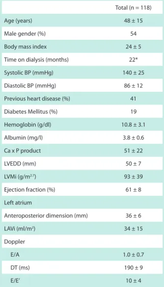
Table 1 - Primary baseline characteristics of the study population
Total (n = 118)
Age (years) 48 ± 15
Male gender (%) 54
Body mass index 24 ± 5
Time on dialysis (months) 22*
Systolic BP (mmHg) 140 ± 25
Diastolic BP (mmHg) 86 ± 12
Previous heart disease (%) 41
Diabetes Mellitus (%) 19
Hemoglobin (g/dl) 10.8 ± 3.1
Albumin (mg/l) 3.8 ± 0.6
Ca x P product 51 ± 22
LVEDD (mm) 50 ± 7
LVMi (g/m2.7) 93 ± 39
Ejection fraction (%) 61 ± 8
Left atrium
Anteroposterior dimension (mm) 36 ± 6
LAVi (ml/m2) 34 ± 15
Doppler
E/A 1.0 ± 0.7
DT (ms) 190 ± 9
E/E’ 10 ± 4
Dada presented in mean ± SD, percentages, or median with variation. BP - blood pressure; Ca x P - calcium x phosphorus; LVEDD - left ventricular end-diastolic diameter; LVMi - left ventricular mass index; LAVi - left atrial volume index; E - early transmitral illing velocity; A - transmitral low velocity during atrial contraction; DT - deceleration time; E/E’ - ratio
between E and early diastolic mitral annular velocity. *median. Fig. 1 - Percentage of main abnormalities in LV anatomy and function on echocardiogram. LV - left ventricle.
dilation
hypertrophy
systolic dysfunction
LV change abnormalities
0.001; and 13 ± 4 vs 8 ± 4, p < 0.001, respectively). The frequency of deceleration time (DT) < 140 ms was similar in the subgroups (p = 0.2), but there was a statistical trend toward shorter DT in patients who reached endpoints (175 ± 51 vs 197 ± 62 ms, p = 0.05). After the most signiicant predictors were selected (Table 2), the basic clinical model included age, male gender, previous heart disease, diabetes, time on HD, and albumin. Subsequently, we added ejection fraction, LVMi, and DT to the model, followed by E/E’ and, inally, LAVi. In order to prevent statistical colinearity, we sought not to use
correlated variables, such as LV end-diastolic diameter, which plays a role in ejection fraction and LVMi calculation.
Independent prognostic predictors, with respective hazard ratios and 95% conidence interval (CI), are listed in Table 3. After LAVi was added to the analysis, ejection fraction, LVMI and E/E’ were no longer independent predictors of the study endpoint. In the inal model, LAVi was the sole independent echocardiographic predictor of combined death and nonfatal cardiovascular events, with hazard ratio of 1.03 per ml/m2
(95% CI, 1.01-1.05, p = 0.014) from 32 ml/m2. Among the Table 2 - Comparison of demographic, clinical, biochemical and Doppler echocardiographic data of patients with and without the study endpoint
Without endpoint (n=81)
With endpoint
(n=37) p value
Age (years) 45 ± 13 56 ± 15 <0.001
Male gender (%) 48 67 0.047*
BMI 24 ± 6 25 ± 5 0.5
Time of dialysis (months) 11 24 0.014†
Diabetes mellitus (%) 12 35 0.004*
Previous heart disease (%) 12 73 <0.001*
Systolic BP (mmHg) 152 ± 26 148 ± 24 0.7
Diastolic BP (mmHg) 88 ± 12 86 ± 11 0.8
Hemoglobin (g/dl) 11 ± 3.3 10±2.1 0.2
Ca x P product 49 ± 22 56 ± 18 0.2
Albumin (mg/l) 3.9 ± 0.6 3.4 ± 0.4 0.02
LVEDD (mm) 48 ± 7 53 ± 6 <0.001
LV dilation (%) 15 30 0.05*
Ejection fraction (%) 64 ± 6 57 ± 9 <0.001
LVMi (g/m2.7) 85 ± 34 109 ± 44 0.002
E/A 0.98 ± 0.4 1.5 ± 1 0.01
DT (ms) 197 ± 62 175 ± 51 0.05
DT < 140 ms (%) 14 24 0.18*
E’/A’ 0.79 ± 0.3 0.80 ± 0.3 0.7
E/E’ 8 ± 4 13 ± 4 <0.001
E/E’ > 15 (%) 17 33 0.02*
LAD (mm) 34 ± 6 39 ± 6 0.001
LA dilation on M-mode (%) 22 38 0.08*
LAVi (ml/m2) 30 ± 14 42 ± 15 <0.001
LAVi > 32 ml/m2 (%) 36 81 <0.001*
other variables, the presence of previous heart disease and time on HD (over 21 months) reached statistical signiicance. The incremental value provided to the model by LAVi is shown in Figure 2 (chi-square increased from 69 to 77 in the inal step, p = 0.02).The endpoint rate was signiicantly higher in patients with LAVi > 32 ml/m2, compared with the subgroup
with LAVi ≤ 32 ml/m2 (51% vs 12%, respectively, p < 0.001).
Figure 3 shows adjusted Kaplan-Meier curves for endpoint-free survival using the partition value of 32 ml/m2. Annual
event-free survival was estimated at 98% for patients with LAVi ≤ 32 ml/m2 vs 79% for those with LAVi > 32 ml/m2 (p < 0.001).
Table 4 depicts the main clinical, Doppler echocardiographic, and biochemical features according to this partition value. Patients with LAVi > 32 ml/m2 had more heart failure and
LV echocardiographic abnormalities, such as hypertrophy, systolic dysfunction, and increased E/E’; in addition they were predominantly male. Conversely, no diferences were found in age, biochemical parameters and prevalence of hypertension, diabetes, and obesity. Death rate was higher among those with LAVi > 32 ml/m2 (30.5% vs 3.4%, p < 0.001). Among
59 patients with LAVi ≤ 32 ml/m2, only two died, in contrast
with 18 death among 59 patients with LAVi > 32 ml/m2.
Discussion
In recent years, echocardiographic assessment of the left atrium and its relationship with cardiovascular risk have been
Table 3 - Independent predictors of prognosis according to Cox multivariate analysis
HR 95% CI p value
Previous heart disease 1.91 1.79-1.96 < 0.001
Time on dialysis (months) 1.03 1.01-1.04 0.006
LAVI (per ml/m2) 1.03 1.01-1.05 0.014
HR - hazard ratio; CI - conidence interval; LAVI - left atrial volume index.
revalued. The most relevant inding of the present study is that LAVi may provide independent prognostic and incremental information to traditional clinical and echocardiographic data of HD patient population. Of all echocardiographic parameters assessed, LAVi was shown to be the best predictor of clinical course in this group, known to be at high cardiovascular risk. Left ventricular mass, recognized as a strong marker of mortality in this population4,6, was no longer an independent
predictor of endpoint after LAVi was included in the analysis. The presence of increased LAVi was also more prevalent than other risk indicators, such as ejection fraction and DT. This inding corroborates previous reports indicating that LAVi is a more effective parameter than systolic function analysis in predicting death in patients with acute myocardial infarction13,14 and dilated cardiomyopathy15,16.
Some speculations may be made to understand LAVi’s remarkable prognostic power. It is known that a positive correlation exists between LA size and LV passive properties32.
The progressive deterioration of myocardial diastolic properties causes an elevation in LV illing pressures. Consequently, LA pressure rises, causing it to dilate32. Therefore, it is highly likely
that LA size acts as a marker of diastolic dysfunction chronicity. In keeping with this proposition, we recently reported that LAVi is less sensitive to acute changes in preload than pulsed Doppler-derived parameters of mitral flow33. Additionally,
we have demonstrated that LAVi is correlated with diastolic dysfunction severity in HD patients34 and is more efective in
detecting pseudonormalization of mitral low, compared with various previously tested indices35. Thus, unlike mitral
low-derived Doppler indices, known to be volume-dependent and provide transient information about ventricular illing33,36, LAVi
appears to be a more stable marker of diastolic function, related to the cardiovascular “track record” built over time. This may also explain its predictive superiority over the E/E’ ratio, the most reliable non-invasive index for estimating LV end-diastolic pressure31,37, but responsive to acute preload changes.
It is likely that in clinical cohorts, with signiicant prevalence of cardiovascular disease, left atrial dilation may result not only from the long-standing diastolic dysfunction but also
0 10 20 30 40 50 60 70 80 90
Desfecho
Q
u
i-q
u
a
d
ra
d
o
Clínicos Clínicos+Eco tracicionais Clínicos+Eco tradic.+E/E' Clínicos+Eco tradic.+E/E'+IVAE
Fig. 2 - Increase in the prognostic predictive power of left atrial volume index (LAVi). Traditional echocardiographic data include left ventricular mass index, ejection fraction, and deceleration time. E/E’, ratio between early transmitral illing velocity and early diastolic mitral annulus velocity.
Traditional clinical and echo
Endpoint
C
h
i-s
q
u
a
re Clinical
Trad. clinical and echo + E/E’ Trad. clinical and echo + E/E’ + LAVi
Fig. 3 - Kaplan-Meier curves for endpoint-free survival based on a partition value of left atrial volume index (LAVi) of de 32 mL/m2 (p < 0.001).
% survival
follow-up (months)
%
Su
rv
iv
a
l
Follow-up (months)
IVAE < 32 ml/m2
Table 4 - Primary clinical, biochemical, and Doppler echocardiographic characteristics according to LAVi
Variable LAVi ≤ 32 ml/m2 LAVi > 32 ml/m2 p value
Number of patients 59 59
Age (years) 46 ± 14 51 ± 15 0.09
Male gender (%) 44 67 < 0.05
Diabetes Mellitus (%) 14 27 0.1
Hypertension (%) 50 50 0.7
Previous heart disease (%) 12 56 < 0.001
Myocardial infarction (%) 1.5 9.6 0.09
Angina (%) 1.5 5.8 0.4
Heart failure (%) 14 56 < 0.001
Obesity (%) 12 11 0.9
LV hypertrophy (%) 82 100 < 0.001
Systolic dysfunction (%) 6 30 < 0.01
Diastolic dysfunction (%) 68 73 0.68
Hemoglobin (g/dl) 11.3 ± 3.6 10.2 ± 2 0.1
Albumin (mg/l) 3.9 ± 0.6 3.7 ± 0.6 0.9
Ca x P product 48 ± 14 55 ± 29 0.2
E/A 0.93 ± 0.4 1.44 ± 0.9 < 0.01
DT (ms) 192 ± 53 188 ± 67 0.7
E’ (cm/s) 9.3 ± 2.8 7.6 ± 1.6 < 0.01
E/E’ 7.9 ± 2.6 12 ± 4 < 0.01
Dada presented in mean ± SD, percentages or median with variation. LV - left ventricle; Ca x P - calcium x phosphorus; E - early transmitral illing velocity; A - transmitral low velocity during atrial contraction; DT - deceleration time; E - ealy diastolic mitral annular velocity.
from other factors associated with worse prognosis. In a study using an animal model of heart failure, Khan and colleagues38
have demonstrated LA remodeling similar to that found in the ventricular chamber. The neurohumoral activation causes changes in the extracellular matrix, atrial myocites hypertrophy, fibrosis, and progressive dilation of the left atrium38. Based on accumulated evidence, it is reasonable to
interpret a chronically dilated LA as a result from both diastolic dysfunction burden and remodeling in patients with sinus rhythm but without signiicant mitral valve disease.
Recently, Tripepi and colleagues39 published a similar
prospective study evaluating 199 patients on HD and 50 on peritoneal dialysis, and found an independent association of LAVi with diastolic function, as well as higher all-cause mortality. This study suggested that the indexation of LA volume by height raised to the 2.7th power added prognostic information and was superior to the indexation by body surface area. We used the body surface approach because a previous population-based study (over 2000 patients) showed that the adjustment to body size using height failed to nullify
the gender inluence on atrial size, unlike the adjustment to body surface area. Furthermore, the indexation by height to the 2.7th power was described by Simone and colleagues22
taking LV geometry into account, and was not validated to the LA. Finally, it must be remembered that the indexation method based on body height was superior to that based on body surface area to prevent all-cause mortality, but not cardiovascular event incidence39.
Limitations - No patient included in this study had changes in segmental contractility in LV basal regions, but 8% had history of coronary artery disease. To optimize accuracy in assessing global diastolic function using tissue Doppler, we used a mean E’ value measured at both sides of the annulus29. Ejection fraction was calculated based on linear
measurements, rather than the planimetric two-dimensional technique. However, as other studies failed to demonstrate the superiority of ejection fraction by planimetry over LAVi in the prognostic evaluation14,16, and given the relatively
References
1. Sarnak MJ, Levey AS, Schoolwerth AC, Coresh J, Culleton B, Hamm LL, et al. Kidney disease as a risk factor for development of cardiovascular disease: a statement from the American Heart Association Councils on Kidney in Cardiovascular Disease, High Blood Pressure Research, Clinical Cardiology, and Epidemiology and Prevention. Circulation. 2003;108:2154-69.
2. London GM. Cardiovascular disease in chronic renal failure: pathophysiologic aspects. Semin Dial. 2003;16:85-94.
3. Stenvinkel P, Pecoits-Filho R, Lindholm B. Coronary artery disease in end-stage renal disease: no longer a simple plumbing problem. J Am Soc Nephrol. 2003;14:1927-39.
4. Foley RN, Parfrey PS, Harnett JD, Kent GM, Martin CJ, Murray DC, et al. Clinical and echocardiographic disease in patients starting end-stage renal disease therapy. Kidney Int. 1995;47:186-92.
5. London GM. Left ventricular alterations and end-stage renal disease. Nephrol Dial Transplant. 2002;17 (Suppl 1):29-36.
6. Silberberg JS, Barre PE, Prichard SS, Sniderman AD. Impact of left ventricular hypertrophy on survival in end-stage renal disease. Kidney Int. 1989;36:286-90.
7. Harnett JD, Foley RN, Kent GM, Barre PE, Murray D, Parfrey PS. Congestive heart failure in dialysis patients: prevalence, incidence, prognosis and risk factors. Kidney Int. 1995;47:884-90.
8. Zoccali C, Benedetto FA, Mallamaci F, Tripepi G, Giacone G, Cataliotti A, et al. Prognostic value of echocardiographic indicators of left ventricular systolic function in asymptomatic dialysis patients. J Am Soc Nephrol. 2004;15:1029-37.
9. Lang RM, Bierig M, Devereux RB, Flachskampf FA, Foster E, Pellikka PA, et al, Chamber Quantification Writing Group; American Society of Echocardiography’s Guidelines and Standards Committee; European Association of Echocardiography. Recommendations for chamber quantiication: a report from the American Society of Echocardiography’s Guidelines and Standards Committee and the Chamber Quantification Writing Group, developed in conjunction with the European Association of Echocardiography, a branch of the European Society of Cardiology. J Am Soc Echocardiogr. 2005;18:1440-63.
10. Simek CL, Feldman MD, Haber HL, Wu CC, Jayaweera AR, K aul S. Relationship between left ventricular wall thickness and left atrial size: comparison with other measures of diastolic function. J Am Soc Echocardiogr. 1995;8:37-47.
11. Tsang TS, Barnes ME, Gersh BJ, Bailey KR, Seward JB. Left atrial volume as a morphophysiologic expression of left ventricular diastolic dysfunction and relation to cardiovascular risk burden. Am J Cardiol. 2002;90:1284-9.
12. Pritchett AM, Jacobsen SJ, Mahoney DW, Rodehefer RJ, Bailey KR, Redield MM. Left atrial volume as an index of left atrial size: a population-based study. J Am Coll Cardiol. 2003;41:1036-43.
13. Møller JE, Hillis GS, Oh JK, Seward JB, Reeder GS, Scott Wright R, et al. Left atrial volume: a powerful predictor of survival after acute myocardial
infarction. Circulation. 2003;107:2207-12.
14. Beinart R, Boyko V, Schwammenthal E, Kuperstein R, Sagie A, Hod H, et al. Long-term prognostic signiicance of left atrial volume in acute myocardial infarction. J Am Coll Cardiol. 2004;44:327-34.
15. Rossi A, Cicoira M, Zanolla L, Sandrini R, Golia G, Zardini P, et al. Determinants and prognostic value of left atrial volume in patients with dilated cardiomyopathy. J Am Coll Cardiol. 2002;40:1425.
16. Sabharwal N, Cemin R, Rajan K, Hickman M, Lahiri A, Senior R. Usefulness of left atrial volume as a predictor of mortality in patients with ischemic cardiomyopathy. Am J Cardiol. 2004;94:760-3.
17. Pecoits-Filho R, Goncalves S, Barberato SH, Bignelli A, Lindholm B, Riella MC, et al. Impact of residual renal function on volume status in chronic renal failure. Blood Purif. 2004;22:285-92.
18. DuBois D, DuBois DF. A formula to estimate the approximate surface area if height and weight be known. Arch Int Med. 1916;17:863-71.
19. Parfrey PS, Foley RN, Harnett JD, Kent GM, Murray D, Barre PE. Outcome and risk factors of ischemic heart disease in chronic uremia. Kidney Int. 1996;49:1428-34.
20. Devereux RB, Reichek N. Echocardiographic determination of left ventricular mass in man. Anatomic validation of the method. Circulation. 1977;55:613-8.
21. Devereux RB, Alonso DR, Lutas EM, Gottlieb GJ, Campo E, Sachs I, et al. Echocardiographic assessment of left ventricular hypertrophy: comparison to necropsy indings. Am J Cardiol. 1986;57:450-8.
22. de Simone G, Daniels SR, Devereux RB, Meyer RA, Roman MJ, de Divitiis O, et al. Left ventricular mass and body size in normotensive children and adults: assessment of allometric relations and impact of overweight. J Am Coll Cardiol. 1992;20:1251-60.
23. Savage DD, Garrison RJ, Kannel WB, Levy D, Anderson SJ, Stokes J 3rd, et al. The spectrum of left ventricular hypertrophy in a general population sample: the Framingham Study. Circulation. 1987;75:I26-33.
24. Teichholz LE, Kreulen T, Herman MV, Gorlin R. Problems in echocardiographic volume determinations: echocardiographic-angiographic correlations in the presence or absence of asynergy. Am J Cardiol. 1976;37:7-11.
25. Q u i n o n e s MA , O t to C M , S to d d a rd M , Wa g g o n e r A , Zo g h b i WA . Recommendations for quantification of Doppler echocardiography: a report from the Doppler Quantiication Task Force of the Nomenclature and Standards Committee of the American Society of Echocardiography. J Am Soc Echocardiogr. 2002;15:167-84.
26. Rakowski H, Appleton C, Chan KL, Dumesnil JG, Honos G, Jue J, et al. Canadian consensus recommendations for the measurement and reporting of diastolic dysfunction by echocardiography: from the Investigators of Consensus on Diastolic Dysfunction by Echocardiography. J Am Soc Echocardiogr. 1996;9:736-60.
27. Sohn DW, Song JM, Zo JH, Chai IH, Kim HS, Chun HG, et al. Mitral annulus velocity in the evaluation of left ventricular diastolic function in atrial
compromised. Patients with arrhythmias and significant valvular heart disease were excluded from our study, thereby eliminating confounding factors; yet, this may have rendered our sample less representative of the HD population.
Conclusion
Concluding, LAVi was an independent predictor of prognosis in patients on renal replacement therapy with hemodialysis, providing additional information to traditional clinical and echocardiographic data. We propose that LAVi be incorporated in the routine assessment of this group of
patients to improve the stratiication of cardiovascular risk and, thereby, reduce morbidity and mortality rates.
Acknowledgements
We thank Alexandre Bignelli, Simone Gonçalves, and Marcio Misocami for their help in recruiting patients.
Potential Conlict of Interest
ibrillation. J Am Soc Echocardiogr. 1999;12:927-31.
28. Nagueh SF, Middleton K J, Kopelen HA, Zoghbi WA, Quinones MA. Doppler tissue imaging: a noninvasive technique for evaluation of left ventricular relaxation and estimation of illing pressures. J Am Coll Cardiol. 1997;30:1527-33.
29. Khouri SJ, Maly GT, Suh DD, Walsh TE. A practical approach to the echocardiographic evaluation of diastolic function. J Am Soc Echocardiogr. 2004;17:290-7.
30. Sahn DJ, DeMaria A, Kisslo J, Weyman A. Recommendations regarding quantitation in M-mode echocardiography: results of a sur vey of echocardiographic measurements. Circulation. 1978;58:1072-83.
31. Sharma R, Pellerin D, Gaze DC, Mehta RL, Gregson H, Streather CP, et al. Mitral peak Doppler E-wave to peak mitral annulus velocity ratio is an accurate estimate of left ventricular illing pressure and predicts mortality in end-stage renal disease. J Am Soc Echocardiogr. 2006;19:266-73.
32. Pagel PS, Kehl F, Gare M, Hettrick DA, Kersten JR, Warltier DC. Mechanical function of the left atrium: new insights based on analysis of pressure-volume relations and Doppler echocardiography. Anesthesiology. 2003;98:975-94.
33. Barberato SH, Mantilla DE, Misocami MA, Goncalves SM, Bignelli AT, Riella MC, et al. Efect of preload reduction by hemodialysis on left atrial volume and echocardiographic Doppler parameters in patients with end-stage renal
disease. Am J Cardiol. 2004;94:1208-10.
34. Barberato SH, Riella MC, Pecoits Filho R. Relação entre a função diastólica do ventrículo esquerdo e o índice do volume de átrio esquerdo em pacientes de hemodiálise. Rev Bras Ecocardiogr. 2005;3:15-22.
35. Barberato SH, Pecoits Filho R. Usefulness of left atrial volume for the diferentiation of normal from pseudonormal diastolic function pattern in hemodialysis patients. J Am Soc Echocardiogr. 2007; 20(4):359-65.
36. Barberato SH, Pecoits Filho R. Influência da redução da pré-carga sobre o índice de desempenho miocárdico e outros parâmetros Doppler ecocardiográficos da função ventricular esquerda. Arq Bras Cardiol. 2006;86:425-31.
37. Ommen SR, Nishimura RA, Appleton CP, Miller FA, Oh JK, Redfield MM, et al. Clinical utility of Doppler echocardiography and tissue Doppler imaging in the estimation of left ventricular illing pressures: a comparative simultaneous Doppler-catheterization study. Circulation. 2000;102:1788-94.
38. Khan A, Moe GW, Nili N, Rezaei E, Eskandarian M, Butany J, et al. The cardiac atria are chambers of active remodeling and dynamic collagen turnover during evolving heart failure. J Am Coll Cardiol. 2004;43:68-76.