Comparative differences between T1a/b and T1e/m as
substages in T1 urothelial carcinoma of the bladder
_______________________________________________
Turgay Turan
1, Özgür Efilo
ğ
lu
1, Bilal Günaydin
1,
Ş
eyma Özkanli
2, Emrah Nikerel
3, Gökhan Ati
ş
1, Turhan
Ça
ş
kurlu
1, Asif Yildirim
11 Department of Urology, Istanbul Medeniyet University, Faculty of Medicine, Istanbul, Turkey; 2 Istanbul
Medeniyet University, Faculty of Medicine, Department of Pathology, Istanbul, Turkey; 3 Yeditepe Universitesi, Genetics and Bioengineering Istanbul, Turkey
ABSTRACT
ARTICLE
INFO
______________________________________________________________ ______________________
Objective: To evaluate the prognostic value of the depth of lamina propria invasion in patients with T1 bladder cancer and to display comparative differences between the T1a/b and T1e/m substaging systems.
Patients and Methods: This study included 106 patients with primary stage T1 uro-thelial bladder tumours who underwent surgery between January 2009 and December 2014. Pathologic specimens were re-evaluated to confirm the diagnosis of T1 and substaging by the same pathologist using two systems: T1a and T1b, and T1m and T1e. Age, tumour size, multiplicity, associated carcinoma in situ, tumour grade, and T1 substaging system were investigated to detect the relation between disease progression and recurrence.
Results: The recurrence rate was 52% for T1a (n=42) vs. 76% for T1b (n=20) (p=0.028) and 55% for T1m (n=32) vs. 62% for T1e (n=30), respectively (p=0.446). There was no significant difference between the substaging groups for disease progression: T1a (n=12, 15%) vs. T1b (n=7, 27%), and T1m (n=8, 13.8%) vs. T1e (n=11, 23%) (p>0.05). In the multivariate analysis, tumour size >3 cm (p=0.008), multiplicity (p=0.049), and substaging T1b (p=0.043) were independent predictive factors for tumour recurrence. According to the Kaplan-Meier actuarial method, recurrence-free survival was signifi-cantly different in patients with pT1a tumours compared with those with pT1b tumours (p=0.033).
Conclusions: Substaging T1 provides a prediction of disease recurrence. Regarding recurrence, T1a/b substaging can provide better knowledge of disease behaviour be-cause it is predicted as more superior than T1 m/e, and it can help in determining the requirement for early cystectomy.
INTRODUCTION
Bladder cancer is the 7th most common cancer in men; around 79.000 new cases of bla-dder cancer are estimated to be diagnosed in the
percent of these cases are confined to the muco-sa or submucomuco-sa. The management of stage T1 urothelial bladder tumours can be considered as a therapeutic challenge. Disease progression and death may result from conservative treatment, but
Keywords:
Carcinoma; Urinary Bladder; Urinary Bladder Neoplasms
Int Braz J Urol. 2017; 44: 267-72
_____________________
Submitted for publication: July 19, 2017
_____________________
Accepted after revision: November 03, 2017
_____________________
can be overtreatment for patients with no disease progression. As a result of conservative therapy, disease progression will develop in 20-40% of pa-tients within 5 years (2). Radical cystectomy can be considered as a primary therapy that can be overtreatment for half of the total number of pa-tients (3).
Grade, tumour size, early recurrence, mul-tiplicity, and presence of carcinoma in situ (CIS) are the prognostic factors in T1 urothelial bladder tumours (4). Lamina propria invasion stratifica-tion has a high potential risk of disease progres-sion (5). T1 bladder cancer sub-staging has not yet been recommended in clinical guidelines (6). The World Health Organization (WHO) recommended its use according to the recent 2016 classification (7). In addition, there is no consensus as to how to perform sub-staging.
The purpose of the present study was to evaluate the prognostic value of the depth of la-mina propria invasion in patients with T1 bladder cancer and to display comparative differences be-tween the T1a/b and T1e/m sub-staging systems.
PATIENTS AND METHODS
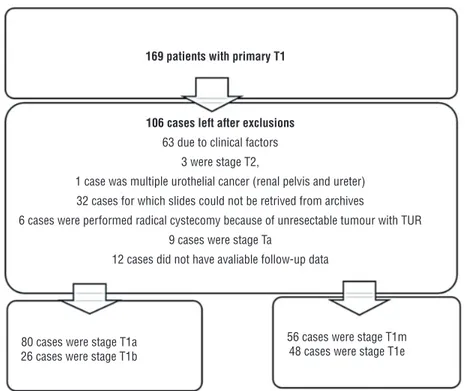
All patients with primary stage T1 urothe-lial bladder tumours who underwent surgery in our institution between January 2009 and December 2014 were included in the study. To the original study, 169 patients were registered as having pri-mary Tl BC. After re-evaluation and review of the hospital records, a total of 106 patients remained to be included in the analysis (Figure-1). Patho-logic specimens were re-evaluated so as to prove the diagnosis of T1 and sub-staging by the same pathologist (SO). The pathologist classified the pa-tients using two systems: T1a (the tumour does not infiltrate the muscularis mucosae-vascular plexus [MM-VP]) and T1b (the tumour infiltrates and/or invades the [MM-VP]), and T1m (micro--invasive- a single focus of lamina propria inva-sion with a maximum diameter of 0.5mm) and T1e (extensive-invasive, >0.5mm) (8).
All patients underwent macroscopically complete TUR-BT. A second TUR-BT was performed 2-6 weeks after the initial resection in accordance
Figure 1 - The studied cohort after re-evaluation.
169 patients with primary T1
106 cases left after exclusions
63 due to clinical factors 3 were stage T2,
1 case was multiple urothelial cancer (renal pelvis and ureter) 32 cases for which slides could not be retrived from archives
6 cases were performed radical cystecomy because of unresectable tumour with TUR 9 cases were stage Ta
12 cases did not have avaliable follow-up data
80 cases were stage T1a 26 cases were stage T1b
with the recommendations of the current Europe-an Association of Urology (EAU) guidelines. De-trusor muscle was present in 96 cases (90.5%) in the first TUR. 10 patients underwent re-TUR and after the evaluation of specimens, it was repor-ted that there was detrusor muscle in all of them. All patients underwent intravesical treatment with bacillus Calmette-Guerin or mitomycin C (BCG or MMC). The patients’ follow-up included cystos-copy and cytology every 3 months in the first 2 years. If no recurrence was found, patients were requested to visit every six months thereafter for 5 years. Disease progression was accepted as the detection of a muscle-invasive BC.
Age, tumour size, multiplicity, associa-ted CIS, second TUR-BT, tumour grade, and T1 sub-staging system were investigated to detect the relation between disease progression and recurrence.
SPSS software version 21.0, was used to perform statistical analyses (IBM, Armonk, NY,
USA). Categorical and continuous variables were determined using the t-test, Mann-Whitney U test, and Pearson’s Chi-square test. The clinical results, such as disease recurrence and progression, were analysed using univariate statistical analysis ac-cording to Kaplan-Meier and multivariate analyses using Cox regression models. A two-sided P<0.05 was considered to indicate statistical significance.
RESULTS
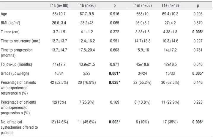
The mean age of the patients was 67.9±10 years. The mean follow-up time was 54 months. The evaluation of the patients according to staging systems is shown in Table-1. During follow-up, 62 patients experienced recurrence and 19 had disease progression. According to the sub-staging systems, the recurrence rate was 52% for T1a (42 cases) vs. 76% for T1b (20 cases) (p=0.028), and 55% for T1m (32 cases) vs. 62% for T1e (30 cases), respectively (p=0.446) (Table-1). Thus, with
regar-Table 1 - Characteristics of the analyzed patients according to the two sub-staging systems.
T1a (n= 80) T1b (n=26) p T1m (n=58) T1e (n=48) p
Age 68±10.7 67.7±9.5 0.916 668±10 69.4±10.2 0.203
BMI (kg/m2) 26.6±3.4 28.2±43 0.065 26.9±3.2 27±4.2 0.879
Tumor (cm) 3.7±1.9 4.1±1.2 0.372 3.38±1.6 4.38±1.8 0.005*
Time to recurrence (mo.) 12.7±13.7 12.4±16.2 0.951 14.7±13.8 10.3±14.6 0.227
Time to progression (months)
13.7±14.7 17.5±20.4 0.603 15.9±16 14±17,2 0.781
Follow-up (months) 44±17.7 43.9±21.5 0.971 45±18.6 42±18.5 0.546
Grade (Low/High) 46/34 3/23 0.001* 34/24 15/33 0.005*
Percentage of patients who experienced recurrence n (%)
42 (52.5%) 20 (76.9%) 0.028* 32 (55.2%) 30 (62.5%) 0.446
Percentage of patients who experienced progression n (%)
12(15%) 7(26.9%) 0.169 8 (13.8%) 11 (22.9%) 0.223
No. of radical
cystectomies offered to
ds disease progression, there was no significant di-fference between sub-staging groups: T1a (n=12, 15%) vs. T1b (n=7, 27%), and T1m (n=8, 13.8%) vs. T1e (n=11, 23%) (p>0.05).
During follow-up, 17 (16%) (T0:2, Ta:1, T1:1, T2:10, T3:3) patients underwent radical cys-tectomy. Radical cystectomy rates for the sub-sta-ging groups were 12.5% vs. 27% for T1a vs. T1b (10 cases vs. 7 cases p=0.082), and 8.6% vs. 25% for T1m vs. T1e (5 cases vs. 12 cases, p=0.022), respec-tively. Despite our recommendations, six patients refused radical cystectomy surgery due to surgical and/or anaesthetic risks. Nine patients died during follow-up; 7 patients died of tumour progression and 2 died of other causes.
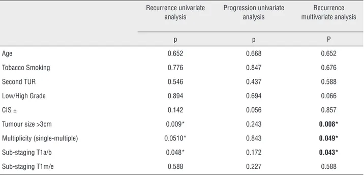
The formulated model of parameters rela-ted with recurrence and progression is shown in Table-2. In the univariate analysis, no effect of the sub-staging systems could be determined for disease progression. In multivariate analysis, tu-mour size >3cm (p=0.008), multiplicity (p=0.049) and sub-staging T1b (p=0.043, OR 0.407 [5-95% CI: 0.173-0.896]) were independent predictive factors for tumour recurrence (Table-2).
According to the Kaplan-Meier actuarial method, recurrence-free survival was significantly different in patients with pT1a tumours compared
with those with pT1b tumours (p=0.033). Recurren-ce-free survival was not significantly different in patients with pT1m tumours compared with those with pT1e tumours (p=0.232). Curves for recurren-ce-free survival are plotted in Figures 1 and 2.
DISCUSSION
The management of stage T1 urothelial bladder tumours can be considered a therapeutic challenge. In stage T1 urothelial bladder tumours, progression risk has been reported at different rates. In addition, recurrence following treatment (TURB, second TUR and intravesical treatment) and disease progression cause the necessity for radical cystec-tomy. In the treatment of high-risk non-muscle invasive bladder cancer, early radical cystectomy is recommended by the EAU guidelines (6). In our study, disease progression was observed in 19 pa-tients and radical cystectomy was recommended to 23 patients. Tumour size >3cm, multiplicity, and T1b were seen as independent risk factors for the prediction of disease recurrence. Sub-staging using T1m/e has no effect on either recurrence or pro-gression. In the multicentre study of Rouprêt et al., it was reported that T1a/b sub-staging was signifi-cantly associated with recurrence-free (p=0.03),
Table 2 - Prognostic factors of tumur recurrence and progression.
Recurrence univariate analysis
Progression univariate analysis
Recurrence multivariate analysis
p p P
Age 0.652 0.668 0.652
Tobacco Smoking 0.776 0.847 0.676
Second TUR 0.546 0.437 0.588
Low/High Grade 0.894 0.694 0.066
CIS ± 0.142 0.056 0.857
Tumour size >3cm 0.009* 0.243 0.008*
Multiplicity (single-multiple) 0.0510* 0.843 0.049*
Sub-staging T1a/b 0.048* 0.172 0.043*
progression-free (p<0.001) and cancer-specific (p=0.02) survival as a consequence of multivariate analysis (9). In our study, similar results of recur-rence-free survival were obtained.
Skoup et al. evaluated the prognostic value of the depth of lamina propria invasion in patients with T1 bladder cancer (10). In that study, during 3.13 years follow-up of 128 patients, the recur-rence and progression rates were 61% (n=101) and 16.3% (n=27), respectively. T1 sub-staging and grade also acted as independent predictors of tumour progression in their multivariate analysis (p<0.001). According to the results observed in that study, like in our study, there was no difference in sub-staging using T1a/b and T1m/e (11) on disease behaviour. De-Marko et al. reported that there was no significant diversity between tumour progres-sion and disease-specific survival after 9.5 years of follow-up. However, in classification, the use of T1e/m was recommended, but they did not evalu-ate recurrence-free survival in their study.
Van Rhijn et al. assessed the T1a/T1b/T1c and T1m/T1e sub-staging systems, which show a higher prognostic value for disease progression and disease-specific survival (8). In their study, sub-staging with T1m/T1e was significant for pro-gression in multivariate analysis. In the present study, however, there was no relation between
dis-Patriarca et al. analyzed three sub-staging systems for T1 bladder cancers that exhibited dif-ferent kinds of clinical behavior (T1a/b: 0.5 - 1mm invasion) (12). The authors reported that the 1mm invasion system predicted progression (p<0.04). In the same study, they reported on patients who un-derwent reTUR; the survival rate without recurrence was satisfactory or more. However, in our study, there appeared no difference because of the performance of nearly standard second TUR.
In the study of Orsola et al., all patients had T1 high grade urothelial carcinoma of the bladder. The authors studied treatment strategies according to sub-staging using depth of lamina propria invasion (5). They showed that sub-staging remained signifi-cant for progression on multivariate analysis. In our study, there was no relation between disease progres-sion and the two sub-staging systems. The T1a/b sys-tem had a tendency for predicting progression; there-fore, a long follow-up period was needed.
In most studies about sub-staging of T1, there have been deficiencies and even failures in reporting. However, we did not encounter such an obstacle. Our study has several limitations. First, the design of the study was retrospective. Secondly, several treatments are available after TURBT (BCG induction therapy, BCG induction, and maintenance therapy, mitomy-cin C or device-assisted mitomymitomy-cin C). Our patients
Figure 2 - Association of substaging T1m/e and time to recurrence.
120.0
100.0
80.0
60.0
40.0
20.0
0.0
0 10 20 30 40 50 60 70 80 90
p= 0,232
Per
centage of patients free of
recurrence
Time to recurrence from diagnosis
cause there was currently a worldwide shortage of BCG. Thirdly, the limited number of patients and fol-low-up period may have played a role in the failure of predictive values of sub-staging of T1 for progres-sion. Another limitation to the study is the relative low number of events (62 recurrences) to perform a reliable multivariable analysis with 9 variables. On the other hand, early progression of the disease can be prevented using second TUR, current intravesical induction, and maintenance treatment protocols. In addition, without progression, early cystectomy was performed to several patients. This intervention could bring about a relative decrease in the numbers of disease progression. In our study, tumour size >3cm, multiplicity, and sub-staging T1b for recurrence was very meaningful in univariate and multivariate anal-yses; however, long-term follow-up might reveal sig-nificant diversity for disease progression. Increased numbers of studies and translation of sub-staging of T1 to molecular pathology can lead to more precise prediction of recurrence and progression.
CONCLUSIONS
Sub-staging T1 in the surveillance of non--muscle-invasive bladder cancer provides the prediction of disease recurrence. Sub-staging of lamina propria invasion depth was designed ac-cording to muscularis mucosa retention and these were found as independent factors. Sub-staging should be reported during the pathologic evalua-tion report because it provides supplementary in-formation for surgeons who undertake the follow--up. Within the assessment of recurrence, T1a/b sub-staging can provide better knowledge about disease behaviour because it is predicted as more superior than T1m/e, and it can also help in deter-mining the requirement for early cystectomy.
CONFLICT OF INTEREST
None declared.
REFERENCES
1. Siegel RL, Miller KD, Jemal A. Cancer Statistics, 2017. CA Cancer J Clin. 2017;67:7-30.
2. Stein JP. Indications for early cystectomy. Urology. 2003;62:591-5.
3. Stein JP, Penson DF. Invasive T1 bladder cancer: indications and rationale for radical cystectomy. BJU Int. 2008;102:270-5. 4. Nieder AM, Brausi M, Lamm D, O’Donnell M, Tomita K, Woo H, et
al. Management of stage T1 tumors of the bladder: International Consensus Panel. Urology. 2005;66(6 Suppl 1):108-25. 5. Orsola A, Werner L, de Torres I, Martin-Doyle W, Raventos
CX, Lozano F, et al. Reexamining treatment of high-grade T1 bladder cancer according to depth of lamina propria invasion: a prospective trial of 200 patients. Br J Cancer. 2015;112:468-74. 6. Babjuk M, Böhle A, Burger M, Capoun O, Cohen D,
Compérat EM, et al. EAU Guidelines on Non-Muscle-invasive Urothelial Carcinoma of the Bladder: Update 2016. Eur Urol. 2017;71:447-61.Available at. <https://uroweb.org/guideline/ non-muscle-invasive-bladder-cancer/>
7. Moch H, Humphrey PA, Ulbright TM, Reuter VE. WHO Classification of Tumours of the Urinary System and Male Genital Organs. 4th ed. O. H. 2016, Lyon, France. Available at. <http://apps.who.int/bookorders/WHP/detart1.jsp?sesslan=1&c odlan=1&codcol=70&codcch=4008>
8. van Rhijn BW, van der Kwast TH, Alkhateeb SS, Fleshner NE, van Leenders GJ, Bostrom PJ, et al. A new and highly prognostic system to discern T1 bladder cancer substage. Eur Urol. 2012;61:378-84.
9. Rouprêt M, Seisen T, Compérat E, Larré S, Mazerolles C, Gobet F, et al. Prognostic interest in discriminating muscularis mucosa invasion (T1a vs T1b) in nonmuscle invasive bladder carcinoma: French national multicenter study with central pathology review. J Urol. 2013;189:2069-76.
10. Soukup V, Dušková J, Pešl M, Čapoun O, Feherová Z, Zámečník L, et al. The prognostic value of T1 bladder cancer substaging: a single institution retrospective study. Urol Int. 2014;92:150-6. 11. DE Marco V, Cerruto MA, D’Elia C, Brunelli M, Otte O, Minja
A, et al. Prognostic role of substaging in T1G3 transitional cell carcinoma of the urinary bladder. Mol Clin Oncol. 2014;2:575-80.
12. Patriarca C, Hurle R, Moschini M, Freschi M, Colombo P, Colecchia M, et al. Usefulness of pT1 substaging in papillary urothelial bladder carcinoma. Diagn Pathol. 2016;11:6.
_______________________ Correspondence address: