M a i l i n g A d d r e s s : F á b i o V i l a s - B o a s • P r a ç a A l m e i d a C o u t o , 5 0 0 , N a z a r é - 4 0 0 5 0 - 4 1 0 , S a l v a d o r - B A - B r a z i l
E-mail: [email protected] Received on 05/28/06 • Accepted on 06/05/06
Early Results of Bone Marrow Cell Transplantation to the
Myocardium of Patients with Heart Failure due to Chagas
Disease
Fábio Vilas-Boas1, Gilson S. Feitosa1, Milena B.P. Soares2, Augusto Mota1, Joel Alves Pinho-Filho1,
Augusto José Gonçalves Almeida1, Marcus Vinicius Andrade1, Heitor G. Carvalho1,
Adriano Dourado-Oliveira1, Ricardo Ribeiro-dos-Santos2
Hospital Santa Izabel1, Centro de Pesquisas Gonçalo Moniz – Fundação Oswaldo Cruz2 - Salvador, BA - Brazil
O
BJECTIVETo evaluate early effects of bone marrow cell transplantation to the myocardium of patients with heart failure (CHF) due to Chagas disease.
M
ETHODSWe studied 28 patients (mean age 52.2 ± 9.9), of whom 24 were male. Despite optimized treatment, 25 patients were in NYHA class III and three patients, in NYHA class IV. The procedure consisted of aspiration of 50 mL of bone marrow, separation of the mononuclear fraction, and intracoronary injection. Effects on left ventricle ejection fraction (LVEF), distance walked in the six-minute walking test, quality-of-life, NYHA class, arrhythmogenic and biochemical parameters, were all evaluated.
R
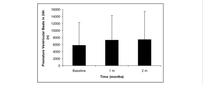
ESULTSThere were no complications directly related to the procedure. Baseline left ventricular ejection fraction was 20.1 ± 6.8%, and 60 days after transplantation it increased to 23.0 ± 9.0%, p = 0.02. Signifi cant improvements were observed in the NYHA class (3.1 ± 0.3 to 1.8 ± 0.5; p < 0.0001); quality-of-life (50.9 ± 11.7 to 21.8 ± 13.4; p < 0.0001); and distance walked in six minutes (355 ± 136 m to 443 ± 110 m; p = 0,003). The number of ventricular premature beats in 24 hours tended to increase (5,322 ± 4,977 to 7,441 ± 7,955; p = 0,062), but without increase in ventricular tachycardia episodes (61 ± 127 to 54 ± 127; p = 0.27).
C
ONCLUSIONOur data demonstrate for the fi rst time that intraco-ronary injection of bone marrow mononuclear cells is feasible and suggest that it may be potentially safe and effective in patients with CHF due to Chagas disease.
K
EY WORDSThe ability of bone marrow stem cells to differentiate into cardiomyocytes has been demonstrated especially in experimental models of myocardial infarction1-3. Although some degree of controversy regarding stem cell plasticity exists, such observations have led investigators to examine, in humans, their role in the management of myocardial infarction and heart failure of ischemic etiology, and the fi rst results are promising4-6.
In Brazil, Chagas disease is one of the main contributors to the nosological description of heart failure. Serological estimates show that eight or nine million Brazilians are infected by Trypanosoma cruzi, and that 30 to 40% of them may have some degree of cardiac involvement7. Patients with heart failure secondary to chronic Chagas heart disease progress to severe systolic dysfunction due to the chronic myocarditis and consequent fi brosis8.
The etiopathogenic and pathophysiological mechanisms involved in Chagas disease make stem cell therapy attractive. Both the presence of persistent myocarditis and increased production of cytokines by the myocardium in patients with CHF due to Chagas disease suggest the existence of an attractive environment for stem cell homing and adhesion9.Therefore, unlike the management of other heart diseases in which little infl ammation is found, in Chagas heart disease it may be assumed that circulating stem cells migrate to the myocardium, recruited by the infl ammatory signals spread throughout the organ.
Experimental studies using stem cells derived from bone marrow to treat chronic Chagas cardiomyopathy demonstrate that, two month after treatment with adult bone marrow cells, chronic chagasic mice showed a signifi cant reduction in infl ammation and fi brosis, compared with control animals10.
The fi rst case of bone marrow stem cell transplantation to the myocardium of a patient with heart failure secondary to Chagas disease was reported by our group, leading to improvement in ventricular function, quality of life, and functional capacity11.
This study was designed to assess the safety and feasibility of autologous bone marrow mononuclear cell (BM-MNC) transplantation in patients with heart failure due to Chagas disease, as well as identify early evidence supporting the effectiveness of the procedure.
M
ETHODSThe study was an open-label, uncontrolled, single-center, phase-1 clinical trial carried out at the Santa Izabel Hospital of the Santa Casa de Misericórdia da Bahia, Brazil, involving selected patients with CHF secondary to Chagas disease.
Patients who met the following inclusion criteria were consecutively included in the study: subjects of both genders, ranging from 20 to 70 years, with CHF due to Chagas disease, left ventricle (LV) ejection fraction less than 40% on echocardiogram, functional class III
and IV (NYHA), receiving optimal chronic treatment to CHF (digoxin, diuretics, angiotensin-converting enzyme inhibitors, with or without beta-blockers or angiotensin-II receptor antagonists), and who remained stable in this condition in the month prior to the experiment.
We excluded patients with associated systemic conditions, such as infections or neoplasias, autoimmune diseases, and neurodegenerative diseases; acute or acutely decompensated heart failure; previous hematologic diseases; coagulopathies; liver failure; moderate renal failure (creatinine above 2 mg/dL); previous history of chronic obstructive pulmonary disease (COPD); implantation of biventricular resynchronization pacemaker in the previous 90 days; women of childbearing potential, and patients with coronary artery disease detected previously or after coronary angiography.
This protocol was approved by CONEP (Brazilian National Committee for Ethics in Research) under No. 4108; Case No. 25000.054219/2002/20). Patients were fully informed about study procedures and, after reading and signing the informed consent, they were included in the study.
All patients underwent a baseline clinical evaluation, and the following clinical data and variables were recorded: a) functional class (NYHA), b) quality-of-life score of the “Minnesota Living With Heart Failure Questionnaire”; c) hematological and biochemical assessment; d) twelve-lead electrocardiogram; e) transthoracic echocardiogram; f) radionuclide ventriculography; g) six-minute walk test; g) 24-hour Holter. After the procedure, patients were referred to the Intensive Care Unit, where they were monitored for at least 24 hours. If everything went uneventfully, they were transferred to the regular ward where remained for a minimum of fi ve days for in-hospital follow-up. All clinical evaluation, laboratory tests and complementary exams were repeated after 30 and 60 days.
To check for any possible myocardial damage caused by bone marrow cell injection, serial measurements (every six hours during the fi rst 24 hours) of markers of myocardial damage (CK-MB and troponin I), as well as electrocardiograms, were performed. To investigate the development of cardiac arrhythmias as a complication of cell injection, patients underwent ambulatory electrocardiography (Holter) 24 hours before the procedure and periodically thereafter (after 24 hours, 30 days and 60 days).
by Ficoll Hystopaque gradient (Amersham Pharmacia, a product licensed for clinical use in humans). The isolated mononuclear cell fraction was then diluted in sterile saline solution and centrifuged again. One sample was used for cell count and viability test. At the end of the process, samples were diluted in 20 ml of saline solution. Immediately before intracoronary injection, patients underwent left heart catheterization by the femoral approach followed by coronary angiography. Patients with evidence of hemodynamically signifi cant coronary disease (stenosis > 50%) were excluded from the study. The cell solution was slowly injected, during 10 minutes, into the right and left coronary system. Ten ml of the solution was injected in the anterior descending artery, 5 ml in the right coronary artery, and 5 ml in the left circumfl ex artery.
Twenty-five days later, patients received daily subcutaneous injections of human G-CSF (Granulokine®) for fi ve days, at a dose of 5 mcg/kg/day, to induce mobilization of bone marrow stem cells to peripheral blood, so that a greater number of stem cells would be available for injury repair. In studies using an experimental model of chagasic myocarditis, combined cell therapy and G-CSF treatment led to a signifi cantly greater decrease in the number of infl ammatory cells than cell therapy alone (unpublished data).
The six-minute walk test was performed according to the protocol used by Bitner et al. in the SOLVD study12. After a fi fteen-minute rest, a new test was carried out, and the average of the distances walked in the two tests was used as the result. Quality of life was assessed by the “Living with Heart Failure Questionnaire” developed by the University of Minnesota13 and validated by Carrara et al. in Brazil14. Echocardiographic studies were conducted by the same observer, blind to previous examinations, on a Hewlett-Packard Sonos 5500 System (Seattle, WA, EUA). Left ventricular ejection fraction was calculated according to the modifi ed Simpson’s rule15,16.Holter monitoring was performed with a Dynamis 3000 ECO recorder and analyzed in a Burdick machine using AltairPC Holter System software.
Statistical analysis - Statistical analysis was performed using SPSS (Statistical Package for Social Sciences) for Windows version 9.0. Continuous variables were presented as mean ± standard deviation. Variable distribution was assessed by the Kolmogorov-Smirnov test. Since variables were not normally distributed, non-parametric tests were used. All comparisons were two-tailed. P values < 0.05 were considered statistically signifi cant.
Funding sources - This study was fi nancially supported by the following institutions: Fundação de Amparo à Pesquisa do Estado da Bahia (FAPESB); Financiadora de Estudos e Projetos do CNPq (FINEP), and Instituto do Milênio de Bioengenharia Tecidual. The authors declare that they have no confl ict of interest.
R
ESULTSThirty patients were selected, but two were excluded after bone marrow collection: one patient due to coronary disease detected by catheterization and the other due to technical problems with the equipment. The primary characteristics of the 28 patients studied are described in Table 1.
An analysis of the clinical profi le reveals a population with advanced stage of the disease and various markers of severity. All patients were extremely limited, in functional class III and IV, despite optimal treatment with a multi-drug regimen and high doses of diuretics. Functional capacity was found to be low, based on the short distance covered during the six-minute walking test, and quality-of-life was severely impaired, as indicated by high scores on the Minnesota questionnaire. Ventricular function was severely depressed, with very low ejection fraction and high left ventricular diastolic diameter on the echocardiogram. The presence of marked hyponatremia and renal dysfunction characterizes the severe condition of this population.
There were no complications directly related to either bone marrow aspiration or cell injection. Viability tests showed that 96 ± 6.5 % of the cells were viable. Neither signifi cant changes in myocardial necrosis markers in 24 hours nor electrocardiographic changes suggestive of ischemia or infarction occurred.
In order to check if the myocardial implantation of stem cells was associated with arrhythmias, the
Table 1 - Clinical and laboratory characteristics of the patients
(n = 28)
Age (years) 52.2 ± 9.9
Male (n) 24
NYHA class ( n)
III 25
IV 4
6 min walking test (m) 355 ± 136
LV ejection fraction (%) 20.1 ± 6.8
LVEDD (mm) 72.6 ± 8.9
QOL score 50.9 ± 11.7
Serum sodium (mEq/L) 131 ± 7.6
Serum urea (mg/dL) 69 ± 36.8
Creatinine (mg/dL) 1.4 ± 0.4
Digoxin (%) 95
Furosemide (%) 85
Hydrochlorothiazide (%) 35
ACEI or ARB (%) 85
Beta-blockers (%) 55
arrhythmogenic profi le was evaluated through the total number of ventricular extrasystoles in 24 hours, as well as their clustering and recurrence pattern. No signifi cant change was found in the arrhythmogenic profi le, despite a non-signifi cant trend for increase in the number of ventricular premature beats in 24 hours (Figure 1), with no change in the number of ventricular tachycardia episodes (Figure 2).
There were three deaths during the two-month follow-up, all during the second month (patient 04, male, age 37, pulmonary hemorrhage and respiratory failure; patient 16, female, age 34, sudden death; patient 27, age 69, male, end-stage heart failure). Although this study was not designed to assess effi cacy, and that mortality cannot be evaluated as a safety endpoint, except by the causal association of immediate temporariness, no direct causal association was found between death and cell transplantation. There were no neoplasias,
hematological diseases, coagulopathies or any other kind of disease that could be attributed to cell injections or their implantation.
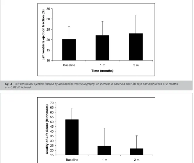
Ventricular function, evaluated by left ventricular ejection fraction showed signifi cant improvement one month after the procedure, which was maintained at two months (Figure 3).
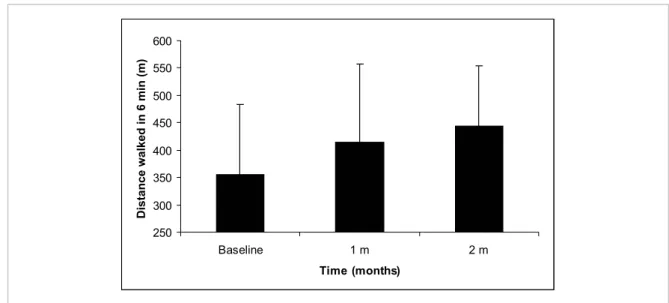
Quality of life assessment by the Minnesota Living with Heart Failure questionnaire revealed signifi cant improvement in the global score, also at one month and was maintained throughout the follow-up period (Figure 4).
Functional capacity, as measured by the distance walked during the six-minute corridor walk test, showed signifi cant improvement, which was maintained for two months (Figure 5).
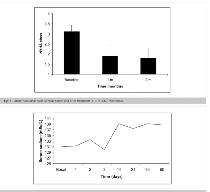
When functional class was analyzed as an ordinal variable, a decrease in mean NYHA functional class was
Fig. 1 - Non-signifi cant trend of elevation in the number of ventricular premature beats in 24 hours. VE = ventricular extrasystoles p = 0.062 (Friedman). Bars represent mean ± standard deviation.
Fig. 2 - No elevation in the number of ventricular tachycardia episodes in 24 hours. T = ventricular tachycardia p = NS (Friedman). Bars represent mean ± standard deviation.
0 2000 4000 6000 8000 10000 12000 14000 16000
Baseline 1 m 2 m
Time (months)
Premature Ventricular Beats in 24h
(n)
0 50 100 150 200
Baseline 1 m 2 m
Time (months)
Ventricular tachycardia episodes in
noted after one month and maintained over the follow-up period (Figure 6).
Biochemical analysis showed correction of serum sodium concentration between 72 hours and three weeks of treatment, which was maintained over the fi rst two-months of study follow-up (fi gure 7).
D
ISCUSSIONThis is the fi rst study to report the implantation of bone marrow mononuclear cells in patients with heart failure due to Chagas disease and the fi rst cell therapy study using a non-ischemic model. Our model involved intracoronary administration of stem cells, followed by bone marrow stimulation with subcutaneous fi lgrastim after 30 days.
Patients in this study are more severe than those previously reported in other stem-cell studies of ischemic
etiology. In the other studies, patients had milder degrees of ventricular dysfunction, renal dysfunction and hyponatremia. The number of injected cells in the present study was greater than that of previously published studies. Whether there is a relationship between the number of cells injected and the degree of clinical response to cell therapy remains unclear, but this hypothesis warrants further investigation.
The absence of adverse events directly related to the procedure indicates that this is feasible, provided that the protocols implemented in this study are followed, as they aim at the patient’s protection and safety.
One of the most relevant aspects analyzed regarding safety was whether bone marrow cell injection into the coronary system could trigger acute myocardial infatction. To ascertain this, troponin I and CK-MB measurements were performed, as well as serial electrocardiograms, which showed no evidence of
Fig. 3 - Left ventricular ejection fraction by radionuclide ventriculography. An increase is observed after 30 days and maintained at 2 months. p = 0.02 (Friedman).
10 15 20 25 30 35
Baseline 1 m 2 m
Time (months)
Left ventricle ejection fraction (%)
Fig. 4 - Quality of life score (Minnesota questionnaire). A signifi cant decrease in the score is noted, indicating improvement in quality of life. p < 0.0001 (Friedman).
15 20 25 30 35 40 45 50 55 60 65 70
Baseline 1 m 2 m
Time (months)
Fig. 5 - Distance covered in the six-minute corridor walk test. A signifi cant increase is noted after 30 days and maintained at 2 months. p = 0.003 (Friedman).
250 300 350 400 450 500 550 600
Baseline 1 m 2 m
Time (months)
Distance walked in 6 min (m)
myocardial injury, thus suggesting that this procedure is safe regarding ischemia inducement.
Another important parameter analyzed was the potential to induce arrhythmias. Previous studies using skeletal muscle stem cells (satellite cells) have shown promising results, with partial recovery of cardiac function; yet, there has been concern regarding the development of arrhythmias in patients that underwent this kind of cell therapy17,18. In our study, the number of isolated ventricular extrasystoles increased signifi cantly, but the number of sustained or nonsustained ventricular tachycardia episodes remained unchanged.
As far as parameters related to effi cacy are concerned, LVEF increments were similar to those described in previous studies. In the BOOST study19, mean LVEF was 52%, and the authors detected an increase of 5%, on average, corresponding to around 10% of relative increase. In the present study, mean LVEF increase was similar; yet, it corresponds to approximately 20% of relative increase, since LVEF of our patients was signifi cantly lower.
One fi nding that calls attention is the improvement in quality of life and functional capacity. Despite being on optimal treatment and multi-drug regimen in optimal doses, patients’ functional capacity and quality of life remained severely impaired. However, their quality of life improved consistently, as evaluated by the Minnesota questionnaire, which refl ects not only dyspnea parameters, but also emotional, psychological, economic, and professional aspects. The same was true with respect to more objective variables, as distance covered during the six-minute walk test, which measures exercise capacity at submaximal levels. Consistent with the variations mentioned above, there was a signifi cant improvement in functional capacity, as assessed by the NYHA classifi cation. While motivational aspects may
be related to these subjective variables, the consistent modifi cation suggests that an underlying clinical effect is likely to exist. All these fi ndings were observed only one month after treatment, too early to be considered an effect of transdifferentiation or cell fusion. In that regard, recent evidence indicates that the mechanism of action associated with stem cell transplantation may involve three different phenomena: transdifferentiation, cell fusion, and paracrine effects. This hypothesis is based on the observation that measurable effects may be demonstrated as early as 72 hours20.
More objective variables, irrespective of motivation or self-suggestion by the patient, as well as investigator interference, indicate that the therapy used produced positive biological effects. Hyponatremia, for example, is one of the most important prognostic markers in advanced CHF, and its pathophysiological mechanism is related to the neurohumoral activation of the syndrome21. The hyponatremia correction achieved within two weeks in this study is uncommon in clinical practice, where it is hardly ever observed in severely ill patients, even after optimal therapeutic interventions.
of subpopulations involved in the repair process is an important issue that warrants investigation, it is essential to evaluate cautiously whether other methods for purifying these subpopulations, indeed more expensive than the simple purifi cation of the mononuclear fraction used today in clinical trials, will bring more benefi t to patients.
The corroboration of our fi ndings in a longer-term follow-up, as well as in randomized controlled clinical trials, may provide a new therapeutic line available worldwide at a very low cost and with no rejection, requiring no specifi c medication and that is more physiological, less aggressive and allows multiple repetitions.
Acknowledgement
The authors would like to express their appreciation to Cristiane Carvalho and Aline Bernardes, nurses of the Núcleo de Pesquisas em Cardiologia of the Hospital Santa Izabel, for the patients assistance throughout the study. The contribution of Daniele Brustolim, of the Centro de Pesquisas Gonçalo Moniz – Fundação Oswaldo Cruz – Bahia, is particularly acknowledged.
Potencial Confl ict of Interest
No potential confl ict of interest relevant to this article was reported.
1. Orlic D. Stem cell repair in ischemic heart disease: An experimental model. Intern J Hematol. 2002; 76 (Suppl. 1): 144–5.
2. Orlic D, Kajstura J, Chimenti S, Jakoniuk I, Anderson SM, Li B, et al. Bone marrow cells regenerate infarcted myocardium. Nature. 2001; 410: 701–15.
R
EFERENCES3. Orlic D, Kajstura J, Chimenti S, Bodine DM, Leri A, Anversa P. Transplanted adult bone marrow cells repair myocardial infarcts in mice. Ann NY Academy Sci. 2001; 938: 221–9.
4. Assmus B, Schachinger V, Teupe C, Britten M, Lehmann R, Dobert N, et al. Transplantation of progenitor cells and regeneration enhancement Fig. 6 - Mean functional class (NYHA) before and after treatment. p < 0.0001 (Friedman).
1 1,5 2 2,5 3 3,5 4
Baseline 1 m 2 m
Time (months)
NYHA clas
s
Fig. 7 - Serum sodium concentration before and after treatment. Sodium concentration was normalized between 72 hours and 2 weeks and maintained up to study conclusion (Friedman, p < 0.0001).
125 127 129 131 133 135 137 139 141
Basal 1 2 3 14 21 30 60
Time (days)
in acute myocardial infarction (TOPCARE-AMI). Circulation. 2002; 106: 3009–17.
5. Perin EC, Dohmann HF, Borojevic R, Silva SA, Sousa AL, Mesquita CT. Transendocardial, autologous bone marrow cell transplantation for severe, chronic ischemic heart failure. Circulation. 2003; 107: 2294–302.
6. Stamm C, Westphal B, Kleine HD, Petzsch M, Kittner C, Klinge H, et al. Autologous bone-marrow stem-cell transplantation for myocardial regeneration. Lancet 2003; 361: 45-6.
7. Dias JCP, Silveira AC, Schofi eld CJ. The impact of Chagas disease control in Latin America: a review. Mem. Inst. Oswaldo Cruz. 2002; 97(5): 603-12.
8. Soares MBP, Pontes-de-Carvalho L, Ribeiro-dos-Santos R. The pathogenesis of Chagas’ disease: when autoimmune and parasite-specifi c immune responses meet. Ann Acad Bras Cienc. 2001; 73: 547-59.
9. Vilas-Boas F, Ribeiro-dos-Santos R, Soares MBP, et al. Identifi cation of regional differences in proinfl ammatory cytokine concentrations in chronic heart failure due to Chagas’ cardiomyopathy: a key element in the comprehension of the disease. J Am Coll Cardiol. 2003; 41: 155A.
10. Soares MBP, Lima RS, Rocha LL, et al. Transplanted bone marrow cells repair heart tissue and reduce myocarditis in chronic chagasic mice. Am J Pathology. 2004 Feb; 164(2): 441-7.
11. Vilas-Boas F, Feitosa GS, Soares MBP, Pinho-Filho JA, Mota A, Almeida AJG. Bone Marrow Cell Transplantation to the Myocardium of a Patient with Heart Failure Due to Chagas’ Disease. Arq Bras Cardiol. 2004; 82: 185-7.
12. Bittner V, Weiner DH, Yusuf S, et al. Prediction of mortality and morbidity with a 6-minute walk test in patients with left ventricular dysfunction. JAMA. 1993; 270: 1702-7.
13. Rector TS, Kubo SH, Cohn JN. Patients self-assessment of their
congestive heart failure: content, reliability and validity of a new measure, the Minnesota Living with Heart Failure questionnaire. Heart Failure. 1987; 3: 198-209.
14. Carrara D. Avaliação prospectiva da qualidade de vida em pacientes com miocardiopatia dilatada submetidos a ventriculectomia parcial esquerda. Dissertação de Mestrado apresentada à Faculdade de Medicina da Universidade de São Paulo, 2001; p.18-21.
15. Schiller NB, Shah PM, Crawford M, et al. Recommendations for quantitation of the left ventricle by two-dimensional echocardiography: American Society of Echocardiography Committee on Standards. Subcommittee on Quantitation of Two-Dimensional Echocardiograms. J Am Soc Echocardiogr. 1989; 2: 385-97.
16. Stewart WJ, Rodkey SM, Gunawardena S, et al. Left ventricular volume calculation with integrated backscatter from echocardiography. J Am Soc Echocardiogr. 1993; 6(6): 553-63.
17. Murry CE, Wiseman RW, Schwartz SM, Hauschka SD. Skeletal myoblast transplantation for repair of myocardial necrosis. J Clin Investigation. 1996; 98, 2512–2523.
18. Menasche P, Hagege AA, Vilquin JT, Desnos M, Abergel E, Pouzet B, et al. Autologous skeletal myoblast transplantation for severe postinfarction left ventricular dysfunction. J Am Coll Cardiol. 2003 Apr 2; 41(7):1078-83.
19. Wollert KC, Meyer GP, Lotz J. Intracoronary autologous bone-marrow cell transfer after myocardial infarction. BOOST trial. Lancet. 2004;364:141-48.
20. GnecchiM, He H, Liang OD, Melo LG, Morello F, Mu H, et al. Paracrine action accounts for marked protection of ischemic heart by Akt-modifi ed mesenchymal stem cells. Nature Medicine. 2005;11:367–68.