Arq Bras Cardiol 2004; 82: 185-7.
Vilas-Boas et al Bone marrow cell transplantation to the myocardium
1 8 5
Hospital Santa Izabel and Centro de Pesquisas Gonçalo Moniz - FIOCRUZ/BA Mailing address: Fábio Vilas-Boas - Av. Juracy Magalhães Jr., 2096/109 Cep 41920-000 - Salvador, BA, Brazil - E-mail: [email protected] Received: 8/11/03
Accepted: 8/26/03
English version by Stela Maris C. e Gandour
Arq Bras Cardiol, volume 82 (nº 2), 185-7, 2004
Fábio Vilas-Boas, Gilson Soares Feitosa, Milena B. P. Soares, Joel Alves Pinho-Filho, Augusto Mota, Augusto José Gonçalves Almeida, Cristiane Carvalho, Heitor Ghissoni de Carvalho,
A driano Dourado de O liveira, Ricardo Ribeiro dos Santos
Salvador, BA - Brazil
Bone Marrow Cell Transplantation to the Myocardium of a
Patient with Heart Failure Due to Chagas’ Disease
Case Report
We report the first case of bone marrow cell transplan-tation to the myocardium of a patient with heart failure due to chagas' disease. The patient is a 52-year-old man with chronic heart failure, NYHA functional class III, despite the optimized clinical therapy. The procedure con-sisted of aspiration of 50 mL of bone marrow through puncture of the iliac crest, followed by filtration, separa-tion of the mononuclear cells, resuspension, and intraco-ronary injection. The left ventricular ejection fraction at rest, measured using radionuclide ventriculography with labeled red blood cells prior to transplantation, was 24%, and, after 30 days, it increased to 32% with no change in the medicamentous schedule. The following measurements were assessed before and 30 days after transplantation: left ventricular end diastolic diameter (82 mm and 76 mm, respectively); Minnesota living with heart failure questio-naire score (55 and 06, respectively); and distance wal-ked in the 6-minute walking test (513 m and 683 m, respec-tively). Our findings show that intracoronary injection of bone marrow cells may be performed, suggesting that this is a potentially safe and effective procedure in patients with due to Chagas' disease heart failure.
Heart failure is epidemic at the beginning of this centu-ry. In Latin America, an endemic region for Chagas’ disease with approximately 11 million people suffering from the di-sease, the situation is even worse 1. In chagasic patients, no
specific etiologic treatment after heart failure syndrome is in place has yet been proven efficient. Therefore, the treat-ment of these patients does not differ from that of patients with heart failure due to other etiologies 2.
Given the limited efficacy of the current pharmacologi-cal therapeutic alternatives for the treatment of heart failure2,
other forms of treatment have been developed 3. The
de-monstration of the capacity of adult bone marrow cells to diffe-rentiate in vitro into various cell types was the initial stimulus for their experimental use in the treatment of heart failure 4.
Preliminary studies on the use of bone marrow cells for the treatment of chronic chagasic cardiomyopathy have been carried out at the Gonçalo Moniz Research Center
(FIOCRUZ-BA) using a mice model infected by
Trypanoso-ma cruzi, which showed a significant reduction in inflam-mation and a regression in fibrosis after 2 months of treat-ment with adult bone marrow cells as compared with that in control animals 5.
This and other findings 6-8 served as a basis for
desig-ning a protocol for the use of bone marrow cell therapy in patients with chagasic cardiomyopathy.
Case report
1 8 6
Vilas-Boas et al
Bone marrow cell transplantation to the myocardium
Arq Bras Cardiol 2004; 82: 185-7.
The procedure was performed with the patient seda-ted, and comprised 5 punctures of the crest of each iliac bone, from where approximately 50 mL of bone marrow where withdrawn. The material was passed through a bone marrow filtering system (Washington University® - USA),
and, then, the mononuclear cells were separated through a Ficol gradient. After resuspension in 20 mL of albumin sali-ne solution (5%), approximately 2.4 x 10⁸ cells were injected
in the right and left coronary system, through an angioplas-ty catheter as follows: 10 mL of the suspension were slowly injected in the anterior descending coronary artery for 10 mi-nutes; 5 mL in the circumflex artery; and 5 mL in the right co-ronary artery. Coco-ronary artery disease was excluded through coronary angiography. The patient experienced neither arrhythmias nor electrocardiographic alterations during the procedure. The vital data remained stable during the entire period of hospitalization (tab. I).
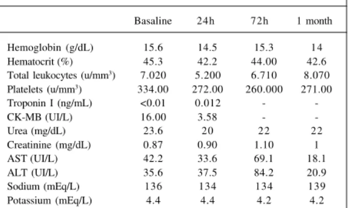
Alterations were not observed in the markers of myo-cardial necrosis nor in the biochemical and hematological parameters (tab. II). After 4 days, the patient was dischar-ged using the same medications used prior to the procedure in addition to ascorbic acid (1g/d). In the initial 30-day follow-up, weekly clinical reassessments were performed, as were laboratory assessments at the end of the period.
A reduction in the ventricular diameters was observed on echocardiography and there was a significant increase in the left ventricular ejection fraction both on echocardio-graphy and radionuclide ventriculoechocardio-graphy with labeled red blood cells. Functional capacity, assessed both with the NYHA functional classification and 6-minute walking test,
showed a significant improvement, as did the quality of life score (tab. III).
Discussion
Chagasic cardiomyopathy is one of the major causes of heart failure in Latin America, and its clinical presentation is si-milar to other forms of dilated cardiomyopathy. The chronic phase of the disease is characterized by a multifocal inflamma-tory infiltration of mononuclear cells. In this phase, no asso-ciation exists between the presence of parasites (Trypanosoma cruzi) and the intensity of inflammation or fibrosis 9.
In recent decades, several therapeutic options have been developed or improved to delay the progression of ven-tricular dysfunction in patients with heart failure. However, plain reversion of the process has not yet been obtained, and the prognosis of these patients remains very limited.
Cell therapy has emerged as a promising option for the treatment of advanced cases of heart failure. Orlic et al6, in a
study with infarcted mice, reported myocardial repair through the formation of new cardiac fibers and neoangio-genesis after bone marrow cell transplantation. Strauer et al7
were the first to demonstrate the feasibility and safety of in-tracoronary infusion of bone marrow stem cells for the treat-ment of patients with ischemic cardiomyopathy. Perin et al 8
showed, for the first time, the implatation of bone marrow mononuclear cells to the myocardium of patients with ischemic heart failure with no possibility of percutaneous or surgical revascularization. Using a technique of elec-tromechanical endomyocardial mapping (NOGA), the cells were injected via the endocardial route in the periphery of the ischemic region, resulting in improvement in clinical and laboratory parameters.
Our's the first report of bone marrow cell transplantation to the heart of a patient with chagasic heart failure. The Cha-gas’ disease model is particularly attractive for the use of stem cell therapy. Theoretically, howing of stem cells in the myo-cardium requires that some cytokine or chemotactic factor be produced, attracting the cells 3. In chronic Chagas’ heart
di-sease, an elevated production of cytokines occurs due to per-sistent multifocal inflammation; therefore, these factors may account for cell attraction, fixation, and differentiation 9 ,10.
In contrast with previous studies, we injected approxi-mately 10 to 20 times more cells (2.4 X 10⁸) than what has Table I - Evolution of vital data after bone marrow
cell transplantation
Basaline 30 min 1h 6h 12h 24h
SBP (mmHg) 120 120 110 100 103 96
DBP (mmHg) 80 70 60 48 71 45
HR (bpm) 68 60 52 65 72 85
RR (bpm) 18 17 18 18 17 15
SBP - systolic blood pressure; DBP - diastolic blood pressure; HR- heart rate; RR - respiration rate.
Table II - Laboratory evolution of hematological and biochemical parameters and of myocardial necrosis markers after bone marrow
cell transplantation
Basaline 24h 72h 1 month
Hemoglobin (g/dL) 15.6 14.5 15.3 14 Hematocrit (%) 45.3 42.2 44.00 42.6 Total leukocytes (u/mm3) 7.020 5.200 6.710 8.070
Platelets (u/mm3) 334.00 272.00 260.000 271.00
Troponin I (ng/mL) <0.01 0.012 -
-CK-MB (UI/L) 16.00 3.58 -
-Urea (mg/dL) 23.6 20 22 22
Creatinine (mg/dL) 0.87 0.90 1.10 1
AST (UI/L) 42.2 33.6 69.1 18.1
ALT (UI/L) 35.6 37.5 84.2 20.9
Sodium (mEq/L) 136 134 134 139
Potassium (mEq/L) 4.4 4.4 4.2 4.2
Table III - Evolution of the ventricular function, functional capacity, and quality of life
Baseline 1 month Variation %
LVEDD – echocardiogram (mm) 82 76 -7.3 LVEF – echocardiogram (%) 28 38 +35.0 LVEF – ventriculography (%) 24 32 +33.0
NYHA functional class III II
-Distance walked in the 513 683 +33 6-minute walking test(m)
Minnesota quality of life score 55 06 -89
Arq Bras Cardiol 2004; 82: 185-7.
Vilas-Boas et al Bone marrow cell transplantation to the myocardium
1 8 7 been reported in patients with ischemic cardiomyopathy 7,8.
The lack of adverse effects related to the infusion of a greater cell concentration may be partially attributed to the following 2 factors: 1) use of a slow-infusion regimen under low pressure; 2) absence of coronary artery disease in epi-cardial vessels or in the microcirculation.
The cell type used is worth noting. We used the te-chnique of bone marrow aspiration and separation of mono-nuclear cells, among which were those recognized as stem cells. Another way of obtaining pluripotent cells is through stimulation of the bone marrow with granulocyte colony-stimulating factor (G-CSF), and removal, through serial aphereses, of populations of stem cells present in the peri-pheral blood. The theoretical disadvantage of removing cells from the peripheral blood lies in the fact that it has not yet been determined which cell lineage is responsible for the differentiation into cardiomyocytes; ie, another mononu-clear cell lineage may differentiate into cardiomyocytes, or even into new vessels, nerves, etc. Another theoretical di-sadvantage of the use of peripheral blood cells is that the
Fig. 1- Radionuclide ventriculography with labeled red blood cells pre (A) and post (B) bone marrow cells transplantation.
mobilized cells may have preferentially followed a hemato-poietic lineage, while the cells that could differentiate into the cardiomyocyte lineage remained in the bone marrow.
The lack of adverse events related to the procedure in-dicates its safety. Improvement in ventricular function was consistently identified through 2 methods, one of which is the gold standard for assessing ejection fraction. In the ab-sence of changes in the therapeutic regimen, and with the patient stable for more than 2 months, we found no explana-tion for the improvement in ventricular funcexplana-tion other than cell therapy. The other parameters of improvement (func-tional capacity, quality of life, func(func-tional class) should be carefully analyzed, especially because they are subjective variables influenced by the patient’s motivation.
The case reported showed that intracoronary injection of transplanted bone marrow cells can be performed in pa-tients with chagasic heart failure, suggesting that the proce-dure is potentially safe and effective. A larger case series is re-quired to confirm whether the results observed can be repro-duced and justify the beginning of phase II clinical trials.
1. Dias JCP, Silveira AC and Schofield CJ. The impact of Chagas disease control in Latin America: a review.Mem. Inst. Oswaldo Cruz. 2002; 97(5): 603-12. 2. Mesquita ET, Bocchi EA, Vilas-Boas F, Batlouni M. Revisão das II Diretrizes da
Sociedade Brasileira de Cardiologia para o Diagnóstico e Tratamento da Insufi-ciência Cardíaca. Arq Bras Cardiol. 2002; 79: S-IV.
3. Körbling M and Estrov Z. Adult Stem Cells for Tissue Repair - A New Therapeu-tic Concept? N Engl J Med. 2003; 349: 570-82.
4. Pittenger MF, Mackay AM, Beck SC et al. Multilineage potential of adult human mesenchimal stem cells. Science.1999; 284: 143.
5. Soares MBP, Lima RS, Rocha LL et al. Transplanted bone marrow cells repair heart tissue and reduce myocarditis in chronic chagasic mice. Am J Pathology (in press). 6. Orlic D, Kajstura J, Chimenti S, et al. Bone marrow cells regenerate infarcted
myocardium. Nature. 2001; 410: 701-05.
References
7. Strauer BE, Brehm M, Zeus T, et al. Repair of infarcted myocardium by autologous intracoronary mononuclear bone marrow cell transplantation in humans. Circu-lation. 2002; 106: 1913-18.
8. Perin EC, Dohmann HFR, Borojevic R et al. Transendocardial, autologous bone marrow cell transplantation for severe, chronic ischemic heart failure. Circula-tion.2003; 107: 2294-2302.
9. Soares MBP, Pontes-de-Carvalho L, Ribeiro-dos-Santos R. The pathogenesis of Chagas’ disease: when autoimmune and parasite-specific immune responses meet. An Acad Bras Cienc. 2001; 73: 547-59.