Systolic Function of Patients with Myocardial Infarction Undergoing
Autologous Bone Marrow Transplantation
Fernanda Belloni dos Santos Nogueira
1,2;Suzana Alves Silva
1;Andrea Ferreira Haddad
1;Cintia M. Peixoto
1;Rodrigo
Moreira de Carvalho
1; Fabio Antonio A. Tuche
1; Vinício Elia Soares
3; André Luiz Silveira Sousa
1; Arnaldo Rabischoffsky
1;
Claudio Tinoco Mesquita
1,2; Radovan Borojevic
4,5; Hans Fernando Rocha Dohmann
1,5Hospital Pró-Cardíaco1, Rio de Janeiro; Universidade Federal Fluminense2, Niterói; Hospital Municipal Miguel Couto3, Rio de Janeiro; Universidade
Federal do Rio de Janeiro4, Rio de Janeiro; Excellion Serviços Biomédicos S/A, Petrópolis5, RJ - Brazil
Summary
Background: Several studies have been published on the effect of bone-marrow stem cells on the left ventricle when acting on post- acute myocardial infarction remodeling. However, the results have been controversial.
Objective: To carry out an echocardiographic analysis of the systolic function of patients with acute myocardial infarction after autologous mononuclear bone marrow cell transplantation (AMBMCT) as performed via the intracoronary and intravenous routes.
Methods: This is an open-label, prospective, randomized study. Inclusion criteria: patients admitted for ST-elevation acute myocardial infarction (MI) who had undergone mechanical or chemical reperfusion within 24 hours of the onset of symptoms and whose echocardiogram showed decreased segmental wall motion and fixed perfusion defect related to the culprit artery. Autologous bone marrow was aspirated from the posterior iliac crest under sedation and analgesia of the patients randomly assigned for the treatment group. After laboratory manipulation, intracoronary or intravenous injection of 100 x 106 mononuclear cells was performed. Echocardiography (Vivid 7) was used to assess ventricular function before and three and six months after cell infusion.
Results: A total of 30 patients were included, 14 in the arterial group (AG), 10 in the venous group (VG), and six in the control group (CG). No statistical difference was found between the groups for the echocardiographic parameters studied.
Conclusion: Autologous mononuclear bone marrow cell transplantation did not improve the echocardiographic parameters of systolic function. (Arq Bras Cardiol 2009; 93(3) : 347-352)
Key words: Ventricular function, left; myocardial infarction; transplantation, autologous; bone marrow.
Mailing address: Fernanda Belloni dos Santos Nogueira •
Rua Dona Mariana, 192 – Botafogo - Rio de Janeiro, RJ - Brazil E-mail: [email protected];
Manuscript received November 12, 2007; revised manuscript received June 24, 2008; accepted July 16, 2008.
Introduction
Treatment of patients with heart failure increasingly involves social and economic issues and the requirements for this treatment grow increasingly more complex1. Left ventricular
remodeling following AMI represents one of the major causes of heart failure2. The phases that precede global left ventricular
dilatation (late remodeling) are related to the process of infarct expansion, neurohumoral activation and myocardial hypertrophy, and represent the initial phases of remodeling1.
The advantages obtained with reperfusion of the viable tissue are unquestionable. However, despite the growing use of hemodynamic interventions and pharmacotherapeutics in the acute phase of AMI, these do not prevent the loss of
cardiomyocytes - and once cell death has startedthey cannot be regenerated1,2.
Experimental studies demonstrated that bone marrow cells injected in the border zone of acute infarcts promoted the generation of new myocytes and vascular structures3.
These findings prompted several studies and further publications. A study conducted by our team demonstrated that transendocardial autologous mononuclear bone marrow cell transplantation (AMBMCT) was safe in a population with heart failure and severe left ventricular systolic dysfunction, and improvement in the parameters of myocardial function was observed4. Strauer et al2 used the intracoronary route
to inject autologous mononuclear bone marrow cells in a population of 10 patients after AMI. This procedure proved to be safe and efficient, since it optimizes hemodynamic and echocardiographic parameters of the group treated in relation to the control group2. Other manuscripts reporting success with
the intracoronary approach have been published5-7.
Original Article
that microvascular obstruction may play a significant role in the pathophysiology of AMI, cell injection in patients without microvascular obstruction contributes not only for an elevation in the left ventricular ejection fraction (LVEF), but also to a reduction in the AMI size8. As such, intravenous cell injection could cross
the microcirculation barrier, since the passage of blood cells to the tissues occurs in the venous side of the microcirculation9,10.
Objective
To assess left ventricular (LV) systolic function using echocardiographic parameters in patients with acute myocardial infarction before and after their undergoing intracoronary or intravenous AMBMCT.
Methods
Inclusion and exclusion criteria
Patients admitted to Hospital Pró-Cardiaco or Hospital Municipal Miguel Couto (from which they were later transferred to Hospital Pró-Cardíaco) in the period from January 2005 to January 2006 were included in the study. Inclusion criteria:
1) age between 18 and 80 years;
2) hospital admission for ST- elevation AMI meeting criteria for reperfusion with thrombolytic therapy, using primary angioplasty performed up to 24 hours after the onset of symptoms;
3) echocardiography showing contractile dysfunction of the culprit vessel-related wall;
4) sublingual nitrate-enhanced 99mTc-MIBI myocardial scintigraphy showing a fixed perfusion defect > 10% of the LV mass 72 hours after the AMI.
Exclusion criteria:
1) indication for coronary artery bypass grafting; 2) creatinine > 2.0mg/dL or hemodialysis patients; 3) TIMI flow < 3 in the AMI-related artery after thrombolysis at the moment of cell infusion;
4) sepsis;
5) cardiogenic shock persisting after 72 hours; 6) significant heart valve disease;
7) mechanical complications of the AMI; 8) hepatic failure;
9) severe pulmonary disease; 10) left bundle branch block; 11) permanent pacemaker; 12) blood disease;
13) neoplasia;
14) coagulation disorders or conditions affecting life expectancy.
Study design and randomization
This is a randomized controlled trial, open-label in relation to the clinical analysis and blind in relation to the
echocardiographic analysis. Within the third and sixth day after successful reperfusion of the AMI-related artery, the eligible patients were randomized and assigned to three groups: intracoronary route (AG), retrograde intravenous coronary route (VG), and control group (CG), in a 2:2:1 ratio, respectively. Random assignment was made in blocks according to the AMI size (≥ 25% or < 25%), by means of sealed envelopes.
Bone marrow cell harvesting
Autologous bone marrow (approximately 80 mL) was aspirated from the posterior iliac crest under sedation, analgesia and local anesthesia in the morning of the procedure. The mononuclear bone marrow cells (MBMC) were isolated and centrifuged in a Ficoll-Paque Plus (Amerham Biosciences, Sao Paulo, Brazil) and handled under aseptic conditions. The cells were washed and suspended in saline solution with 5% human serum albumin. The cells were resuspended and filtered to remove cell aggregates prior to transplantation. A small cell suspension sample was set aside for cell count and viability control. Blood cultures (BactAlert/Biomerieux, Rio de Janeiro) of the cells used were performed, all with negative results.
Cell transplantation technique
MBMC transplantation was performed 8.5±1.44 hours after baseline harvesting. For the arterial access, the femoral or radial arteries were used. Coronary angiography and ventriculography to identify the coronary flow prior to transplantation were performed in both groups, AG and VG.
In AG, after a TIMI flow 3 was ensured, a balloon catheter (Maverick® Over-The-Wire balloon, Boston Scientific, Natick,
MA) was positioned inside the previously placed stent (in the acute phase of AMI). The antegrade flow of the culprit vessel was temporarily interrupted. At this moment, 10 mL of the solution containing 100 x 106 MBMC were injected
through the central lumen of the catheter. A total of three occlusions were performed, with duration of two to three minutes each, followed by two minutes of deflated balloon in the intervals.
In the retrograde VG, in addition to the arterial approach, the internal jugular vein was used as the venous access. The same type of catheter used in AG was used in VG; it was inserted through the cardiac vein corresponding to the culprit artery and positioned side by side with the balloon in the artery where the stent was located. Total occlusion of the AMI-related cardiac vein was performed for 12 minutes. The pattern of arterial occlusion and volume of cell infusion were similar to those of AG.
Echocardiographic analysis
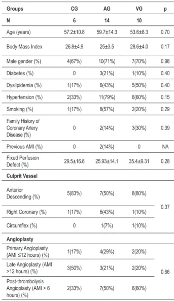
Table 1 – Baseline patient demographics
Groups CG AG VG p
N 6 14 10
Age (years) 57.2±10.8 59.7±14.3 53.6±8.3 0.70
Body Mass Index 26.8±4.9 25±3.5 28.6±4.0 0.17
Male gender (%) 4(67%) 10(71%) 7(70%) 0.98
Diabetes (%) 0 3(21%) 1(10%) 0.40
Dyslipidemia (%) 1(17%) 6(43%) 5(50%) 0.40
Hypertension (%) 2(33%) 11(79%) 6(60%) 0.15
Smoking (%) 1(17%) 8(57%) 2(20%) 0.29
Family History of Coronary Artery Disease (%)
0 2(14%) 3(30%) 0.39
Previous AMI (%) 0 2(14%) 0 NA
Fixed Perfusion
Defect (%) 29.5±16.6 25.93±14.1 35.4±9.31 0.28
Culprit Vessel
Anterior
Descending (%) 5(83%) 7(50%) 8(80%)
0.37 Right Coronary (%) 1(17%) 6(43%) 1(10%)
Circumlex (%) 0 1(7%) 1(10%)
Angioplasty
Primary Angioplasty
(AMI ≤12 hours) (%) 1(17%) 4(29%) 2(20%)
0.66 Late Angioplasty (AMI
>12 hours) (%) 3(50%) 3(21%) 2(20%)
Post-thrombolysis Angioplasty (AMI > 6 hours) (%)
2(33%) 7(50%) 6(60%)
AMI - Acute Myocardial Infarction; AG - Arterial Group; VG - Venous Group; CG - Control group
out on an outpatient basis.
The echocardiographic studies were carried out by physicians of the Echocardiography Laboratory of Hospital Pró-Cardíaco in Vivid 7 instruments (GE Medical Systems, WI), with a 3-MHz transducer and harmonic imaging. In all studies, the instrument itself provided electrocardiographic monitoring. Thus, a complete echocardiographic study of the patients was performed in the first follow-up visit and at three and six months, using long-axis parasternal, 4 and 2-chamber apical, long-axis apical, subcostal, suprasternal, and short-axis views, whenever the acoustic window permitted. By the end of the test, the images were filed and sent to the central workstation, where the tests were available for further analysis, carried out by other echocardiographers who had not had contact with the patient or were blind to the randomization group which the patient had been assigned to.
Parameters of left ventricular systolic function were obtained in a further analysis. The following parameters were studied: ejection fraction (EF) using the modified Simpson’s rule, end-diastolic volume (EDV), end-systolic volume (ESV), and wall motion index score (WMIS). These measurements were obtained as follows: the apical window was used for acquisition of ventricular volumes, by tracing the LV endocardial border in the 4 and 2-chamber view. EDV was obtained at end-diastole and ESV at end-systole, with the exclusion of the papillary muscles. Ejection fraction was calculated using the formula: EF = (EDV – ESV) / EDV x 100, automatically provided by a report generated by the instrument11. Regional function, as reflected by the analysis
of WMIS, was acquired according to the 17-segment model of the American Heart Association12. Therefore, the heart was
divided into a basal portion with six segments, mid-cavity portion with 6 segments, and apical region with 5 segments, including the apex. A degree of contractility was attributed to each segment: 1 for normal contractility of hyperkinesia; 2 for hypokinesia; 3 for akynesia (absence of thickening); 4 for dyskinesia (paradoxical systolic motion) and 5 for aneurismatic diastolic deformation. WMIS was calculated by the sum of all the points divided by the number of segments visualized.
Statistical analysis
Continuous variables were described as mean ± standard deviation and compared using the Kruskal-Wallis test. Categorical variables were compared using the chi-square test or Fisher’s exact test, as appropriate. Comparisons inside and between the groups were performed using the ANOVA test with Bonferroni correction. Tow-tailed P values < 0.05 were considered statistically significant. The SPSS software (version 13.0, SPSS Inc.) was used for the statistical analysis.
The protocol was approved by the Ethics Committee of Hospital Pró-Cardíaco, Rio de Janeiro, and by the National Council of Human Research Ethics (CONEP, Brasília). A written informed consent was obtained from all patients.
Results
A total of 30 patients were included in the study: 14 in AG, 10 in VG and six in GC. No significant difference was found between the groups in relation to clinical and
Original Article
Table 2 - Characteristics of the bone marrow mononuclear cells injected
Groups AG VG
% N. Cells x 106 N % N. Cells x 106 N p
Concentration 10x106 / ml 10x106 / ml
Viability 93.69±2.77% 93.69±2.77% 14 92.64±0.03% 92.64±3.07 10 0.17
Hematopoietic progenitors
(CD45loCD34+) 3.01±0.94% 2.82±0.86 14 3.12±1.32% 2.88±1.20 10 0.98
Lymphocytic progenitors
(CD45loCD34+HLA-DR+) 2.27±1.00% 2.16±1.00 10 0.29±1.13% 0.27±0.12 8 0.45
Early hematopoietic progenitors
(CD45loCD34+HLA-DR-) 0.12±0.76% 0.11±0.71 11 0.14±0.45% 0.13±0.04 9 0.17
Endothelial and mesenchymal
progenitors (CD45loCD34+CD105+) 7.04±2.84% 6.92±2.84 10 0.29±0.17% 0.27±0.16 8 0.59
Hematopoietic cells (CD45+CD34+) 95% 89.27±2.65 12 95% 88.20±3.28 8 0.19
T Cell CD4+ (CD45+CD3+CD4+) 18.67±8.51% 17.49±7.93 14 15.78±6.17% 14.64±5.89 10 0.35
T Cell CD8+ (CD45+CD3+CD8+) 13.09±5.81% 12.20±5.34 13 10.27±3.38% 9.5±3.07 10 0.26
B Cell (CD45+CD19+) 8.02±3.34% 7.53±3.20 11 7.59±4.54% 6.96±4.14 8 0.45
NK Cell (CD45+CD56+) 1.19±0.63% 1.11±0.59 13 1.28±0.86% 1.18±0.79 10 0.93
Monocytes (CD45+CD14+) 10.90±5.90% 10.22±5.23 12 11.34±6.21% 10.45±5.64 10 0.69
Functional Analysis No. of CFU No. of CFU
x 106 x 106
Fibroblast colony-forming units
(CFU-F) 16.84±16.99 10 17.43±9.03 8 0.7
Granulocyte-macrophage
colony-forming units (CFU-GM) 11.83±6.35 13 12.63±7.52 9 0.97 AG - Arterial Group; CFU - Colony-forming Units; VG - Venous Group.
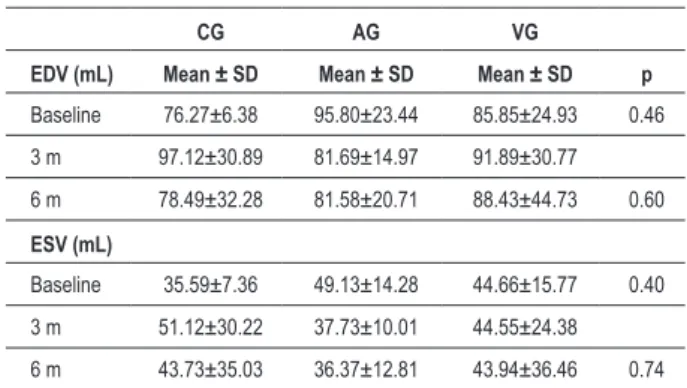
No statistically significant difference was found between the groups after six months of follow-up as regards the systolic function parameters assessed. Although a reduction (improvement) in WMIS (Table 3) had been detected in AG and VG and elevation (worsening) in CG (1.49 ± 0.30; 1.51 ± 0.26; 1.81 ± 0.41; respectively), no statistically significant difference was observed. EF improved (increased) in all groups after six months. EDV values (Table 4) decreased in AG and increased in CG and VG (81.58 ± 20.71 mL; 78.49 ± 32.28 mL and 88.43 ± 44.73 mL, respectively), however without statistical significance. ESV decreased in all groups after six months, with no statistical difference between the groups.
Discussion
This study did not show significant improvement of the echocardiographic parameters after a 6-month follow-up of patients with AMI, regardless of the route used for cell transfer. Some studies in the literature used the intracoronary access to transfer MBMC to the infarct region. The TOPCARE-AMI6
study published in 2002 used the antegrade intracoronary route to approach the AMI area in 20 patients. Two different cell types were transferred, one for each group. One group
Table 3 - EF and WMIS values at baseline and comparison with
values at six months of follow-up
CG AG VG
EF (%) Mean ± SD Mean ± SD Mean ± SD p
Baseline 47.59±14.31 48.26±10.38 48.62±7.08 0.95
3 m 50.19±12.59 54.51±8.38 53.18±13.93
6 m 49.60±17.53 55.17±8.08 55.07±14.13 0.53
WMIS
Baseline 1.76±0.53 1.68±0.24 1.81±0.44 0.78
3 m 1.81±0.46 1.45±0.25 1.67±0.41
6 m 1.81±0.41 1.49±0.30 1.51±0.26 0.15 AG - Arterial group; VG - Venous group; CG - Control group; EF -
Ejection fraction; WMIS - Wall motion index score; SD - Standard deviation.
Table 4 - EDV and ESV values at baseline and comparison with values at six months of follow-up
CG AG VG
EDV (mL) Mean ± SD Mean ± SD Mean ± SD p
Baseline 76.27±6.38 95.80±23.44 85.85±24.93 0.46
3 m 97.12±30.89 81.69±14.97 91.89±30.77
6 m 78.49±32.28 81.58±20.71 88.43±44.73 0.60
ESV (mL)
Baseline 35.59±7.36 49.13±14.28 44.66±15.77 0.40
3 m 51.12±30.22 37.73±10.01 44.55±24.38
6 m 43.73±35.03 36.37±12.81 43.94±36.46 0.74 EDV - End-diastolic volume; ESV - End-systolic volume; AG - Arterial group; VG - Venous group; CG - Control group; SD - Standard
deviation.
stress-echocardiography and magnetic resonance imaging. The procedure was considered safe, since no cell transfer-related complications occurred.
The analysis carried out by the BOOST study5 included 60
patients, half of whom were assigned to the control group. Intracoronary MBMC injection was also used in this study. After six months of follow-up, significant improvement of EF, as measured by ventriculography, could be demonstrated in the intervention group.
In our study, the method used to measure systolic function parameters was resting transthoracic echocardiography. The imaging modality adopted in the different studies is heterogeneous. While studies such as TOPCARE6 and
REPAIR-AMI7 used ventriculography, studies such as BOOST5 and
Janssens et al8 used magnetic resonance imaging. All these
studies demonstrated improved ejection fraction within a maximum period of six months, except for Janssens’s study8, in
which no benefit related to the ejection fraction was found, but rather a reduction in the infarct area in the group treated.
The ASTAMI study13 used echocardiography as well as
SPECT and magnetic resonance imaging to evaluate the effect of bone marrow cells on a post-acute myocardial infarction population. In a six-month follow-up, no difference was found in ejection fraction as measured by the three methods used. Although echocardiography is less accurate than methods such as magnetic resonance imaging14, the discrepant results
found in the different studies do not seem to be related to a specific methodology.
The profile of bone-marrow cells, as well as the amount of cells used in this and other studies aiming at myocardial regeneration is variable, although there is evidence suggesting that there is no preferable bone-marrow cell population in the setting of intracoronary injection13.
A recently published review15 showed significant
improvement in left ventricular ejection fraction and cardiac remodeling in patients receiving cell therapy in comparison with the control group. This systematic review included studies
using several cell types: mesenchymal cells, mononuclear cells, or circulating progenitor cells. The mechanism by which these cells exert their action remains unknown. However, some possibilities such as transdifferentiation in cardiomyocytes, paracrine secretion of growth factors or cytokines, angiogenesis promoting neovascularization and immunomodulation13,16
have been studied.
In the REPAIR-AMI study7, in which 204 patients were
evaluated, there was a statistically significant increase in ejection fraction in the treatment group, based on parameters obtained from angiography. In this case, patients with the lowest LVEF were those who benefited the most from the therapy. The literature demonstrated that patients with left ventricular systolic dysfunction may show a better response to cell therapy. These are the patients who most need therapeutic approaches that provide additional benefit to conventional therapy. This population has the most favorable risk-benefit ratio in relation to the use of new therapies, and studies have demonstrated that this subgroup is the most likely to benefit16.
In 2006, Hendrikx et al17 published a study in which cell
transplantation was performed during elective coronary artery bypass grafting following acute myocardial infarction. The injection was made directly in the border of the infarct area. Magnetic resonance imaging was used to evaluate ejection fraction and ventricular volumes. The results obtained were negative in relation to the improvement of systolic function parameters after cell therapy. However, there was a tendency to improved regional contractility after four months of follow-up, represented by improvement in wall thickening. This benefit was observed in a subgroup named responders, which was comprised of patients who presented a higher number of transplanted progenitor cells and CD34 positive surface marker.
In the present study, the inclusion of patients with mean ejection fraction values higher than 35% may have made it difficult to demonstrate a beneficial effect in this population. Additionally, the small sample size may have impaired the demonstration of a statistically significant improvement between the groups, since for some of the parameters that improved, no statistical significance was found.
Further analyses with a higher number of patients are necessary so that subanalyses comparing the performance of myocardial segments in the infarct area of patients treated with the infarct area of patients of the control group can be carried out. Also, in the long term, reduction of endpoints such as mortality and improved quality of life may not be directly related to EF gain in six months – and this has already been observed in previous studies on ischemic heart disease such as those on the use of betablockers and angiotensin-converting enzyme inhibitors18-20.
Conclusion
References
1. Yosef ZR, Redwood SR, Marber MS. Postinfarction left ventricular remodelling: where are the theories and trials leading us? Heart. 2000; 83: 76-80. 2. Strauer BE, Brehm M, Zeus T, Kostering M, Hernandez A, Sorg RV, et al. Repair
of infarcted myocardium by autologous intracoronary mononuclear bone marrow cell transplantation in humans. Circulation. 2002; 106: 1913-8. 3. Orlic D, Kajstura J, Chimenti S, Limawa F, Jakoniuk I, Quairi F, et al. Mobilized
bone marrow cells repair the infarcted heart, improving function and survival. Proc Natl Acad Sci USA. 2001; 98: 10344-9.
4. Perin EC, Dohmann HF, Borojevic R, Silva SA, Sousa AL, Mesquita CT, et al. Transendocardial, autologous bone marrow cell transplantation for severe, chronic ischemic heart failure. Circulation. 2003; 107: 2294-302. 5. Wollert KC, Meyer GP, Lotz J, Ringes-Lichtenberg S, Lippoet P, Briedenbach
C, et al. Intracoronary autologous bone-marrow cell transfer after myocardial infarction: the BOOST randomised controlled clinical trial. Lancet. 2004; 364 (9429): 141-8.
6. Schachinger V, Erbs S, Elsasser A, Haberbosch W, Hambrecht R, Holschermann H, et al. Intracoronary bone marrow–derived progenitor cells in acute myocardial infarction (REPAIR-AMI). N Engl J Med. 2006; 355: 1210-21. 7. Janssens S, Dubois C, Bogaert J, Theunissen K, Deroose C, Desmet W, et
al. Autologous bone marrow-derived stem-cell transfer in patients with ST-segment elevation myocardial infarction: double-blind, randomised controlled trial. Lancet. 2006; 367: 113-21.
8. Vyrenkov Iu E, Morozova VV, Gofman Kh I, Gofman Kh I. Microcirculatory bed of the heart during peripheral arteriovenous shunting. Arkh Anat Gistol Émbriol. 1986; 91(11): 25-34.
9. Liu L, Kubes P. Molecular mechanisms of leukocyte recruitment: organ-specific mechanisms of action. Thromb Haemost. 2003; 89 (2): 213-20. 10. Lang RM, Bierig M, Devereux RB, Flashskampf FA, Foster E, Pellikka PA, et al.
Recommendations for chamber quantification: a report from the American
Society of Echocardiography’s Guidelines and Standards Committee and the Chamber Quantification Writing Group, developed in conjunction with the European Association of Echocardiography, a branch of the European Society of Cardiology. J Am Soc Echocardiogr. 2005; 18: 1440-63.
11. Cerqueira M, Weissman NJ, Dilsizian V, Jacobs AK, Kaul S, Laskey WK, et al. Standardized myocardial segmentation and nomenclature for tomographic imaging of the heart. Circulation. 2002; 105: 539-42.
12. Bellenger NG, Burgess MI, Ray SG. Comparison of left ventricular ejection fraction and volumes in heart failure by echocardiography, radionuclide ventriculography and cardiovascular magnetic resonance: are they interchangeable? Eur Heart J. 2000; 21: 1387-96.
13. Assmus B, Schachinger V, Teupe C, Britten M, Lehmann R, D�bert N, et al. Transplantation of progenitor cells and regeneration enhancement in acute myocardial infarction (TOPCARE-AMI). Circulation. 2002; 106 (24): 3009-17.
14.Hendrikx M, Hensen K, Clijsters C, Jongen H, Koninckx R, Bijnens E, et al. Recovery of regional but not global contractile function by the direct intramyocardial autologous bone marrow transplantation: results from a randomized controlled clinical trial. Circulation. 2006; 114 (1 Suppl): I-101-7.
15. Lunde K, Solheim S, Aakhus S, Arnesen H, Abdelnoor M, Egeland T, et al. Intracoronary injection of mononuclear bone marrow cells in acute myocardial infarction (ASTAMI). N Engl J Med. 2006; 355: 1199-209. 16. Rosenzweig A. Cardiac cell therapy — mixed results from mixed cells. N Engl
J Med. 2006; 355 (12): 1274-7.
17. Dargie HJ. Effect of carvedilol on outcome after myocardial infarction in patients with left-ventricular dysfunction: the CAPRICORN randomised trial. Lancet. 2001;357(9266):1385-1390.
18. Maggioni AP, Fabbri G. VALIANT (VALsartan In Acute myocardial iNfarcTion) trial. Expert Opin Pharmacother. 2005;6(3):507-512.