1
Original Article
Texture of Mitral Bioprosthesis, Ventricular
Function and Formation of Thrombus. Analysis
through Transesophageal Echocardiography
and Use of Bioscore
Henry Abensur, Max Grinberg, José A. F. Ramires
São Paulo, SP - Brazil
Heart Institute of Hospital das Clínicas - FMUSP Mailing address: Henry Abensur - Rua Gaivota, 222/21 Cep 04522-030 - São Paulo, SP - Brazil
E-mail: [email protected] Sent for publishing on 07/13/2004 Accepted on 12/02/2004
Objective
Facing the hypothesis of participation of mechanical stress as a cause of mitral bioprosthesis dysfunction, we decided to assess the relation of preservation of the texture of the mitral bioprosthesis leaflets with left ventricular function, in addition to the casual formation of thrombus in left atrium in patients with left ventricular dysfunction from the implant of mitral bioprosthesis.
Methods
Forty 40 patients with mitral bioprosthesis through multiplane transesophageal echocardiogram were studied and divided in two groups: with left ventricular dysfunction (FE=0.40±0.09) since the bioprosthesis implant (20 patients: age 47.75±11.10 years old and surgery time 5.3±2.6 years) and with normal left ventricular function (FE=0.73±0.06) since the implant (20 patients: age 49.75±13.59 years old and surgery time 5.7±3 years). The texture of bioprosthesis leaflets was analyzed through a transesophageal echocardiographic score (FACIMT Bioscore): 1) Fusion of leaflets (score 1 to 3); 2) Apposition of tissues (score 1 to 3); 3) Calcium in leaflets (score 1 to 5); 4) Integrity of leaflets (score 1 to 3); 5) Motility of leaflets (score 1 to 4) and 6) Thickness of leaflets (score 1 to 3). The presence of thrombi in left atrium was assessed through multiplane scanning of the left atrium and left atrial appendage in the transesophageal study.
Results
There was no significant difference in the texture of bioprosthesis mitral position between the groups, for the total score (8.7±2.4 vs. 7.9±2.1, p=0.259), and for each analyzed item. A greater incidence of thrombi in left atrium and left atrial appendage was detected in patients with ventricular dysfunction (65% vs. 20%, p=0.004).
Conclusion
The left ventricular dysfunction was not a protecting factor of the texture of bioprosthesis leaflets in mitral position in the tardive post-surgery period. The patients with left ventricular dysfunction showed a more favorable environment for the formation of thrombi in left atrium.
Key words
mitral bioprosthesis, left ventricular function, transesophageal echocardiography, formation of thrombi
It is acknowledged that the a pre-implant cardiac index of mitral valve bioprosthesis greater than 2.0 L/min/m2 is an accele-ration factor of the structural degeneaccele-ration process of valve leaflets1; such hypothesis suggests the participation of mechanical stress as a cause of bioprosthesis dysfunction2-4.
Observations from our clinical practice suggest a possible re-lation between the preservation of the texture of mitral bioprosthesis leaflets and the left ventricular dysfunction. So, left ventricular dysfunction carriers, when submitted to a lower leaflet closing stress, from the implant of mitral bioprosthesis, would somehow show a lower development o degenerative process. Likewise, those mitral bioprosthesis and left ventricular dysfunction carriers, when submitted to a greater stasis environment due to left ventricular dysfunction, could be more predisposed to the formation of intra-cavitary thrombi.
The introduction of transesophageal modality allowed for over-coming technical difficulties presented to transthoracic exam, es-pecially thanks to the closeness of the esophagus to the left atrium; it allows for the use of higher frequency transducers, under absence of structures that obstruct the heart, which results in cardiac images with better quality of signal and better level of resolution5,6,7. The method has a better definition concerning the texture and motility of leaflets, presence of aberrant masses, or a discontinuity of suture of the prosthesis ring8-10. Currently we count on the multiplane technology, which offers transducers with the capacity of perform all Doppler modalities, being also multifrequency that produce a continuous of transversal and longitudinal images through the rotation of crystal displaying, which makes easier the view of intermediary images out of the axle among the primary, transversal and longitudinal plans. So, there is an increase in the quality of tomographic images obtained in comparison with those obtained by the biplane transducers11-14.
The transesophageal method is also relevant in the detection of intracardiac thrombi. The image through transthoracic echo-cardiography has a limited sensitivity for the atrial thrombus. This is mostly because the thrombus usually forms in the left atrial appendage, which is well viewed through the transesopha-geal modality15-17.
2
Methods
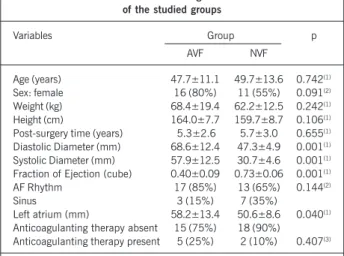
Forty mitral bioprosthesis carriers for more than two years were studied and followed up at the Day Unit of the Clinical Unit of Valve Cardiopathies of Instituto do Coração. Twenty consecutive patients (average age of 47.7±11.1 years old) were selected for showing left ventricular dysfunction since the implant, which was verified by the fraction of ejection lower than 0.45 (unidimensional MODE – cube method) in the follow-up echocardiograms, and constituted the abnormal ventricular function (AVF) group. Other 20 patients (average age of 49.7±13.6 years old), with normal left ventricular function (fraction of ejection greater than 0.65) since the implant, were gauged and composed the control group of normal left ventricular function (NVF). Patients with fraction of ejection between 0.45 and 0.65 were excluded from the study. The clinical data, including functional class, cardiac rhythm, use of medication and laboratory controls of the anticoagulation level were obtained through the analysis of records (tab. I).
The cardiac rhythm (12-derivation electrocardiogram), the use of anticoagulation therapy (anamnesis data and records) and international normalization ratio (INR – turbidimetric, automated method) were additionally marked down.
The patients were submitted to an echocardiogram using the Toshiba Power Vision 7000. From the transthoracic modality the following variables were obtained: final diastolic dimension of left ventricle (FDDLV), final systolic dimension of left ventricle (FSDLV), FE through the Cube method18 and dimension of left atrium
perfor-med at the end of the ejection of the left ventricle, in accordance to the recommendations of the American Society of Echocardio-graphy19, having been obtained in the unidimensional mode and guided through the bidimensional mode. With the assistance of color flow mapping, the presence of regurgitation of aortic and tricuspid valves was verified. In the presence of regurgitation, it was quantified qualitatively through the color flow mapping. With the use of continuous Doppler in the patients showing tricuspid insufficiency, we obtained the pressure difference between the right ventricle and the right atrium which, added by the estimated pres-sure of the right atrium, reflects the systolic prespres-sure of the right ventricle and, consequently, the systolic pressure of the pulmonary artery (PPA)20. The mitral prosthesis area was calculated using the Continuous Doppler and the methodology of pressure half-time21. The gradient through the aortic valve was calculated through the continuous Doppler, by using the modified equation of Bernoulli22.
At the end of the transthoracic echocardiogram, the patient, who had already been told to be fasting for 4h, was signing the post-information consent term for the performance of the multi-plane transesophageal echocardiogram. The examination was per-formed according to an already acclaimed and established method in the clinical practice11,23,24. All patients had topic anesthesia, with lidocaine at 10% - based local anesthetic solution, applied in the oropharinx, hard and soft palate.
After the insertion of the transducer in the esophagus and the location of the mitral bioprosthesis, the multiplane (0 to 180º) prosthesis scanning was performed.
Thrombi and the presence of formation of spontaneous contrast in the atria and the left atrial appendage were observed through multiplane scanning.
Stimulated by the well-known applicability of echocardiographic score for decisions of mitral valvoplasty through percutaneous balloon-catheter25, we developed a transesophageal echocardio-graphy assessment of bioprostheses in mitral position, consisting of six items, which we called FACIMT, (an acronym of fusion of leaflets, apposition of tissues, calcium in leaflets, integrity of leaflets, motility of leaflets and thickness of leaflets – chart I).
The presence of central insufficiency of bioprosthesis or peri-prosthetics was analyzed through the color flow mapping.
Thrombi and presence of formation of spontaneous contrast in atria and the left atrial appendage were observed through multiplane scanning.
The transthoracic and transesophageal echocardiograms were recorded in VHS videotapes. The FACIMT Bioscore was used in the reading of the videotape and the echocardiographic study items descri-bed above were analyzed. The reading of the videotape was done by two observers singly, and the discordant data were solved through a consensus in a third reading done by both observers together.
Chart I - FACIMT Bioscore
Variable levels 1 2 3 4 5
Fusion of leaflets Normal Fusion of 2 leaflets Fusion of 3 leaflets
Apposition of tissue Normal Through filiform image Through thrombus,
vegetation or pannos
Calcium in leaflets Normal 1 - 2 calcium points > 2 calcium points Segmentar in leaflet Segmentar in more than 2 leaflets
Integrity of leaflets Normal Perforation of 1 Rupture of 1 or
or more leaflets more leaflets
Motility of leaflets Normal Diminished in 1 leaflet Diminished in 2 leaflets Diminished in 3 leaflets
Thickness of leaflets Normal 2-4 mm > 4 mm
Table I - Clinical and echocardiograhic characteristics of the studied groups
Variables Group p
AVF NVF
Age (years) 47.7±11.1 49.7±13.6 0.742(1)
Sex: female 16 (80%) 11 (55%) 0.091(2)
Weight (kg) 68.4±19.4 62.2±12.5 0.242(1)
Height (cm) 164.0±7.7 159.7±8.7 0.106(1)
Post-surgery time (years) 5.3±2.6 5.7±3.0 0.655(1)
Diastolic Diameter (mm) 68.6±12.4 47.3±4.9 0.001(1)
Systolic Diameter (mm) 57.9±12.5 30.7±4.6 0.001(1)
Fraction of Ejection (cube) 0.40±0.09 0.73±0.06 0.001(1)
AF Rhythm 17 (85%) 13 (65%) 0.144(2)
Sinus 3 (15%) 7 (35%)
Left atrium (mm) 58.2±13.4 50.6±8.6 0.040(1)
Anticoagulanting therapy absent 15 (75%) 18 (90%)
Anticoagulanting therapy present 5 (25%) 2 (10%) 0.407(3)
3
Initially all variables were descriptively analyzed. For the con-tinuous variables that analysis was performed through the obser-vation of maximum and minimum values, and the calculation of means and standard deviations and medians. The absolute fre-quencies were calculated for the classificatory variables.
For the analysis of the hypothesis of equality of proportions between the two groups, the chi-square test or exact test of Fisher26 was used. The hypothesis of equality between the two means was verified using the t test of Student26. For the total score variable, the non-parametric test of Mann-Whitney26 was used. The non-parametrical test of Kruskal-Wallis26 was used in the comparison of the total score among many groups.
The level of significance used for the tests was 5%.
Results
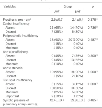
Both groups were similar in relation to the area of mitral bioprosthesis, presence of central and periprosthetic insufficiency, presence of aortic stenosis, presence of tricuspid insufficiency and the estimated value of systolic pressure of pulmonary artery (tab. II). In AVF group, two cases of periprosthetic insufficiency were detected and they were only visible in the transesophageal study. The use of the FACIMT Bioscore did not identify any significant difference concerning the total score (8.70±2.39 and 7.95±2.14, p=0.259) between the AVF and NVF groups. An individual analysis of the items of FACIMT Bioscore did not show significant differen-ces (tab. III), as well.
Thrombi in left atrium were found in AVF group in a greater proportion than in NVF. The AVF group showed thrombi in 10, 20 and 35%, respectively, in left atrial appendage, left atrium and left atrial appendage and left atrium simultaneously. Thrombi were found in the AVF group in 10, 5 and 5% distribution (tab. IV).
The spontaneous contrast was present in 85% of AVF group patients and in 60% of NVF group, showing a more favorable statistic tendency to the formation of spontaneous contrast in the AVF group (tab. IV).
The use of anticoagulant therapy was similar in both groups. In the AVF group, 75% of the patients were not using oral anti-coagulants and in the NVF group, 90% were not either. Only three had updated international normalization rate (INR) at the time of the study, and none of them was properly anticoagulated. In the NVF group, only two patients had oral anticoagulant therapy, with an INR of 3.4 and 4.7, respectively.
The rhythm of atrial fibrillation was present in 85 and 65% in the patients of the AVF and NVF groups, respectively. There were no statistic differences between those values.
The dimension of the left atrium was significantly larger in the AVF group in relation to the NVF (58.2±13.4 vs. 50.6± 8.6mm, p=0.040).
Table III - FACIMT Bioscore - Results
Bioscore items Score AVF Group NVF Group p
Fusion of leaflets 1 18 (90%) 20 (100%) 0.487(3)
2 2 (10%) 0 (0%)
3 0 (0%) 0 (0%)
Apposition of tissues 1 17 (85%) 18 (90%) 0.605(3)
2 2 (10%) 0 (0%)
3 1 (5%) 2 (10%)
Calcium in leaflets 1 6 (30%) 5 (25%) 0.837(3)
2 11 (55%) 13 (65%)
3 1 (5%) 0 (0%)
4 1 (5%) 0 (0%)
5 1 (5%) 2 (10%)
Integrity of leaflets 1 20 (100%) 20 (100%)
-2 0 (0%) 0 (0%)
3 0 (0%) 0 (0%)
Motility of leaflets 1 11 (55%) 17 (85%) 0.166(3)
2 5 (25%) 2 (10%)
3 4 (20%) 1 (5%)
4 0 (0%) 0 (0%)
Thickness of leaflets 1 9 (45%) 12 (60%) 0.693(3)
2 7 (35%) 4 (20%)
3 2 (25%) 3 (15%)
Total bioscore 8.7±2.4 7.9±2.1 0.259(4)
(3) Descriptive level of probability of the exact of Fisher; (4) descriptive levels of probability of the test of Mann- Whiney; AVF - abnormal ventricular function; NVF - normal ventricular function.
Table II - Echocardiographic characteristics of the series
Variables Group p
AVF NVF
Prosthesis area - cm2 2.6±0.7 2.4±0.4 0.378(1)
Central insufficiency
Absent 13 (65%) 14 (70%) 0.736(2)
Discreet 7 (35%) 6 (30%)
Periprosthetic insufficiency
Absent 18 (90%) 20 (100%) 0.487(3)
Discreet 1 (5%) 0 (0%)
Moderate 1 (5%) 0 (0%)
Aortic insufficiency
Absent 9 (45%) 7 (35%) 0.300(3)
Discreet 9 (45%) 13 (65%)
Moderate 2 (10%) 0 (0%)
Aortic stenosis
Absent 19 (95%) 18 (90%) 1.000(3)
Discreet 1 (5%) 2 (10%)
Tricuspid insufficiency
Absent 3 (15%) 3 (15%) 1.000(3)
Discreet 10 (50%) 10 (50%)
Moderate 5 (25%) 6 (30%)
Important 2 (10%) 1 (5%)
Systolic pressure of 42.4±10.7 39.8±10.1 0.485(1)
pulmonary artery - mmHg
(1) Descriptive level of probability of the t test of Student; (2) descriptive level of probability Chi-square test; (3) descriptive level of probability of the exact of Fisher; AVF - abnormal ventricular function; NVF - normal ventricular function.
Table IV - Echocardiographic aspects related to left atrial thrombosis
Variable Group p
AVF NVF
Thrombus Absent 7 (35%) 16 (80%) 0.012(3)
Thrombus Present
LAA 2 (10%) 2 (10%)
LA 4 (20%) 1 (5%)
LA - LAA 7 (35%) 1 (5%)
Total 13 (65%) 4 (20%) 0.004(2)*
Spontaneous contrast
Absent 3 (15%) 8 (40%)
Present 17 (85%) 12 (60%) 0.077(2)
4
Discussion
The evolution of bioprostheses is well characterized by means of actuarial curves. The structural changes of clinical repercussion of bioprostheses happen, on average, between 5 and 10 years after their implant, with stenosis due to calcification and inspis-sation of leaflets, and incompetence due to inappropriate juxtapo-sition of commissures in calcium infiltration sites27-30. It seemed important to us to elaborate a score, in which we could quantita-tively document that biological phenomenon of degeneration of bioprostheses, by placing emphasis on the aspects that are more analyzed in the literature, related to leaflets of bioprostheses. We used the FACIMT acronym, which are six distinct letters with which we intend to facilitate the memorization of bioscore items: F for fusion of leaflets, A for apposition of tissue, C for calcium of leaflets, I for integrity of leaflets, M for motility of leaflets and T for thickness of leaflets. So, this way we hope to make use of a suitable diagnosis method that is a homogeneous communication language between clinicians and surgeons.
The average time of implant of bioprostheses was 5.3±2.6 and 5.7±3 years, respectively, for the AVF and NVF groups. There were 10 and 8 patients, respectively, for the AVF and NVF groups with more than five years of bioprostesis implant.
The left ventricular dysfunction was not a protection factor of the texture of leaflets of mitral bioprosthesis, as both the total score and each FACIMT Bioscore item, showed similar in both groups. In the studied literature, there is only one reference on left ventricular function and texture of leaflets1, in which the acceleration of the dysfunction process of bioprostheses in patients with cardiac index greater than 2.0L/min/m2 was observed, in an attempt of explaining a lower dura-bility of bioprostheses in younger patients.
The individual analysis of the items of FACIMT Bioscore beco-mes useful: for example, the patient #23 from NVF group and #5 from AVF group had a total score of 12. However, AVF patient #5 had 3 in apposition of tissue item and NVF patient #23 had, in that item, 1. So, AVF patient #5 had thrombosis of one of his leaflets, a situation that determines a specific therapeutic conduct. Therefore, the individual analysis of each item of the score is essential for us to know the real situation of the texture of bio-prosthesis leaflets. That situation was witnessed when the echo-cardiographic score of mitral valve for mitral valvoplasty through balloon-catheter was used. It demonstrated that the subvalve sys-tem had a greater prognostic importance for the success of the procedure. However, such hierarchy does not invalidate the routine use of the score31.
It was only possible to identify the changes in the texture of leaflets with the use of the transesophageal method. The study through transthoracic echocardiography has its own value, espe-cially in the analysis of functional factors related to bioprostheses, such as area, gradients, presence of regurgitations and huge chan-ges of leaflets of prostheses6,9,32-35. The transesophageal echocar-diography, through the use of higher frequency transducers and through a greater closeness of the analyzed structures, allows us for a fine assessment of the texture of leaflets, a more accurate quantification of the regurgitation level of mitral bioprostheses and the detection of complications, such as periprosthetic vege-tations, thrombi and regurgitations5,6,10,36,37. In that casuistry, pe-riprosthetic insufficiency was detected in two AVF group patients,
which were not detected at the transthoracic echocardiogram, a situation demonstrated by Khanderia et al.36.
The mitral position of bioprosthesis favors the earlier primary degeneration of leaflets, probably because of a greater closing stress, which takes place in mitral position during the systole27,38-40. In mi-tral position, the opening time of leaflets is up to three times the opening time of aortic bioprosthesis leaflets41. The rhythm is sinus, there is a double opening movement of the mitral bioprosthesis lea-flets; in a carried out study the relation between the type pf rhythm and the bioprosthesis primary degeneration42 was not demonstrated. Up to the present moment there is no confirmation in relation to the size of bioprosthesis and the prevalence of primary degeneration of the prosthesis. Regarding sex, there is not a consensus either. The left ventricular function factor did not have any influence in the texture of the bioprosthesis, as demonstrated in this study.
Thrombus in left atrium and its appendage was dominating in AVF group (65% vs. 20%, p=0.004), however there was not a significant difference in relation to the type of rhythm (atrial or sinus fibrillation) and concerning the anticoagulating therapy among the groups studied. It was demonstrated that patients with left ventricular dysfunction show a more favorable environment for the formation of thrombus in left atrium and left atrial appendage. In relation to the size of left atrium, the AVF group showed the left atrium a little larger (58.2±13.4 vs. 50.6±8.6 mm, p=0.040). Edmunds et al.43 showed that there was a need for anticoagu-lation in 40 to 60% of the patients with biological prosthesis in mitral position; the incidence of thromoembolic episodes is greater in the first three months of bioprosthesis implant, the atrial fibrilla-tion increases the risk of thromboembolic complicafibrilla-tions, the role of the presence of atrial thrombi, the size of left atrium and the history of previous embolic events is not clear yet in the increase of incidence of thromboembolic events. The atrial fibrillation is the main factor identified in the literature as responsible for the increase of risk of systemic thromboembolism in patients with mitral valvo-pathy44-47. Reports in the literature show a lower incidence of throm-boembolic events in patients with bovine pericardium or dura mater bioprostheses in comparison with porcine biosprostheses48-50.
The size of left atrium, age lower than 60 years old, possibly the left ventricular dysfunction and hypertension add risks to thromboembolism in patients with atrial fibrillation51. Those pa-tients must have prolonged anticoagulating therapy, by keeping an international normalization rate between 2.0 and 3.0. For probably social reasons, most of our patients did not have their coagulation properly controlled.
Spontaneous contrast inleft atrium and left atrial appendage was more frequent in AVF group. However, such datum did not have statistic significance (85% vs. 60%, p=0.077). We must menton that the presence of spontaneous contrast, atrial fibrillation and intra-atrial septum aneurysm are independent positive factors, predictors of the presence of thrombi in left atrium and cerebro-vascular events. In addition, the dilation of left atrium and cere-brovascular events are positive independent predictors of the pre-sence of spontaneous contrast and thrombi in left atrium52.
5
References
1. Magilligan DJ, Lewis JW, Stein P, Alam M. The porcine bioprosthetic heart valva: experience at 15 years. Ann Thorac Surg 1989; 48:324-30.
2. Sabbah HN, Hamid MS, Stein PD. Mechanical stress on closed cusps of porci-ne bioprosthetic valves: correlation with sites of calcification. Ann Thorac Surg 1986; 42:93-6.
3. Thubrikar MJ, Deck JD, Aovad J, Nolan SP. Role of mechanical stress in calcifica-tion of aortic bioprosthetic valves. J Thorac Cardiovasc Surg 1983; 86:115-25.
4. Stein PD, Sabbah HN, Magilligan DJJr. Can we delay the ocurrence of spontaneous
degeneration of bioprosthetic valves? J Thorac Cardiovasc Surg 1988;96:343.
5. Nelessen U, Schnittger I, Appleton CP. Transesophageal two-dimensional
echocar-digraphy and color Doppler flow velocity mapping in the evaluation of cardiac valve prastheses. Circulation 1988; 78:848-55.
6. Chaudhry FA, Herrera C, Defrino PF, Mehlman DJ, Zabalgoitia M. Pathologic and
angiographic correlations of transesophageal echocardigraphy in prosthetic heart valve dysfunction. Am Heart J 1991;122:1057-64.
7. Herrera CJ, Chaudhry FA, Defrino PF. Value limitations of transesophageal
echo-cardiography in evaluating prosthetic or bioprosthetic valve dysfunction. Am J Car-diol 1992; 69:697-9.
8. Khandheria BK. Transesophageal echocardiography in the evaluation of prosthetic
valves. Cardiol Clin 1993; 11:427-36.
9. Daniel WG, Mügge A, Grote J. Comparison of transthoracic and transesophageal
echocardiography for detection of abnormalities of prosthetic and bioprosthetic valves in the mitral and aortic positions. Am J Cardiol 1993; 71: 210-15. 10. Groundstroem RD, Hoffman P, Bloomfield P, Sutherland GR. Additional value of
biblane transesophageal imaging in assesment of mitral valve prostheses. Br Heart J 1993; 70:259-65.
11. Freeman WK, Seward JB, Khanderia BK. Transesophageal echocardiography. Boston, Little Brown, 1993.
12. Pandian NG, Hsu TL, Schwartz SL et al. Multiplane transesophageal echocar-diography. Imaging planes, echocardigraphic anatomy, and clinical experience with a prototype phased array OmniPlane probe. Echocardiography. Echocar-diography 1992; 9:649-66.
13. Roelandt JR, Thomsom IR, Vletter WB. Multiplane transesophageal echocardiogra-phy: latest evolution in imaging revolution. J Am Soc Echocardiogr 1992; 5: 361-7. 14. Seward JB, Khandheria BK, Freeman WK. Multiplane transesophageal echocar-diography: image orientation technique, anatomic correlations, and clinical appli-cations. Mayo Clin Proc 1993; 68:1-29.
15. Aschenberg W, Schluter M, Kremer P. Transesophageal two-dimensional echo-cardiography for the detection of left atrial appendage thrombus. J Am Coll Cardiol 1986; 7: 163-7.
16. Dressler FA, Labovitz AJ. Systemic arterial emboli and cardiac masses: assesment with transesophageal echocardiography. Cardiol Clin 1993; 11: 447-60. 17. Olsen JD, Goldenberg IF, Pederson W. Exclusion of atrial thrombus by
transeso-phageal echocardiography. J Am Soc Echocardiogr 1992; 5: 52-6.
18. Triulzi MO, Wilkins GT, Gillam LD. Normal adult cross-sectional echocardiographic valves: LV volumes. Echocardiography 1985; 2:153-70.
19. Sahn DJ, Demaria A, Kisslo J, Weyman A. The commitee on M-mode standar-tization of the American Society of Echocardiography. Recommendations re-garding quantitation in M-mode echocardiographic measurements. Circulation 1978; 58: 1072.
20. Yock PG, Popp RL. Non–invasive estimation of right ventricular systolic pressure by Doppler ultrasound in patients with tricuspid regurgitation. Circulation 1984; 70: 657-62.
21. Hatle L, Angelsen B, Tromsdal B. Nom-invasive assesment of pressure half-time by Doppler ultrasound. Circulation 1980; 60:1096.
22. Hatle L, Angelsen B, Tromsdal B. Noninvasive assesment of aortic stenosis by Dop-pler ultrasound. Br Heart J 1980; 43: 284.
23. Seward JB, Khandheria BK, Edwards WD. Biplanar transesophageal echocardio-graphy: anatomic correlations, image orientation, and clinical applications. Mayo Clin. Proc., v.65, p.1193-213, 1990.
24. Seward JB, Khandheria BK, Freeman WK. Multiplane transesophageal echocar-diography: image orientation, examination technique, anatomic correlations, and clinical applications. Mayo Clin Proc 1993, 68:1-29.
25. Wilkins GT, Weyman AE, Abascal VM. Percutaneous balloon dilatation of mitral valve. Analysis of echocardiographic variables related to outcome and the mecha-nism of dilatation. Br Heart J1988; 60: 299-308.
26. Rosner B. Fundamentals of biostatistics. 2nd ed. Boston, PWS Publishers, Second
edition, 1986.
27. Jones EL, Weintraub WS, Craver JM et al. Tem-year experience with the porcine bioprosthetic valve: interrelationship of valve survival and patient survival in 1,050 valve replacements. Ann Thorac Surg 1990; 49: 370-84.
28. Pelletier LC, Carrier M, Leclerc Y, Lepage G, Deguise P, Dyrda I. Porcine versus peri-cardial bioprostheses: a comparison of late results in 1,583 patients. Ann Thorac Surg 1989; 47: 352-61.
29. Bortolotti U, Milano A, Thiene G. Early mechanical failures of the Hancock pericar-dial xenograft. J Thorac Cardiovasc Surg 1987; 94: 200-07.
30. Cohn LH, Allred EN, Disesa VJ. Early and late risk of aortic valve replacement: a 12 year concomitant comparison of the porcine bioprosthetic and tilting disc prosthe-tic aorprosthe-tic valves. J Thorac Cardiovasc Surg 1984; 88:695-705.
31. Medeiros CCJ, Moraes AV, Cardoso LF. São os componentes do aparelho valvar mitral de mesmo valor preditivo na valvoplastia mitral por cateter-balão? Estudo ecocardiográfico. Arq Bras Cardiol 1991; 57:11-20.
32. Cooper DM, Stewart WJ, Schiavone WA et al. Evalation of normal prosthetic valve function by Doppler echocardiography. Am Heart J 1987; 114: 576 –82. 33. Alam M, Lakier JB, Pickard SD, Goldstein S. Echocardiographic evaluation of
por-cine bioprosthetic valves: experience with 309 normal and 59 dysfunctioning val-ves. Am Heart J 1983; 52:309-15.
34. Formann MB, Phelan BK, Robertson RM, Virmani R. Correlation of two-dimen-sional echocardiography and pathologic findings in porcine valve dysfunction. J Am Coll Cardiol 1985; 5: 224-30.
35. Almeida J, Sepúlveda F, Gomes MR. Valor diagnóstico da ecocardiografia tran-sesofágica no estudo das disfunções das próteses mitrais. Rev Port Cardiol 1993; 12: 155-61.
36. Khandheria BK, Seward JB, Oh JK. Value and limitations of transesophageal echo-cardiography in assesment of mitral valve prostheses. Circulation 1991; 83: 1956-68. 37. Scott PJ, Ettles DF, Wharton GA, Williams GJ. The value of transesophageal echocardiography in the investigation of acute prosthetic valve dysfunction. Clin Cardiol 1990; 13: 541-4.
38. Jamieson WRE, Hayden RI, Miyagishima RT et al. The Carpentier-Edwards standard porcine bioprosthesis: clinical performance to 15 years. J Cardiac Surg 1991; 6: S550-6.
39. O’brien MF, Stafford EG, Gardner MAH, Pohlner PG, Tesar PJ, Kear L, Smith SE. The Medtronic intact xenograft: analysis of 342 patients over a seven-year fol-low-up period. Ann Thorac Surg 1995; 60: S253-7.
40. Akins CW, Carrol DL, Buckley MJ, Daggett WM, Hilgenberg AD, Austen WG. Late results with Carpentier- Edwards porcine bioprosthesis. Circulation 1990; 82(supp 4):IV-65-IV-74.
41. Thurbrikar MJ, Deck DJ, Aouad J. Role of mechanical stress in calcification of aortic bioprosthetic valve. J Thorac Cardiovasc Surg 1983; 86:115.
42. Pansini S, Ottino G, Caimmi F, Del Ponte S, Morea M. Risk factors of primary tissue
failure within the 11th postoperative year in 217 patients with porcine
bioprosthe-ses. J Card Surg 1991; 6: S644-8.
43. Edmunds LH. Thromboembolic complications of current cardiac valvular prosthe-ses. Ann Thorac Surg 1982; 34: 96-106.
44. Garcia-Bengechea JB, González-Juanatey JR, Rubio J, Durán D, Sierra J. Throm-boembolism in patients with pericardial valves in the absence of chronic antico-agulation: 12 years’ experience. Eur J Cardiothorac Surg 1991; 5: 592-7. 45. Askey JM, Bernstein S. The management of rheumatic heart disease in relation to
systemic arterial embolism. Prog Cardiovasc Dis 1960; 3: 220-32.
46. Bannister R. The risks of deferring valvotomy in patients with moderate mitral ste-nosis. Lancet 1960; 2: 329-32.
47. Coulshed N, Epstein EJ, Mckendrick CS, Galloway RW, Walker E. Systemic embo-lism in mitral valve disease. Br Heart J 1970; 32: 26-34.
48. Silverton NP, Tandon AD, Ionescu MI. Trombosis embolism and anticoagulant-rela-ted hemorrhage in mitral valve disease and mitral valve replacement. In: Ionescu MI, Cohn LH. Mitral valve disease diagnosis and treatment. London, Butteworths, 1984, p.337-45.
49. Zerbini EJ, Puig LB. The dura mater allograft valve. In: Ionescu MI. Tissue heart valves. London, Butteworths, 1979. P.253-301.
50. Pomerantzeff EJ, Zerbini EJ, Verginelli G, Jatene AD. Valve replacement in the Heart Institute, University of São Paulo, Brazil. Ann Thorac Surg 1989; 48: S41-44. 51. Heras M, Chesebro JH, Fuster V et al. High risk of thromboemboli early after
bio-prosthetic cardiac valve replacement. J Am Coll Cardiol 1995; 25: 1111-19. 52. Kamensky G, Drahos P, Plevová N. Left atrial spontaneous echo contrast: its