Brain natriuretic peptide based strategy to detect left ventricular
dysfunction in Chagas disease: A comparison with the
conventional approach
B
Antonio Luiz P. Ribeiro
a,b,*, Mauro M. Teixeira
c, Adelina M. Reis
d, Andre Talvani
c,
Amanda A. Perez
a, Ma´rcio Vinicius L. Barros
a, Manoel Ota´vio C. Rocha
aa
Postgraduate Course of Tropical Medicine, Internal Medicine Department, School of Medicine, Federal University of Minas Gerais; Av. Alfredo Balena, 190-Campus Sau´de, 30130-100, Belo Horizonte, Brazil
bCardiology Service, Hospital das Clı´nicas, Federal University of Minas Gerais; Av. Alfredo Balena, 190-Campus Sau´de, 30130-100, Belo Horizonte, Brazil cDepartment of Biochemistry and Immunology, Institute of Biological Sciences, Federal University of Minas Gerais, Av. Antoˆnio Carlos,
6627-Pampulha, 31270-910. Belo Horizonte, Brazil
dDepartment of Physiology and Biophysics, Institute of Biological Sciences, Federal University of Minas Gerais, Av. Antoˆnio Carlos, 6627-Pampulha,
31270-910. Belo Horizonte, Brazil
Received 5 December 2004; received in revised form 5 April 2005; accepted 21 May 2005 Available online 14 July 2005
Abstract
Background: Left ventricular dysfunction (LVd) is the main predictor of mortality in Chagas disease (ChD).
Aims: To compare the diagnostic performance of the conventional approach (ECG and chest X-ray) in the recognition of LVd in ChD, with a new strategy, in which BNP is measured in patients with an abnormal ECG.
Methods: Consecutive ChD patients recruited at an Outpatient Reference Center in Belo Horizonte, Brazil, without other systemic diseases, in 1998 – 99 (sample 1,n= 165) and in 2001 – 02 (sample 2,n= 62) underwent ECG, chest X-ray, BNP measurement and echocardiography.
Results: The prevalence of LVd (ejection fraction0.40) was 9.1% in the sample 1. The conventional strategy recognized all patients with LVd (sensitivity: 100%, 95% CI: 79.6 – 100% and negative predictive value PV 100%, 92.1 – 100%), but with low specificity (30%, 95% CI: 23.2 – 37.8) and + PV (12.5%, 95% IC: I7.7 – 19.6). The BNP/ECG strategy showed significantly better specificity (96.0%, 95% CI: 91.5 – 98.2,p< 0.001) and + PV (66.7%, 95% CI: 43.7 – 83.7,p< 0.001), and non-significantly lower sensitivity (80.0%, 95% CI: 54.8 – 93.0,
p= 0.25) and PV (98.0%,95% CI: 94.2 – 99.3,p= 0.08). Overall accuracy was improved with the new strategy. (94.5%,95% CI: 90.0 – 97.136.4%, 95% CI: 29.4 – 43.9,p< 0.001). Similar results were obtained for the sample 2.
Conclusions: The BNP-based strategy was more accurate than the conventional approach in the detection of LVd in ChD patients and should be considered as a valid option.
D2005 Elsevier Ireland Ltd. All rights reserved.
Keywords:Natriuretic peptides; Cardiomyopathy; Diagnosis; Ventricles; Sensitivity and specificity; Chagas disease
1. Introduction
Chagas disease is a major health challenge in Latin America, where recent estimates indicate an infection prevalence of 13 million, with 3.0 – 3.3 million symptomatic cases [1]. Left ventricular (LV) systolic dysfunction, which is the main predictor of mortality in Chagas disease, [2] occurs in nearly 15% of this population[3]. In other clinical settings, treatment of LV systolic dysfunction (LVSD) may
0167-5273/$ - see front matterD2005 Elsevier Ireland Ltd. All rights reserved. doi:10.1016/j.ijcard.2005.05.048
Abbreviations: LV, left ventricular; LVSD, LV systolic dysfunction; ECG, electrocardiogram; BNP, brain natriuretic peptide; + PV, positive predictive value; PV, negative predictive value.
iThis study was supported by grants from Conselho Nacional do
Desenvolvimento Cientı´fico e Tecnolo´gico (CNPq), Fundac¸a˜o de Amparo a` Pesquisa do Estado de Minas Gerais (FAPEMIG), Coordenadoria de Aperfeic¸oamento do Ensino Superior (CAPES).
* Corresponding author. Rua Campanha, 98/101, Belo Horizonte, 30310-770. MG, Brazil. Tel.: +55 31 32879213; fax: +55 31 32847298.
reduce the risk of heart failure by as much as 37% in asymptomatic patients[4]and the risk of death in about one fifth [5]. Since LVSD is asymptomatic in approximately 50% of cases,[6]screening for LVSD has been considered to be highly useful by some authors,[7]although this issue is still a matter of debate[8].
Echocardiography is the best non-invasive technique used in the assessment of left ventricular function in Chagas disease. However, there are limitations for its widespread use, especially the difficulty in performing the echocardio-gram in rural areas where the disease is endemic, and the need for an experienced examiner. Therefore, the develop-ment of alternative screening methods for the detection of LV dysfunction is desirable. ECG and chest X-ray are usually recommended as first-line methods in the recog-nition of LV dysfunction in Chagas disease[9]. Although the ECG has been recognized as a highly sensitive test, the diagnostic accuracy of a chest X-ray is considered poor[10] and the overall diagnostic performance of this strategy has not been studied. Recently, we demonstrated that an elevation of brain natriuretic peptide (BNP) concentration measured by radioimmunoassay (RIA) in blood, a reliable indicator of systolic left ventricular dysfunction, might be a promising screening method in Chagas disease [11]. This excellent diagnostic performance were confirmed in a subsequent study using a simple and reliable point-of- care commercial kit for BNP measurement, which could be easily used in such distant rural areas[12].
In the present study we compared, using the STARD initiative patterns, the diagnostic accuracy of abnormalities in ECG and/or chest X-ray in the recognition of LV dysfunction (conventional approach) with a new strategy, in which BNP is measured in patients with an abnormal ECG.
2. Methodology
The study protocol was approved by the Ethics Commit-tee of the Federal University of Minas Gerais and was conducted at the Chagas Disease Outpatient Center of the University Hospital, a regional reference center for blood banks and primary care units in Belo Horizonte, Minas Gerais, Brazil. The study was planned before data collection (prospective design) and complies with STARD initiative [13]. All examinations were interpreted by investigators blinded to the results of the other diagnostic tests and they were generally performed within the same week. No adverse effect resulting from the diagnostic procedures occurred.
2.1. Study design
The two different strategies are described in Fig. 1and were applied to all study subjects, with no kind of verification bias. The conventional strategy [9]consists of the simultaneous evaluation of the patient by
electrocardio-gram and Chest X-ray (ECG/X-ray); those patients with abnormal results in one or both tests are considered to have the cardiac form of the disease and would be candidates to the echocardiographic study. In the new BNP-based strategy (ECG/BNP), a normal ECG obviates the need for further testing for global LV systolic dysfunction; those patients with an abnormal ECG and elevated BNP levels might have LV systolic dysfunction and should undergo an echocar-diography study.
2.2. Patients
The study population consisted of two distinct samples: a first sample recruited in 1998–99 and a second sample studied during the 2001–2 period, both including consecutive patients (20–70 years of age) with a definite diagnosis of Chagas disease. The diagnosis of Chagas disease was based on the presence of at least two positive serological examinations using distinct techniques (ELISA, indirect hemagglutination or indirect immunofluorescence) in an individual with a relevant epidemiological history. The recruited patients signed an informed consent term and underwent a standardized protocol that included extensive clinical, ECG, laboratory and chest X-ray examinations, echocardiogram and BNP measure-ment. Exclusion criteria were other significant systemic diseases, alcoholism, or pregnancy.
The study design and the number of patients submitted to the diagnostic procedures are displayed in the flow diagram [13]inFig. 2. The first sample was selected from a group of 222 consecutive Chagas disease patients without other apparent systemic diseases who underwent a routine medical visit at the Chagas Disease Outpatient Center of the University Hospital. Twenty-nine patients were excluded during the recruitment procedures due to con-comitant systemic diseases (hypertension, diabetes or thyroid dysfunction, n= 18), alcoholism (n= 1) or impossi-bility of following the study protocol (n= 10). For the remaining 193 patients, incomplete data precluded the analysis of 28 subjects, since 19 samples of BNP were lost, seven patients did not undergo a chest X-ray or ECG and two echocardiographic studies were not performed for technical reasons. These patients had general features similar to those of patients included in the study, with a prevalence of left ventricular dysfunction, defined by LV ejection fraction of 40% or less of 15.4% (4 / 26).
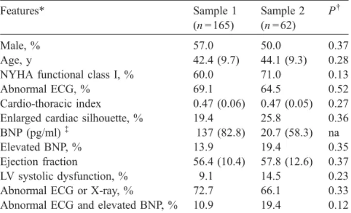
The second sample was selected using a similar recruit-ment procedure. Of 75 patients that satisfied the criteria for inclusion and did not present exclusion criteria, 62 (82.3%) had completed all required tests. The second sample had a clinical profile similar to that of the first, with a 14.5% prevalence of LV systolic dysfunction (nine patients). The general features of both samples patients are shown inTable 1. The 165 subjects in the first study sample were predominantly males (57%), with a mean age of 42.4T9.7 years; the second sample was not significantly different. Most patients in both samples are asymptomatic and only few patients (six in sample 1 and five in sample 2) were in NYHA functional class III or IV.
2.3. Procedures
The reference standard of LV systolic function was the LV ejection fraction (LVEF), estimated using the Simpson rule and obtained by a standardized echocardiogram. All studies were performed by an experienced examiner (MVLB) using an ATL HDI 5000 device (Bothell, Washington, USA). Significant left ventricular systolic
dysfunction is considered to be present if LV ejection fraction is0.40. This cut-off point was chosen since it has been previously used in several clinical trials performed on patients with LV dysfunction[14].
In all participants, a standard 12 leads ECG was recorded at a paper speed of 25 mm/s with a portable Fukuda Denshi Model FX-2111 electrocardiograph (Fukuda Co., Tokyo, Japan). The tracing was analyzed blindly by a cardiologist experienced in electrocardiology (ALPR) using the Buenos Aires code, specifically designed to be used in Chagas disease, [15] and normal and abnormal ECG were identified. Chest radiography was performed according to the conventional technique used at the Radiological Unit of the University Hospital of UFMG and analyzed by one observer (AAP) supervised by an experienced researcher (MOCR). The cardiac silhouette and the cardiothoracic ratio were assessed as previously reported [10]. Chest radiog-raphy with a cardiothoracic ratio greater than 0.50 or an enlarged cardiac silhouette, or both, was considered abnormal.
BNP measurement was performed in venous blood after a supine 20 min rest by different methods in both samples. In the first sample, BNP measurement was performed by a researcher experienced in the natriuretic peptide field (AMR), in the Department of Physiology and Biophysics of the Federal University of Minas Gerais, as previously described [11]. Samples were placed in chilled tubes containing protease inhibitors (EDTA, PMSF and Pepstatin A, all 10 5M, all purchased from Sigma Chemical Co., St Louis, MO, USA). After centrifugation at 1500g for 15 min at 4 -C, plasma was separated and stored at 80-C. For BNP extraction, plasma was allowed to pass slowly through a Sep-Pak C18 column (Waters Corp., USA) activated by sequential washes with acetonitrile and 0.2% ammonium acetate, pH 4, and eluted with an acetonitri-le : ammonium acetate solution (60 : 40). The sampacetonitri-les were dried in a Speed-Vac apparatus and redissolved in buffer for
Table 1
General features of Chagas disease patients
Features* Sample 1
(n= 165)
Sample 2 (n= 62)
P.
Male, % 57.0 50.0 0.37
Age, y 42.4 (9.7) 44.1 (9.3) 0.28
NYHA functional class I, % 60.0 71.0 0.13
Abnormal ECG, % 69.1 64.5 0.52
Cardio-thoracic index 0.47 (0.06) 0.47 (0.05) 0.27 Enlarged cardiac silhouette, % 19.4 25.8 0.36 BNP (pg/ml)- 137 (82.8) 20.7 (58.3) na
Elevated BNP, % 13.9 19.4 0.35
Ejection fraction 56.4 (10.4) 57.8 (12.6) 0.37 LV systolic dysfunction, % 9.1 14.5 0.23 Abnormal ECG or X-ray, % 72.7 66.1 0.33 Abnormal ECG and elevated BNP, % 10.9 19.4 0.12 *Data are means (SD), unless otherwise indicated,.P value refers to the comparison between the two samples. - median (interquartile range), na = not applicable.
Chagas disease n = 222
Incomplete tests n = 28
n = 2
LVSD n = 4
No LVSD n = 22
LVSD
n = 15 n = 105 No LVSD
LVSD n = 12 n = 6
No LVSD LVSD
n = 3 n = 144 No LVSD LVSD
n = 0 n = 45 No LVSD Excluded patients (n=29)
Concomitant disease Alcoholism
Non adherence to protocol No echo
Echo, n = 26
ECG / Xray n = 165
ECG / BNP n = 165 Abnormal tests
n = 120
Normal tests n = 45
Normal tests n = 147 Abnormal tests
n = 18
Echo, n = 120 Echo, n = 45
Echo, n = 147 Echo, n = 18
assay. BNP was measured by RIA using hBNP as standard, tyr0 -BNP for iodination and specific hBNP antibody (all purchased from Peninsula Laboratories, USA). Each sample was tested in duplicate and the final result was the mean value; all samples were measured in the same assay. In the second sample, [12] a small amount of blood (5 ml) collected into tubes containing potassium EDTA (1 mg/mL of blood), were centrifuged at 3000 g for 10 min and plasma was obtained and frozen at 80 -C until further analysis, performed at Southwestern Medical School, Dallas, Texas. BNP was measured using the Triage BNP test (Biosite Inc., USA), which is a fluorescence immuno-assay for the quantitative determination of BNP in plasma specimens. The precision, analytical sensitivity and stability characteristics of the system have been previously described [16]. Each sample was tested in triplicate to minimize variation from single observations and for internal controls and the final results were reported as the mean of the 3 samples.
The diagnostic performance of the BNP values in the diagnosis of LV systolic dysfunction in the Chagas disease population was evaluated using the receiver – operator – characteristic (ROC) curve. The area under the curve was 0.89T0.04 in the first sample [11] and 0.92T0.03 in the second sample[12]. Different cut-off points were obtained for different samples, since the BNP methods used were also different (RIA and fluorescence immunoassay)[17]. Based on the analysis of the ROC curve and aiming at obtaining specificity 80%, a BNP cut-off value of 210 pg/ml was chosen for the first sample and a cut-off value of 75 pg/ml was chosen for the second sample. Other cut-off values were tested considering also the ECG results without further improvements of the diagnostic performance.
3. Statistical analysis
The number of subjects needed to be examined (NNE) by echocardiography to detect one case of LV systolic dysfunction in both strategies was calculated as 1/preva-lence of LV dysfunction in abnormal screening tests [18]. The sensitivity, specificity, accuracy, positive and the negative predictive values and positive and negative
like-lihood ratios (LR) were calculated for each strategy of detection of abnormal LV systolic function[19]. Confidence intervals (95%) were calculated by the method of Wilson [20]using CIA software, version 2.0.0 (Confidence Interval Analysis for Windows, London, 2000). The statistical significance of the differences in diagnostic measurement strategies was assessed by the McNemar test for paired data. All measurements of diagnostic indexes were performed for both strategies also in the sample 2 and compared to those obtained in the sample 1 by the ztest. Ap-value of < 0.05 was considered significant.
4. Results
4.1. First study
ECG abnormalities and an enlarged heart silhouette were found in 69.1 and 19.4% of patients of the first sample, respectively (Table 1). The median BNP value was 137.0 pg/ml (Q1 – Q3: 97.7 – 180.7 pg/ml); in the same original study, normal subjects had median values of 144.7 pg/ml (Q1 – Q3: 103.9 – 163.6 pg/ml) [11]. The wide range of distribution of BNP values and the relative low number of patients with systolic dysfunction (15 patients, 9.1%) hinders the interpretation of these numbers. The median BNP value was 131.0 pg/ml (Q1 – Q3: 95.4 – 169.7 pg/ml) in patients with LV ejection fraction > 0.40 and 228.7 pg/ml (Q1 – Q3: 176.2 – 360.8 pg/ml,p< 0.001) in those with LV ejection fraction0.40.
An abnormal ECG or X-ray was observed in 120 patients (72.7%) while elevation of BNP in patients with abnormal ECG occurred in 41 subjects (10.9%). The NNE, calculated as 1 / prevalence of LV dysfunction in the abnormal screen-ing test subgroup, was 8.0 [1 / (15 / 120)] by the conventional strategy and 2 [1 / (12 / 18)] by the new strategy.
The conventional strategy (ECG/X-ray) recognized all patients with LV systolic dysfunction (sensitivity: 100%, 95% CI: 79.6 – 100% and negative predictive value PV 100%, 92.1 – 100%), but with low specificity (30%, 95% CI: 23.2 – 37.8) and positive predictive value (12.5%, 95%C I7.7 – 19.6), yielding an overall accuracy of 36.4% (95% CI: 29.4 – 43.9, see Table 2).
Table 2
Diagnostic accuracy of two strategies in the detection of LV systolic dysfunction in Chagas disease patients
Sample 1 (n= 165) Sample 2 (n= 62)
The BNP/ECG-based strategy was significantly better than the conventional approach (Table 2) in terms of specificity (96.0, 95% CI: 91.5 – 98.2,p< 0.001) and positive predictive value (66.7, 95% CI: 43.7 – 83.7, p< 0.001), displaying non-significantly lower sensitivity (80.0, 95% CI: 54.8 – 93.0,p= 0.25) and negative predictive value (98.0, 95% CI: 94.2 – 99.3, p= 0.08). The overall accuracy was much improved with the new strategy (94.5, 95% CI: 90.0 – 97.1,p< 0.001).
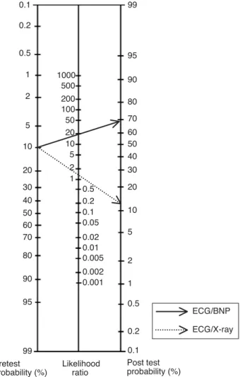
The superiority of using the new strategy can be graphically displayed using the likelihood ratios and a Fagan normogram (Fig. 3). Considering the positive like-lihood ratio obtained for the sample 1 (conventional approach, 1.4, 95% CI: 1.37 – 1.61; new strategy, 20.0, 95% CI: 8.77 – 45.58) and an estimated 10% pre-test prevalence of left ventricular dysfunction in an outpatient Chagas disease population, an elevated BNP in the presence of an abnormal ECG increases the post-test probability to 68.9%, although the post-test probability after an abnormal ECG or X-ray would be only 13.4%. The negative likelihood ratio was zero (95% CI: 0.00 – 0.69) in the conventional approach and 0.21 (95% CI: 0.07 – 0.57) in the
new one, indicating that a negative test in both strategies leads to a very low post-test probability of LV dysfunction, i.e., 0% and 2.3%, respectively.
4.2. Second study
The frequency of abnormal ECG, X-ray and BNP measurement is displayed in Table 1. BNP values were significantly different, reflecting the different methodologies used for BNP measurement. The median BNP value was 11.7 pg/ml (Q1 – Q3: 5.6 – 30.7 pg/ml) in patients with LV ejection fraction > 0.40 and 93.5 pg/ml (Q1 – Q3: 66.6 – 534.5 pg/ml, p< 0.001) in those with LV ejection fraction
0.40.
Both strategies were applied to the second sample and the indexes obtained, displayed in Table 2, were closely similar when compared to those of the first sample. All comparisons between measurements obtained in the two samples revealed absence of statistically significant differ-ences, although a slight degeneration of the diagnostic performance of the new strategy is suggested by a lower positive likelihood ratio (8.24, 95% CI: 3.33 – 20.36).
5. Discussion
This study describes the comparison of two strategies for the detection of left ventricular systolic dysfunction in two different groups (first and second samples) of patients with positive serology for Chagas disease, showing that measur-ing BNP in patients with abnormal ECG is more accurate than performing ECGs and chest X-rays in all patients. To our knowledge, this is the first study that compares these two strategies in patients with Chagas disease and it is one of first reports in tropical medicine field that complies with the STARD initiative recommendations.
The electrocardiogram and chest X-ray have been used in Chagas disease since the original studies by Carlos Chagas [21] and have an established role in the management of Chagas disease patients, with both diagnostic and prognos-tic importance[9]. Nonetheless, major improvements in the diagnostic capabilities and therapeutic options have occurred in the last decades and it is now clear that left ventricular systolic dysfunction, evaluated non-invasively by echocardiography, is the best predictor of an elevated risk of death in Chagas disease. Moreover, it is now indisputable that pharmacological treatment may halt the progression of LV dysfunction in studies performed at other settings, indicating the urgent need to redefine strategies to recognize and treat these patients.
When the accuracy of this classical strategy was scruti-nized using modern evidence-based diagnostic tools, we observed that it was able to detect all patients with significant LV systolic dysfunction, with a sensitivity and negative predictive value of 100% in both samples. However, the specificity, the positive predictive value and
ECG/BNP
ECG/X-ray
Pretest probability (%)
Post test probability (%) Likelihood
ratio 0.1
0.2
0.5
1 1000
500
200 100 50 20 10 5 2 1
0.5 0.2 0.1 0.05
0.02 0.01 0.005
0.002 0.001 2
5
10
20
30
40 50 60 70
80
90
95
99
99
95
90
80
70
60 50 40
30
20
10
5
2
1
0.5
0.2
0.1
the overall accuracy were relatively low, indicating that a large number of patients would need to be examined by echocardiography in order to detect one that could benefit from pharmacological treatment. We hypothesize that this relatively low accuracy can be attributed to two factors: the use of two diagnostic tests in parallel, which can lower the specificity and positive predictive values,[22]and the poor diagnostic performance of the chest X-ray[10].
Few other studies have addressed the issue of screening to recognize Chagas patients with LV systolic dysfunction [3,23]. Bestetti studied a hospital cohort of 74 Chagas disease patients and found a prevalence of 59% of left ventricular systolic dysfunction, defined as ejection fraction below 55% [23]. The combination of New York Heart Association functional class (2 or more) and a systolic blood pressure below 120 mmHg predicted left ventricular dysfunction with a sensitivity of 59% and a specificity of 77%. ECG variables were not predictive of LV dysfunction and BNP measurement was not performed. Although potentially useful in the hospital setting, this strategy may not be applicable to screening of ambulatory patients, since different clinical profiles and LV dysfunction prevalence may impair the transferability of the diagnostic strategy [24]. It should also be pointed out that the sensitivity of the strategy in the latter study is relatively low, leading to the withholding of the echocardiogram in a large number of patients (41%) that would benefit from the formal evalua-tion of LV systolic funcevalua-tion. More recently, Salles and colleagues, [3] after studying 738 adult outpatients in the chronic phase of Chagas disease, reported that a QT dispersion above 60 ms was associated with LV systolic dysfunction and could be used to predict asymptomatic dysfunction in chronic Chagas’ disease. A model consider-ing QT dispersion, the presence of cardiomegaly, frequent PVCs, and male sex had 90% specificity and 71% sensitivity in this population, in which LV systolic dysfunction (ejection fraction < 45%) was observed in 14.7% of cases. Although the diagnostic performance of this strategy was quite good, limitations related to QT dispersion measurements may hamper its use in general practice[3,25].
We evaluated a new strategy, based on sequential tests and the use of a new and promising simple method for the diagnosis of LV systolic dysfunction, i.e., the measurement of BNP in blood samples[11,12]. In the present study, that BNP-based approach was more accurate and superior to the conventional one in terms of specificity and positive predictive value in both samples. A marked and significant increment of accuracy, odds ratio and positive likelihood ratio was detected, with a reduction of the number of patients to be examined by echocardiography from 8 to 2. Since the echocardiogram is not widely available in the public health system in many areas of the Latin America, patients candidate to the echocardiogram by the conven-tional approach are not actually being submitted to the exam. The marked reduction of the number of cases needed
to be examined by echocardiography using this new strategy may permit that all screened patients would be examined by the echocardiogram, optimizing the use of limited resources. In fact, the association of ECG and BNP measurement was successful in the detection of LV systolic dysfunction when tested in other clinical settings [18,22,26] and was recommended by the recently released NICE guideline for
Management of Chronic Heart Failure in Adults in Primary
and Secondary Care[27]. Nonetheless, the use of BNP as a
diagnostic tool is a recent conquest and there are unsolved issues and problems related to the implementation of its use in clinical practice [28]. Different commercial and exper-imental measurement techniques are available, with differ-ent cut-off points and clinical determinants [29]. In our study, a different methodology was applied to the first and second samples (RIA and fluorescence immunoassay), leading to markedly different cut-off points[17]. More than a limitation, this fact indicates that the excellent perform-ance obtained in the first sample was transferable to the second sample, in which a commercial kit was used. The cost-effectiveness of this strategy is still controversial, although studies have suggested that screening high-risk subjects by BNP before echocardiography could reduce the cost per detected case of LV systolic dysfunction by 26% [18]and may be economically attractive for patient groups with at least 1% prevalence of moderate or greater LV systolic dysfunction [30]. Additionally, commercial BNP measurement was not yet available in many Latin America cities by the time of the writing of this paper (September 2004) and this strategy could be applied only if, as expected, measuring BNP becomes a widespread and low-cost laboratory method in the near future, what could be anticipated by the simplicity and the reliableness of the point-of-care measurement system used in the second sample.
A major challenge in global diseases is ‘‘to transform new knowledge into cost-effective, equitably affordable interven-tions and to guarantee their access to the patients and populations of endemic countries’’[1]. Effectively screening for LV systolic dysfunction is a major issue in Chagas disease, since it can significantly reduce mortality and morbidity. A BNP-based strategy proved to be accurate and should be considered as an alternative option in the detection of LV systolic dysfunction in Chagas disease. Nonetheless, further studies should be performed to define the best strategy in the management of Chagas disease cardiopathy.
Acknowledgments
Conselho Nacional do Desenvolvimento Cientı´fico e Tecnolo´gico (CNPq), Fundac¸a˜o de Amparo a` Pesquisa do Estado de Minas Gerais (FAPEMIG), Coordenadoria de Aperfeic¸oamento do Ensino Superior (CAPES).
References
[1] Chagas disease. Disease watch 2003. TDR|Nature Reviews Micro-biology Available at: http://www.who.int/tdr/dw/chagas2003.htm. Acessed April 10, 2004.
[2] Mady C, Cardoso RH, Barretto AC, da Luz PL, Bellotti G, Pileggi F. Survival and predictors of survival in patients with congestive heart failure due to Chagas’ cardiomyopathy. Circulation 1994;90: 3098 – 102.
[3] Salles GF, Cardoso CR, Xavier SS, Sousa AS, Hasslocher-Moreno A. Electrocardiographic ventricular repolarization parameters in chronic Chagas’ disease as predictors of asymptomatic left ventricular systolic dysfunction. Pacing Clin Electrophysiol 2003;26:1326 – 35. [4] SOLVD Investigators. Effect of enalapril on mortality and the
development of heart failure in asymptomatic patients with reduced left-ventricular ejection fractions. N Engl J Med 1992;327:685 – 91. [5] Pfeffer MA, Braunwald E, Moye LA, et al. Effect of captopril on mortality and morbidity in patients with left ventricular dysfunction after myocardial infarction: results of the survival and ventricular enlargement trial: the SAVE Investigators. N Engl J Med 1992; 327:669 – 77.
[6] Davies M, Hobbs F, Davis R, Kenkre J, Roalfe AK, Hare R, et al. Prevalence of left-ventricular systolic dysfunction and heart failure in the echocardiographic heart of England screening study: a population based study. Lancet 2001;358(9280):439 – 44.
[7] McMurray JV, McDonagh TA, Davie AP, Cleland JG, Francis CM, Morrison C. Should we screen for asymptomatic left ventricular dysfunction to prevent heart failure? Eur Heart J 1998 (Jun);19(6): 842 – 6.
[8] Wang TJ, Levy D, Benjamin EJ, Vasan RS. The epidemiology of ‘‘asymptomatic’’ left ventricular systolic dysfunction: implications for screening. Ann Intern Med 2003 (Jun 3);138(11):907 – 16.
[9] Marin Neto JA, Simoes MV, Sarabanda AV. Forma croˆnica cardı´aca. In: Brener Z, Andrade ZA, Barral-Neto M, editors.Trypanosoma cruzi e doenc¸a de Chagas, 2nd ed. Rio de Janeiro’ Guanabara Koogan; 2000. p. 266 – 96.
[10] Perez AA, Ribeiro AL, Barros MV, et al. Value of the radiological study of the thorax for diagnosing left ventricular dysfunction in Chagas’ disease. Arq Bras Cardiol 2003;80:208 – 13.
[11] Ribeiro AL, Reis AM, Barros MV, et al. Brain natriuretic peptide in the diagnosis of systolic left ventricular dysfunction in Chagas disease. Lancet 2002;360:461 – 2.
[12] Talvani A, Rocha MO, Cogan J, et al. Brain natriuretic peptide and left ventricular dysfunction in human chagasic cardiomyopathy. Mem Inst Oswaldo Cruz 2004;99(6):645 – 9.
[13] Bossuyt PM, Reitsma JB, Bruns DE, et al. Standards for reporting of diagnostic accuracy. Towards complete and accurate reporting of studies of diagnostic accuracy: the STARD Initiative. Ann Intern Med 2003;138:40 – 4.
[14] Effect of metoprolol CR/XL in chronic heart failure: Metoprolol CR/XL Randomised Intervention Trial in Congestive Heart Failure (MERIT-HF). Lancet 1999;353:2001 – 7.
[15] Lazzari JO, Pereira M, Antunes CM, et al. Diagnostic electro-cardiography in epidemiological studies of Chagas’ disease: multi-center evaluation of a standardized method. Rev Panam Salud Publica 1998;4:317 – 30.
[16] Vogeser M, Jacob K. B-type natriuretic peptide (BNP)-validation of an immediate response assay. Clin Lab 2001;47(1 – 2):29 – 33.
[17] Ribeiro AL, Reis AM, Teixeira MM, Rocha MO. Brain natriuretic peptide in Chagas disease: further insights. Lancet 2003;362:333. [18] Nielsen OW, McDonagh TA, Robb SD, Dargie HJ. Retrospective analysis of the cost-effectiveness of using plasma brain natriuretic peptide in screening for left ventricular systolic dysfunction in the general population. J Am Coll Cardiol 2003;41:113 – 20.
[19] Habbema JDF, Eijkemans R, Krijnen P, Knottnerus JA. Analysis of data on the accuracy of diagnostic tests. In: Knottnerus JA, editor. The evidence base of clinical diagnosis. London’BMJ Publishing Group; 2002. p. 117 – 44.
[20] Altman DG, Machin D, Bryant TN, Gardner MJ. Statistics with confidence. 2nd Edition. London’BMJ Books; 2000. 240 pp. [21] Chagas C, Villela E. Forma cardı´aca da Trypanosomiase Americana.
Mem Inst Oswaldo Cruz 1922;14:3 – 54.
[22] Nielsen OW, Hansen JF, Hilden J, Larsen CT, Svanegaard J. Risk assessment of left ventricular systolic dysfunction in primary care: cross sectional study evaluating a range of diagnostic tests. BMJ 2000 (Jan 22);320(7229):220 – 4.
[23] Bestetti RB. Predictors of unfavourable prognosis in chronic Chagas’ disease. Trop Med Int Health 2001 (Jun);6(6):476 – 83.
[24] Irwig L, Bossuyt P, Glasziou P, Gatsonis C, Lijmer J. Designing studies to ensure that estimates of test accuracy are transferable. BMJ 2002 (Mar 16);324(7338):669 – 71.
[25] Coumel P, Maison-Blanche P, Badilini F. Dispersion of ventricular repolarization: reality? Illusion? Significance? Circulation 1998 (Jun 30);97(25):2491 – 3.
[26] Ng LL, Loke I, Davies JE, et al. Identification of previously undiagnosed left ventricular systolic dysfunction: community screen-ing usscreen-ing natriuretic peptides and electrocardiography. Eur J Heart Fail 2003;5(6):775 – 82.
[27] Management of chronic heart failure in adults in primary and secondary care. (NICE guideline). Available at:http://www.nice.org.uk. Accessed April 10, 2004.
[28] de Lemos JA, McGuire DK, Drazner MH. B-type natriuretic peptide in cardiovascular disease. Lancet 2003 (July 26);362(9380): 316 – 22.
[29] Clerico A, Iervasi G, Mariani G. Pathophysiologic relevance of measuring the plasma levels of cardiac natriuretic peptide hormones in humans. Horm Metab Res 1999 (Sep);31(9):487 – 98.
[30] Heidenreich PA, Gubens MA, Fonarow GC, Konstam MA, Stevenson LW, Shekelle PG. Cost-effectiveness of screening with B-type natriuretic peptide to identify patients with reduced left ventricular ejection fraction. J Am Coll Cardiol 2004 (Mar 17); 43(6):1019 – 26.
[31] Vasan RS, Benjamin EJ, Larson MG, et al. Plasma natriuretic peptides for community screening for left ventricular hypertrophy and systolic dysfunction: the Framingham heart study. JAMA 2002 (Sep 11); 288(10):1252 – 9.


