Comparative Study of Clinical and Doppler
Echocardiographic Evaluations of the
Progression of Valve Diseases in Children and
Adolescents with Rheumatic Fever
Zilda Maria Alves Meira, Eugênio Marcos Andrade Goulart, Cleonice de Carvalho Coelho Mota
Faculdade de Medicina da Universidade Federal de Minas Gerais - UFMG - Belo Horizonte, MG - BrazilMailing Address: Zilda Maria Alves Meira • Rua Roquete Mendonça, 184/401 - 31275-030 – Belo Horizonte, MG - Brazil
E-mail: [email protected] Received on 08/05/04 • Accepted on 04/29/05
O
BJECTIVECompare clinical and Doppler echocardiographic evaluations in assessing valvular diseases in children and adolescents with rheumatic fever, as well as assess the progression of the disease in light of these assessments.
M
ETHODSThis is a longitudinal study of 258 children and adolescents diagnosed with rheumatic fever according to Jones’ criteria. The follow-up period ranged from 2 to 15 years. The presence and quantifi cation of valve diseases were determined by means of clinical and Doppler echocardiographic evaluations performed during the acute and chronic phases. The Kappa statististics method was used to estimate the degree of agreement between clinical and Doppler echocardiographic evaluations. Comparisons between clinical and Doppler echocardiographic fi ndings on the progress of carditis and valvulitis, respectively, were made using chi-square test or Fisher’s exact test, p< 0.05.
R
ESULTSOf the 109 patients who underwent Doppler echocardiographic evaluation during the acute phase, 31 did not present clinical evidence of carditis, but the Doppler echocardiograms of 17 (54.8%) of them showed valve lesions (subclinical valvulitis). During the chronic phase, 153 of the 258 patients had normal cardiovascular examination results; however, Doppler echocardiograms showed that 81 of them (52.9%) had valve lesions (subclinical chronic valvular diseases). Involution of the valvular lesions, as shown by Doppler echocardiographic evaluations, was less frequent and occurred in 10 (25.0%) patients with mild valvulitis, in only one (2.5%) patient with moderate valvulitis, and in none of the patients with severe valvulitis.
C
ONCLUSIONThe identifi cation of rheumatic fever valve lesions can be enhanced when clinical evaluations are supplemented by Doppler echocardiographic examinations; also, clinical examinations are not as suitable to detect valvular lesion regression as the echocardiography. The diagnosis of subclinical valvulitis and valvulopathy influences the secondary prophylaxis of rheumatic fever and endocarditis.
K
EY WORDSRheumatic fever (RF) is a leading cause of acquired cardiopathy among school children, adolescents and young adults in underdeveloped countries1. RF increases
expenditures with healthcare as it is a chronic disease requiring clinical follow-up and many times cardiac catheterization in order to approach the valvar lesions for balloon valvuloplasty, or surgical treatment to repair or replace the valve.
In the absence of carditis and/or recidivism of the disease, the prognosis is good with a smaller rate of progression to chronic valvulopathy. Generally speaking, patients without carditis will not develop signifi cant chronic cardiopathy. However, the prognosis worsens according to the severity of carditis, and in the presence of heart failure in the initial episode of the disease, 60% showed evidence of cardiopathy after ten years2. In view of the
fact that the diagnosis of RF is the result of a composite of clinical-laboratorial manifestations, the rheumatic origin of subclinical valvulitis or chronic valvular disease should be analyzed in light of such criteria, not including Doppler echocardiographic fi ndings3. Moreover, Doppler
echocardiography, although extremely helpful in evidencing and quantifying cardiac abnormalities even in the absence of clinical signs, does not allow one to establish the etiology of the lesions4, even though it can suggest the etiology,
since rheumatic valvular involvement abnormalities detected by Doppler echocardiogram are typical.
The advance in diagnostic methods with the introduction of Doppler echocardiography provided better means of evaluating orovalvular lesions (valve disease secondary to oral foci of infection). M-mode and two-dimensional images allow for a safe determination of the degree of ventricular dysfunction, presence of pericardial involvement, and alterations in the texture of valve tissue. Doppler analysis (pulsed, continuous, and color fl ow scanning,) enables the detection of valve dysfunction even without clinical evidence, as well as the classifi cation of type and degree of valve lesion according to the regurgitation jet magnitude and valve area estimation5.
Bearing in mind the difference between clinical examinations and Doppler echocardiographic fi ndings in defi ning the presence and degree of valve involvement, this study intends to quantify the degree of cardiac involvement in the acute and chronic phases of RF and to study the progression of the disease according to both assessments.
M
ETHODS
A longitudinal study was conducted, with retrospective data on 258 patients diagnosed with RF, as per Jones’ criteria (1992)6, who were seen and treated at our
institution between August 1983 and December 1998. Subjects were selected among the 392 patients being followed in outpatient clinics specifi c for the treatment of RF. The presence and quantifi cation of valve diseases
in acute and chronic phases were determined by means of clinical and Doppler echocardiographic assessments. Of the 258 patients, 109 underwent Doppler echocardiography during the acute phase. During the chronic phase, considered as the persistence of the disease after at least two years, all patients had the degree of their cardiac involvement determined by clinical and Doppler echocardiographic evaluations.
The determination of the degree of cardiac involvement was made in the acute and chronic phases within a period of three months, at the most, between the clinical classifi cation and Doppler echocardiography. According to the patient’s clinical history, the onset of clinical manifestations and the characterization of major and minor manifestations were defi ned, as per Jones’ criteria. The acute phase was defi ned as the period between the onset of symptoms and signs and the end of the 12th week of the disease, or earlier, with the normalization of laboratory tests that represent acute-phase serum reactions.
From 1994 on, all patients with acute rheumatic fever (ARF) treated at our institution, even without clinical manifestations of cardiac involvement, were also submitted to Doppler echocardiography, besides clinical, radiological and electrocardiography examinations. Doppler echocardiographic examinations, including the four modes – M-mode, two-dimensional, pulsed, continuous and color fl ow mapping – were performed with Siemens CF PLUS and Hewlett-Packard (HP) echocardiography equipment, version 1.500, with 2.5, 3.5 and 5.5 MHZ waves.
The left chambers size determination was obtained in M-mode via the parasternal short axis view. Values obtained were compared to normal reference values, according to the weight. The ejection fraction (EF) was evaluated according to the Teichholz method (1964)5.
The criteria used to quantify valve regurgitations were based on the color fl ow mapping. The mitral regurgitation jet area was used to determine the degree of mitral regurgitation. Mitral regurgitation was considered physiological under the following conditions: valves with normal morphology and texture; normal-sized cardiac chambers; and small regurgitation jet of less than 1 cm of the valve closing level, according to Doppler color fl ow mapping analysis; regurgitation jet occupying less than 50% of the systole according to Doppler continuous analysis. Aortic regurgitation was quantifi ed by the ratio between the extent of the regurgitation jet and the left ventricular outfl ow tract width7. In obstructive lesions,
mitral valve area was determined by means of a Doppler technique based on pressure half-time (PHT), absence of signifi cant aortic regurgitation, and by two-dimensional planimetry. Mitral stenosis was quantifi ed based on the valve area, estimated by the PHT and planimetry, and maximum and medium diastolic gradients between the left atrium (LA) and the left ventricle (LV).
involvement were made during the acute and chronic phases. During the acute phase, classifi cation of the cardiac involvement, known as carditis, was conducted according to institution protocol based on the classifi cation described by Décourt8 and Markowitz & Gordis9. Four
degrees of carditis were considered: absent, mild, moderate and severe. In the chronic phase, the presence and degree of cardiac involvement, chronic cardiopathy, were determined by parameters similar to those of the acute phase, and stratifi ed in absent, mild, moderate, and severe categories. For the Doppler classifi cation of the degree of cardiac involvement in the acute and chronic phases, the valve lesion degree and heart chambers size according to previously established echocardiographic patterns were the only aspects considered5,7,10,11. Doppler
echocardiographic classifi cations of the degree of valve involvement in the acute and chronic phases, valvulitis and chronic valvulopathy, respectively, were also divided into four degrees: absent, mild, moderate and severe.
The term ‘valvulopathy’ used here is restricted solely to the exclusive Doppler echocardiographic analysis of mitral and/or aortic lesions in the chronic phase of RF. In this study, the determination of lesions in the tricuspid and pulmonary valves was not performed. The valve lesions classifi ed as mild-to-moderate were included in ‘moderate involvement’, and those classifi ed as moderate-to-severe were analyzed as severe.
The research protocol was approved by the hospital’s Research Ethics Committee. Data obtained were analyzed using EPI-Info 6 software. A comparative study of the clinical progression of patients with RF was performed in light of clinical evaluations, including electrocardiography and chest X-ray fi ndings, as well as the four classifi cations of valvar lesions according to Doppler echocardiographic criteria. To estimate the agreement among the classifi cations, Kappa statistics were used, with calculations of non-weighted Kappa (K) values. The conventional interpretation of K values is as follows: Kappa above 75% – excellent level of agreement; between 40% and 75% – reasonable-to-good level of agreement; under 40% – poor level of agreement. The progression of the disease according to clinical and Doppler echocardiographic findings for carditis and valvulitis, respectively, was compared by means of the chi-square test (χ2) or Fisher’s exact test. The statistical
signifi cance level was set at 5% (p < 0.05).
R
ESULTS
Of the 258 patients enrolled in the study, 132 (51.2%) were men and 126 (48.8%) were women; patient age at the fi rst bout varied from 3.2 years and 16.5 years, mean age 9.0 ± 2.6 years and median age 9.0 years. Taking into account only the follow-up period of 258 patients at this institution, total patient-years were 1,383.
In the distribution of patients according to Jones’s criteria, arthritis, carditis and chorea were common
manifestations in the ARF medical condition affecting 65.5%, 56.6% and 24.0% of patients, respectively. Among the most common important clinical manifestations related to the presentation of ARF, the association between arthritis and carditis was the most frequent, affecting 29.8% of the 258 patients in the fi rst bout. Only 2.7% of the patients had carditis, arthritis, and chorea combined, and the concomitance of arthritis and chorea was also uncommon (4.3%).
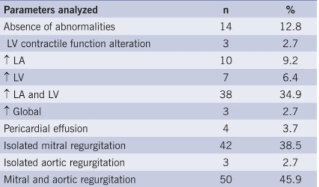
The main Doppler echocardiographic fi ndings of the 109 patients evaluated at our institution during the fi rst bout are listed on Table I. In most cases (99.1%), the left ventricular contractile function was within the limits of normality, and just one patient, with an ejection fraction of 0.52, showed decreased contractility. Fifty-eight patients (53.2%) had one or more enlarged cardiac chambers, and the combined enlargement of LA and LV was more frequent than the isolated or global enlargement of the four chambers. Whether isolated or associated with other lesions, mitral regurgitation was present in 92 (84.4%) patients and aortic regurgitation, in 53 (48.6%). However, aortic regurgitation was recorded separately in just three (2.7%) patients and no obstructive valve lesions were observed. Pericardial fl uid effusion was uncommon, having only been observed in four (3.7%) examinations.
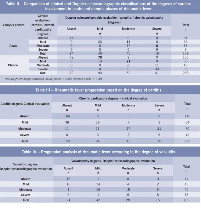
The ‘diagonal’ reading analysis of Table II shows that both in the acute phase and in the chronic phase the clinical evaluation tended to underestimate the degree of cardiac involvement, as compared to the valve lesion degree classification by Doppler echocardiography
Table I – Distribution of frequency of Doppler echocardiographic fi ndings in the fi rst bout of
rheumatic fever (n = 109)
Parameters analyzed n %
Absence of abnormalities 14 12.8 LV contractile function alteration 3 2.7
↑ LA 10 9.2
↑ LV 7 6.4
↑ LA and LV 38 34.9
↑ Global 3 2.7 Pericardial effusion 4 3.7 Isolated mitral regurgitation 42 38.5 Isolated aortic regurgitation 3 2.7 Mitral and aortic regurgitation 50 45.9
evaluations were in agreement on the diagnosis of the degree of valve lesion in those patients with cardiac involvement classifi ed as moderate or severe, both in the acute and chronic phases.
The analysis of the progression of cardiac involvement from the acute phase to the chronic phase, according to clinical evaluation aided by electrocardiography and chest X-ray studies, showed that the percentage of valve lesion regressions was greater when the carditis degree was mild (59.4%), lower in moderate degree carditis (15.2%), and there was no record of involution in cases of severe carditis. In the group of patients with no clinical evidence of carditis, eight (7.1%) had cardiac alterations in the clinical evaluation performed during the chronic phase (table III).
Taking into consideration the degree of cardiac involvement assessed by Doppler echocardiography both
Table II – Comparison of clinical and Doppler echocardiographic classifi cations of the degrees of cardiac involvement in acute and chronic phases of rheumatic fever
Analysis phases
Clinical evaluation: carditis / chronic
cardiopathy (degrees)
Doppler echocardiographic evaluation: valvulitis / chronic valvulopathy (degrees) Total n Absent n Mild n Moderate n Severe n Acute
Absent 14 17 0 0 31
Mild 0 23 13 0 36
Moderate 0 0 27 6 33
Severe 0 0 0 9 9
Total 14 40 40 15 109
Chronic
Absent 72 79 2 0 153
Mild 0 14 21 0 35
Moderate 0 0 29 11 40
Severe 0 0 0 30 30
Total 72 93 52 41 258
Non-weighted Kappa statistics: acute phase = 0.50; chronic phase = 0.39
Doppler echocardiographic evaluation: valvulitis / chronic valvulopathy (degrees) Absent n Mild n Moderate n Severe n
14 17 0 0
0 23 13 0
0 0 27 6
0 0 0 9
14 40 40 15
72 79 2 0
0 14 21 0
0 0 29 11
0 0 0 30
72 93 52 41
in the acute phase and chronic phase, the absence of valvulitis indicated a greater likelihood of normality in the chronic phase; just one (7.1%) patient without a valve lesion in the acute phase evidenced a mild valve lesion in the chronic phase (Table IV).
As shown, valve lesion involution assessed by Doppler echocardiographic evaluation was less common than in clinical evaluations, occurring in ten (25.0%) patients with mild valve lesion (p= 0.0013) and only one (2.5%) patient with moderate valvulitis (p= 0.0526). Moreover, severe valvulitis did not retreat to normal Doppler echocardiographic values (Table V).
D
ISCUSSION
Considering that the tricuspid and pulmonary valves show (physiological) regurgitation in most normal children
Table III – Rheumatic fever progression based on the degree of carditis
Carditis degrees Clinical evaluation
Chronic cardiopathy degrees – clinical evaluation
Total n Absent n Mild n Moderate n Severe n
Absent 104 5 3 0 112
Mild 38 16 7 3 64
Moderate 11 11 27 21 70
Severe 0 3 3 6 12
Total 153 35 40 30 258
Chronic cardiopathy degrees – clinical evaluation
Absent n Mild n Moderate n Severe n
104 5 3 0
38 16 7 3
11 11 27 21
0 3 3 6
153 35 40 30
Table IV – Progression analysis of rheumatic fever according to the degree of valvulitis
Valvulitis degrees: Doppler echocardiographic evaluation
Valvulopathy degrees: Doppler echocardiographic evaluation
Total n Absent n Mild n Moderate n Severe n
Absent 13 1 0 0 14
Mild 10 24 4 2 40
Moderate 1 16 18 5 40
Severe 0 1 6 8 15
Total 24 42 28 15 109
Valvulopathy degrees: Doppler echocardiographic evaluation
Absent n Mild n Moderate n Severe n
13 1 0 0
10 24 4 2
1 16 18 5
0 1 6 8
and adolescents12,13, and that abnormal regurgitation jets
may be due to pulmonary hypertension, the participation of these valves in RF was not evaluated in this study. Aortic regurgitation was not considered physiological in any of the patients. Only valve lesions were taken into consideration in determining the degree of cardiac involvement, even during the acute phase, bearing in mind that pericardial and/or myocardial involvement in ARF is generally mild and does not determine the severity of the rheumatic cardiac involvement14,15,16.
There is no other study similar to this one in medical literature correlating the progression of RF and the degree of mitral and/or aortic valve lesions evidenced by clinical and Doppler echocardiographic assessments. The Kappa (K) test, commonly employed to measure the agreement between different evaluations made by investigators, has not been used in studies comparing clinical and Doppler echocardiographic classifi cations of valve lesions in RF. Refl ecting the lower sensitivity of clinical examinations as compared to the Doppler echocardiographic analysis for detecting valvulitis and/or chronic rheumatic valvulopathy, Kappa values were interpreted as showing poor and moderate agreement, depending on the degree of cardiac involvement. In serious cases, clinical and Doppler echocardiographic assessments were in agreement.
In the analysis of the clinical presentation profi le of RF documented in medical literature, the distribution of the frequency of the main major manifestations of RF is similar regarding the fi rst bout14,17-19. The distribution of
major clinical manifestations in episodes that occurred in the 1980s and the late 1990s in some states of the United States, such as Utah20,21, was similar to that
observed among the patients enrolled in this study, e.g., chorea that affected 24% of the patients in our study and 28% of those in Utah, as were the low rates of association between Chorea and arthritis (4.0% and 4.3%, respectively). In the presence of carditis the diagnosis of RF is easier, especially if there is another major manifestation. One hundred and
forty-six patients (56.6%) [in this study] were affected by carditis, a percentage similar to that of other studies, regardless of their having been conducted in developed or developing countries17,22,23.
In this investigation, 87.2% of the 109 Doppler echocardiograms performed in the acute phase showed some kind of abnormality, whereas 17 (15.6%) of the patients did not have any abnormality upon cardiovascular examination. Ty & Ortiz24 also documented abnormalities
in 89.0% of 28 patients with ARF, fi ve (17.9%) of them without any clinical evidence of carditis. Similarly, among the 258 patients submitted to Doppler echocardiographic examinations in the chronic phase, 190 (73.6%) were diagnosed with valve abnormalities, whereas in 85 (44.7%) patients the clinical examination was normal. Regarding the results of the Doppler echocardiographic study performed in the acute phase with 109 patients, the left ventricular contractile function was slightly affected in just one patient (0.9%), which coincides with the results found by Ty & Ortiz24 who reported a decrease in the
ejection fraction and the shortening fraction in one (3.6%) of the 28 patients evaluated in this phase. The authors attribute this fact to the use of anti-infl ammatory agents and the low sensitivity of these parameters in detecting mild contractile function abnormalities.
Moreover, the left ventricular dilatation due to mitral and/or aortic valve dysfunctions tends to overestimate these measurements. The end-diastolic left ventricular chambers in M-mode were found to be enlarged in 53.2% of the acute phase echocardiograms. Mitral regurgitation was the predominant lesion, either isolated or in combination with other lesions in 84.4% of the cases, which is in agreement with the reports from medical literature20,25. Aortic regurgitation was observed in 48.6%
of the 109 patients in Doppler echocardiographic studies, but only fourteen (12.8%) patients had auscultation results consistent with aortic regurgitation.
Of the 258 patients evaluated during the chronic phase, 189 showed some type of mitral and/or aortic
Table V – Distribution of patients according to the degree of carditis and valvulitis and frequency of regression of cardiac involvement
Cardiac involvement in the acute phase
Regression of cardiac involvement
p
Yes No
n % n %
Mild carditis 38 59.4 26 40.6 0.0013*
Mild valvulitis 10 25.0 30 75.0
Moderate carditis 11 15.7 59 84.3 0.0526±
Moderate valvulitis 1 2.5 39 97.5
Severe carditis 0 0 12 100
-Severe valvulitis 0 0 15 100
• χ2 = 10.36; ± Fisher’s exact test
Regression of cardiac involvement
Yes No
n % n %
38 59.4 26 40.6
10 25.0 30 75.0
11 15.7 59 84.3
1 2.5 39 97.5
0 0 12 100
valve lesion, with mitral regurgitation (isolate or combined) as the predominant lesion (97.9%), followed by aortic regurgitation combined with mitral lesions (49.2%), and mitral stenosis associated with mitral regurgitation (12.7%); no cases of aortic stenosis or isolated mitral stenosis were identifi ed. These fi ndings are in agreement with those documented in medical literature for investigations of patients in age groups similar to those in this study; and it was noted that obstructive lesions require more time to be established 25,26.
In his clinical experience with mild degree carditis patients, Décourt8 had already described that in
approximately 80% of the cases cardiac manifestations vanished, and clinical progression left no residual valve disease. However, in cases of moderate or severe carditis, residual cardiopathy would be the most likely occurrence. Thomas26 demonstrated that patients with no cardiac
abnormalities during the acute phase continued to have clinical examinations considered normal during the follow-up period, whereas for those patients who had murmurs in the acute phase, the regression rate varied according to the intensity of the murmurs. Similarly, Majeed et al.27 observed the development of residual cardiopathy
in 49% of 29 children with carditis, whereas none of the children without carditis experienced chronic rheumatic cardiopathy (CRC).
The fi ndings of the present investigation, in agreement
with the fi ndings by former authors, showed that clinical progression to CRC varied according to the degree of carditis. Of the 64 patients who had mild carditis, 38 (59.4%) showed no further clinical evidence of valve involvement. However, of the 82 patients with moderate or severe carditis, 86.6% progressed to CRC and all of the cases of severe carditis (100.0%) manifested clinical evidences of CRC. The regression of valve lesions was less frequent, verifi ed by Doppler echocardiographic evaluation in the acute and chronic phases. Only ten (25%) of the patients with a mild degree of valvulitis, one (2.5%) patient with moderate valvulitis, and none of the patients with severe carditis had normal Doppler echocardiographic examinations in the chronic phase.
This study demonstrated that the Doppler echocardio-graphic study is an impor tant supplementar y examination for identifying and classifying valve lesions, both in the acute and the chronic phases of RF. With Doppler echocardiographic assessments, the frequency of valve lesion identifi cation is higher and valve lesion regression is smaller relative to the clinical evaluation. In the acute phase, the identifi cation of subclinical valve lesions has implications for treatment regarding rest and close monitoring of the patient. In the chronic phase, it is important to defi ne the timing for interrupting secondary prophylaxis and the indication of endocarditis prophylaxis.
R
EFERENCES
1. Mota CCC, Meira ZMA, Graciano RN et al. Diagnostic aspects, carditis and other acute manifestations of streptococcal infection. Cardiol Young 1992; 2(3): 222-8.
2. Aziz K, Cheema L, Memon AD. Long-term observations of rheumatic carditis. Cardiol Young 1992; 2(3): 254-60.
3. Ferrieri P, for the Jones Criteria Working Group. Proceedings of the Jones criteria workshop. Circulation 2002; 106: 2521-3. 4. Thevenard RS, Medeiros CCJ. Ecodopplercardiografia na febre
reumática. Rev Soc Cardiol Estado de São Paulo 1993; 3(6): 43-48. 5. Feigenbaum H. Echocardiography. 4th ed. Philadelphia: Lea & Febiger,
1986.
6. American Heart Association (AHA) - Committee on Rheumatic Fever, Endocarditis, and Kawasaki Disease of the Council on Cardiovascular Disease in the Young. Guidelines for the diagnosis of rheumatic fever. JAMA 1992; 268: 2069-73.
7. Perry GJ, Helmcke F, Nanda NC et al. Evaluation of aortic insuffi ciency by Doppler color fl ow mapping. J Am Coll Cardiol 1987; 9: 952-9. 8. Décourt LV. Doença Reumática. 2ª ed. São Paulo: Savier, 1972:
208p.
9. Markowitz M, Gordis L. Rheumatic Fever, 2nd ed. Philadelphia: WB
Saunders, 1972.
10. A s s e f J E , B a r r e t t o R B M , B a r r e t t o S N S M . A v a l i a ç ã o Dopplerecocardiográfi ca das lesões mitrais e aórticas: a prática diária, do modo-M ao transesofágico. Rev Soc Cardiol Estado de São Paulo 1997; 7(5): 547-68.
11. Helmcke F, Nanda NC, Hsuing MC et al. Color Doppler assessment of mitral regurgitation with orthogonal planes. Circulation 1987; 75: 175-83.
12. Folger Jr GM, Hajar R, Robida A et al. Ocurrence of valvar heart diseade in acute rheumatic fever without evident carditis: colour-fl ow Doppler identifi cation. Br Heart J 1992; 67: 434-8.
13. Minich LL, Tani LY, Pagotto LT et al. Doppler echocardiography distinguishes between physiologic and pathologic “silent” mitral regurgitation in patients with rheumatic fever. Clin Cardiol 1997; 20(11): 924-6.
14. Veasy LG, Tani LY, Hill HR. Persistence of acute rheumatic fever in the intermountain area of the United States. J Pediatr 1994; 124: 9-16. 15. Edwards BS, Edwards JE. Congestive heart failure in rheumatic
carditis: valvular or myocardial origin? J Am Coll Cardiol 1993; 22(3): 830-1.
16. Barlow JB, Marcus RH, Pocock WA et al. Mechanisms and management of heart failure in active rheumatic carditis. S Afr Med J 1990; 78: 181-6.
17. Hosier DM, Craenen J M, Teske DW et al. Resurgence of acute rheumatic fever. Am J Dis Child 1987; 141(7): 730-3.
18. Congeni B, Rizzo C, Congeni J et al. Outbreak of acute rheumatic fever in northeast Ohio. J Pediatr 1987; 111(2): 176-9.
19. Stollerman GH. Rheumatic fever. Lancet 1997; 349: 935-42. 20. Veasy LG, Wiedmeier SE, Orsmond GS. Resurgence of acute rheumatic
fever in the intermountain area of the United States. N Engl J Med 1987; 316: 421-7.
22. Vasan RS, Shrivastava S, Vijayakumar M. Echocardiographic evaluation of patients with acute rheumatic fever and rheumatic carditis. Circulation 1996; 94(1): 73-82.
23. Wilson NJ, Neutze JM. Echocardiographic diagnosis of subclinical carditis in acute rheumatic fever. Int J Cardiol 1995; 50(1): 1-6. 24. Ty ET, Ortiz EE. M-mode, cross-sectional and color flow Doppler
echocardiographic fi ndings in acute rheumatic fever. Cardiol Young. 1992; 2(3): 229-35.
25. Taranta A, Markowitz M. A Febre Reumática. 2ª ed. Kluwer Academic
Publishers: Dordrecht, 1989: p 1-55; 1-46;
26. Thomas GT. Five-year follow-up on patients with rheumatic fever treated by bed rest, steroids, or salicylate. Brit Med J 1961; 1: 1635-9.