Assessment of Stent Strut Endothelialization in Iliac Arteries of Rabbits
Celso Kiyochi Takimura
1, Ii-sei Watanabe
2, Francisco Rafael Martins Laurindo
1, Paulo Sampaio Gutierrez
1, Vera
Demarchi Aiello
1, Spero Penha Morato
3, Pedro Alves Lemos Neto
1Instituto do Coração (InCor) do Hospital das Clínicas da Faculdade de Medicina da Universidade de São Paulo1, Instituto de Ciências Biomédicas da Universidade de São Paulo2, Instituto de Pesquisas Energéticas e Nucleares da Universidade de São Paulo e Innovatech Medical Ltda3, São Paulo, SP – Brazil
Mailing Address: Celso Kiyochi Takimura •
Av. Açocê, 92, apto 162, Indianópolis. Postal Code 04075020, São Paulo, SP – Brazil
E-mail: [email protected]
Manuscript received February 9, 2012; manuscript revised July 10, 2012; accepted July 31, 2012.
Abstract
Background: Fast post-implantation stent endothelialization is desirable for theoretically reducing the possibility of stent thrombosis.
Objective: To evaluate the extent of sirolimus-eluting stent strut endothelialization (delivered from the luminal and abluminal aspects or abluminal aspect only) in the iliac arteries of rabbits.
Methods: The iliac arteries of 10 rabbits were implanted with four sirolimus-eluting stents in the luminal and abluminal aspects, three sirolimus-eluting stents in the abluminal aspect, six polymer-coated stents, and four uncoated stents. After four weeks, the rabbits were euthanized and scanning electron microscopy was performed to quantify the area of exposed stent strut as well as the percentage of endothelialization.
Results: The area (mean ± SD) (mm2)of exposed uncoated stent struts, polymer-coated stents, sirulimus-eluting stent
in the abluminal and luminal aspects and sirolimus-eluting stent in the abluminal aspect was 0.12 ± 0.08, 0.09 ± 0.12, 0.60 ± 0.67 and 0.05 ± 0.04, respectively (p = 0.120). The percentage of endothelialization (mean ± SD) (%) of uncoated stents, polymer-coated stents, eluting stents in the luminal and abluminal aspects and sirolimus-eluting stents in the abluminal aspect was 99 ± 01, 99 ± 0. 97 ± 03 and 99 ± 0, respectively (p = 0.133).
Conclusion:After four weeks of implantation in the iliac arteries of rabbits, both the sirolimus-eluting stents in the luminal plus abluminal aspects and those in the abluminal aspect only showed stent strut endothelialization rates similar to those of the other types of non-drug eluting stents. (Arq Bras Cardiol 2012;99(6):1123-1128)
Keywords: Drug eluting stents; arteries; sirolimus; microscopy, electron, scanning.
conceived and developed in Brazil and has recently obtained approval from the National Health Surveillance Agency (Agência Nacional de Vigilância Sanitária – Anvisa) for clinical use. Sirolimus is exclusively delivered from the abluminal surface, i.e., from the stent aspect facing the vessel wall, of biodegradable polymers and in pre-clinical studies with implantation of a previous version of this drug-eluting stent, with sirolimus being delivered from both the luminal (stent aspect facing the vessel lumen) and the abluminal stent aspect in the coronary arteries of pigs. This showed effective inhibition of neointimal proliferation5.
The presence of sirolimus only in abluminal stent aspect is theoretically more advantageous in relation to sirolimus-eluting stents in the luminal and abluminal aspects for promoting faster stent endothelialization6.
The presence of bare-metal stent struts uncovered by endothelium is known to possibly have an important role in predisposition to the occurrence of late thrombosis7-10. Therefore,
in order to estimate the clinical safety of drug-eluting stents, it is important to estimate, in pre-clinical studies, the magnitude of stent endothelialization rates after their implantation7,8.
The objective of the present study was to evaluate Scitech®
sirolimus-eluting stent strut endothelialization (with sirolimus
Introduction
Studies in laboratory animals are fundamental for a better understanding of the pathophysiology of cardiovascular diseases1.
These essays enable the development of new diagnostic and therapeutic methods, in addition to the evaluation of the safety and efficacy of these methods before they are applied in humans2.
The introduction of coronary stents in the therapeutic armamentarium of interventional cardiology has resulted in remarkable advance in the treatment of coronary artery disease, especially after antiproliferative drugs have been added to these stents3.
Arq Bras Cardiol 2012;99(6):1123-1128
delivered from the luminal and abluminal aspects or abluminal aspect only) in the iliac arteries of rabbits by means of scanning electron microscopy in comparison to non-drug eluting stent endothelialization.
Methods
All procedures were in accordance with the guidelines of protection and care of research animals established in the Ethical Principles in Animal Research of the Service of Support to Animal Research of Instituto do Coração da Faculdade de Medicina da Universidade de São Paulo (HCFMUSP), as well as in the Guide for the Care and Use of Laboratory Animals of the Institute of Laboratory Animal Resources, Commission on Life Sciences and National Research Council, National Academy Press (Washington, D.C., 1996) and in the Ethical Principles in Animal Research of the Brazilian College of Animal Research (Colégio Brasileiro de Experimentação Animal – COBEA). This study was approved by the Research Ethics Commission under number SDC 2929/07/004.
Angiography
Ten adult male New Zealand non-atherosclerotic rabbits with mean weight of 3345 g (2860g to 3897 g) from the vivarium of Faculdade de Medicina da Universidade de São Paulo (FMUSP) received pre-anesthetic medication with intramuscular Ketamine hydrochloride (Ketalar®, Cristália) and
xylazine hydrochloride 2% (Rompun®, Bayer), and anesthetic
medication with intravenous sodium pentobarbital (Nembutal®).
The animals received inhaled oxygen 3 l/min and their arterial rate and oxygen saturation was determined by a pulse oximeter whose sensor was placed in their tails.
The right carotid artery of each animal was dissected, and a 4F vascular sheath was inserted within the artery under direct view. Through this sheath, unfractionated heparin 250 IU/kg was administered, and a 0.035” hydrophilic wire was directed toward the descending aorta under fluoroscopic guidance by means of Phillips BV Pulsera® digital angiography equipment.
A 4F-angiography catheter was then advanced up to the distal aorta. Nitroglycerin 200 µg was infused, followed by angiographic imaging of the distal descending aorta and iliac arteries. A 0.014” Choice® PT guidewire (Boston Scientific
Corporation) was then introduced through the lumen of the angiography catheter up to one of the iliac arteries. After removal of the catheter, a 2.5x14-mm pre-mounted stent was introduced over the guide and deployed in the iliac artery by inflating for 10 seconds at a mean pressure of 7 ATM, with a targeted balloon/artery ratio of 1.2:1. The operator was blinded to the type of stent implanted in the iliac arteries (right and left) of each animal.
A total of 20 stents (Innovatech Medical Ltda, São Paulo and Scitech® Medical Products, Goiânia, Brazil) were implanted,
of which four were uncovered Cro-Co stens, six were polymer-covered Cro-Co stents in the luminal and abluminal aspects, three were polymer-covered Cro-Co stents only in the abluminal aspect, four were sirolimus-eluting stents in the luminal and abluminal aspects, and three were sirolimus-eluting stents in the abluminal aspect only. After implantation, the vascular sheath wasremoved, the carotid artery was ligated, and
prophylactic antibiotic therapy with intramuscular gentamicin and benzylpenicillin solution was administered.
The animal was then sent back to the vivarium and kept for four weeks under standard diet and ad libitum water. It would later be restudied by means of dissection of the left carotid artery and control angiography.
Euthanasia
The animal was euthanized after control angiography by means of an overdose of the anesthetic agent sodium pentobarbital, soon after heparinization at a dose of 250 IU/kg. Median laparotomy was then performed with exposure of the abdominal aorta and inferior vena cava; saline solution 0.9% was infused in situ via catheter through the abdominal aorta until the content of the sectioned inferior vena cava was a clear solution. Next, a modified Karnovsky’s fixative solution containing paraformaldehyde 2.0% and glutaraldehyde 2.5% in sodium cacodylate buffer solution 0.1 M and pH = 7.4 was infused at 100 mmHg pressure for 30 minutes9.
Scanning electron microscopy
The iliac artery segment containing the stent was dissected, removed and bisected longitudinally; one of the halves was sent to pathological study and the other to specific processing and further analysis on a scanning electron microscope.
The half-segment containing the stent was immersed into modified Karnovsky’s solution for 12 hours at 4°C, washed in sodium cacodylate buffer solution, post-fixed in osmium tetraoxide 1% buffer solution and dehidrated in titrated alcohol up to absolute alcohol. The specimens were dried to the critical point in a Balzers CPD 030 device, then metalized with gold ions in a Balzers SCD 040 device, and analyzed in a JSM 7401S (JEOL, Japan) scanning electron microscope with 25x magnification. Theimages obtained were filed and analyzed by means of the Adobe® Photoshop® version 7.0
(Adobe Systems Inc.) and ImageJ version 1.42q for Windows (NIH, Bethesda, USA) software programs; the areas of exposed stent strut were measured (mm2), and the percentages of
endothelialization (%) for each type of stent were calculated.
Statistical analysis
Data are described as means ± SD or numbers (percentages). Comparison of the means between the types of stents was made using one-way analysis of variance (One-way ANOVA). If the variance ratio test (F test) were significant, the post-hoc analysis of the difference of the means between the groups would be made using the Dunnett’s test. The significance level was set at 5% (α). The IBM® SPSS® Statistics version
20 statistical program was used for data analysis.
Results
All 10 rabbits initially survived the procedure of stent implantation, and none of them developed any neurological deficit as a result of the carotid artery ligation.
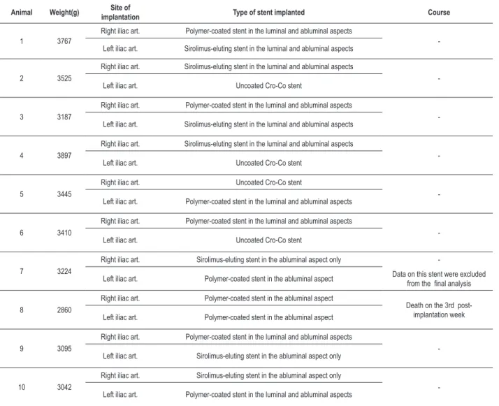
Four weeks after stent implantation, nine rabbits/18 stents underwent control angiography. One animal (rabbit 8), in
which two polymer-coated stents only in the abluminal aspect had been implanted, was found dead (undetermined cause of death) in the 3rd week post stent implantation. This resulted in an insufficient sample size for the analysis of endothelialization of polymer-coated stents only in the abluminal aspect, and data regarding the other stent of the same type (polymer-coated stent only in the abluminal aspect) implanted in the left iliac artery of rabbit 7 were excluded (Table 1).
Control angiography
All stents were patent and without angiographic restenosis or image suggestive of thrombus at the moment of the restudy after four weeks of stent implantation.
Scanning electron microscopy
The areas analyzed (mean ± SD) measured 17.03 ± 4.82; 15.89 ± 3.90; 17.97 ± 3.25 and 10.40 ± 4.54 regarding the uncoated stent, polymer-coated stent in the luminal and
abluminal aspects, sirolimus-eluting stent in luminal and abluminal aspects and sirolimus-eluting stent in the abluminal aspect only, respectively (p = 0.219).
The areas of stent strut exposed (mean ± SD) (mm2)were
0.12 ± 0.08; 0.09 ± 0.12; 0.60 ± 0.67 and 0.05 ± 0.04 (uncoated stent, polymer-coated stent in the luminal and abluminal aspects, sirolimus-eluting stent in luminal and abluminal aspects and sirolimus-eluting stent in the abluminal aspect only, respectively) (p = 0.120).
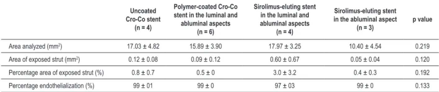
Percentages of endothelialization were 99%; 99%; 97% and 99% (uncoated stent, polymer-coated stent in the luminal and abluminal aspects, sirolimus-eluting stent in luminal and abluminal aspects and sirolimus-eluting stent in the abluminal aspect only, respectively) (p = 0.133) (Table 2 and Figure 1).
Discussion
The results of this study showed that sirolimus-eluting stents in the luminal and abluminal aspects and exclusively in
Table 1 – Data on the stent implantation procedure and post-operative course
Animal Weight(g) implantationSite of Type of stent implanted Course
1 3767
Right iliac art. Polymer-coated stent in the luminal and abluminal aspects
-Left iliac art. Sirolimus-eluting stent in the luminal and abluminal aspects
2 3525
Right iliac art. Sirolimus-eluting stent in the luminal and abluminal aspects
-Left iliac art. Uncoated Cro-Co stent
3 3187
Right iliac art. Polymer-coated stent in the luminal and abluminal aspects
-Left iliac art. Sirolimus-eluting stent in the luminal and abluminal aspects
4 3897
Right iliac art. Sirolimus-eluting stent in the luminal and abluminal aspects
-Left iliac art. Uncoated Cro-Co stent
5 3445
Right iliac art. Uncoated Cro-Co stent
-Left iliac art. Polymer-coated stent in the luminal and abluminal aspects
6 3410
Right iliac art. Polymer-coated stent in the luminal and abluminal aspects
-Left iliac art. Uncoated Cro-Co stent
7 3224
Right iliac art. Sirolimus-eluting stent in the abluminal aspect only
-Left iliac art. Polymer-coated stent in the abluminal aspect Data on this stent were excluded from the inal analysis
8 2860
Right iliac art. Polymer-coated stent in the abluminal aspect
Death on the 3rd post-implantation week Left iliac art. Polymer-coated stent in the abluminal aspect
9 3095
Right iliac art. Polymer-coated stent in the luminal and abluminal aspects
-Left iliac art. Sirolimus-eluting stent in the abluminal aspect only
10 3042
Right iliac art. Sirolimus-eluting stent in the abluminal aspect only
Arq Bras Cardiol 2012;99(6):1123-1128
Table 2 – Quantiication of endothelialization after stent implantation in iliac arteries of rabbits as assessed by scanning electron microscopy
Uncoated Cro-Co stent
(n = 4)
Polymer-coated Cro-Co stent in the luminal and
abluminal aspects (n = 6)
Sirolimus-eluting stent in the luminal and abluminal aspects
(n = 4)
Sirolimus-eluting stent in the abluminal aspect
(n = 3)
p value
Area analyzed (mm2) 17.03 ± 4.82 15.89 ± 3.90 17.97 ± 3.25 10.40 ± 4.54 0.219
Area of exposed strut (mm2) 0.12 ± 0.08 0.09 ± 0.12 0.60 ± 0.67 0.05 ± 0.04 0.120
Percentage area of exposed strut (%) 0.8 ± 0.7 0.5 ± 0 3.0 ± 3.2 0.4 ± 0.3 0.192
Percentage endothelialization (%) 99 ± 01 99 ± 0 97 ± 03 99 ± 0 0.133
abluminal aspect had a rate of stent-strut endothelialization similar to that found in non-eluting stents after four weeks of implantation in the iliac arteries of non-atherosclerotic rabbits. Several coronary stents eluted in antiproliferative drugs showed effective inhibition of neointimal proliferation and reduction of restenosis rates, as well as the need for target-vessel reintervention. One of the concerns related to the use of these drug stents is the probability, albeit rare, of the occurrence of late thrombosis8. Fast post-implantation endothelialization of
drug stents is a desirable phenomenon for theoretically reducing the risk of both early and late thrombosis10.
P r e - c l i n i c a l e v a l u a t i o n o f p o s t - i m p l a n t a t i o n reendothelialization of drug stents is preferably made by implanting these stents in the iliac arteries of rabbits, since the results obtained with these animals are the ones that are most similar to those observed in humans11. The coronary
arteries of pigs reendothelialize very quickly after stent implantation and are more useful for the assessment of implant safety. Additionally, pre-clinical studies with stent implantation in animals with induced atherosclerosis are currently recommended12,13.
When endothelialization data from Inspiron®
sirolimus-eluting stents (Scitech®, Goiânia, Brazil) are compared to the
experimental data from Joner et al’s study14 as regards stent
strut endothelialization, we verify that the Inspiron® stent
endothelialization rate (99%) is higher than that of other drug stents (Cypher®: 64%, Taxus®: 68%, Endeavor®: 76%
and Xience V®: 80%) for the same observation period of four
weeks post-implantation. This difference may result from the sirolimus elution only in the abluminal aspect in the Inspiron®
stent, whereas the antiproliferative drugs are released from both stent aspects in the other stents. Another factor that could explain this difference is that we did not perform denudation prior to the stent implantation, thus minimizing the occurrence of arterial spasm, trauma, and arterial dissection. Thus, using this methodology, we could only analyze endothelialization on the stent struts, but not between the stent struts. Another methodological difference of our study in relation to Joner et al’s study14 is that we used the carotid approach whereas they
used the femoral approach. The use of the femoral approach with artery ligation at the end of stent implantation in iliac arteries may cause the arterial flow to slow down upstream fromthe ligation point, and this would theoretically have an impact on several aspects of coagulation and possibly on stent endothelialization; for this reason we chose the carotid approach15.
Study limitations
Non-atherosclerotic animals were used in the present study. However, because of the atherosclerotic process specificities (multifactorial, chronic disease), an animal model with atherosclerosis reliably reproducing this abnormality as seen in humans has not yet been developed.
The observation period post-stent implantation in iliac arteries of rabbits was 30 days. We observed low standard-deviations (minimum of 0 and maximum of 0.03) for the rates of endothelialization on the stent struts studied; thus, by using a test power of 80% and admitting a beta error of 5%, we calculated that the sample number for each type of stent was sufficient, except for the group of polymer-coated stents in the abluminal aspect, which was removed from this comparative analysis. However, groups of additional animals could have been studied for shorter observation periods (seven and/or 14 days), but this was not the object of this initial investigation. The functional aspects of the neointima recovering the stent struts after four weeks of implantation were not assessed in the present study.
Figure 1 – Images obtained by scanning electron microscopy of the luminal aspect of the iliac arteries of rabbits after four weeks of stent implantation. (A) uncoated Cro-Co stent; (B) polymer-coated Cro-Co stent in the luminal and abluminal aspects; (C) sirolimus-eluting stent in the luminal and abluminal aspects; (D) sirolimus-eluting stent in the abluminal aspect only
Conclusion
After four weeks of implantation in the iliac arteries of rabbits, the drug stents releasing sirolimus from the luminal and abluminal aspects and abluminal aspect only showed almost complete endothelialization on struts (97% to 99%) with no statistically difference from endothelialization occurring in non-drug eluting stents (uncoated stents and polymer-coated stents in the luminal and abluminal aspects) (99%).
Acknowledgements
The authors wish to express their thanks to Mrs. Adriana Palombo Nunes, of the Analytics Center of the Chemistry Institute and to Mr. Sebastião Aparecido Boleta, of the Electronic Microscopy Laboratory of the Anatomy Department of the Institute of Biomedical Sciences of the University of São Paulo for all the help given in the preparation of samples and obtentions of images in electronic scanning microscopy. We extend our thanks to Mmes. Andréa R. de Carvalho and Fernanda Giordano for all their work in the administration of science, technology and innovation projects.
The present study is part of the National Development of Vascular Endoprotheses (stents)-PDNS – created in 2004-2005.
It counts on the support of the Secretariat of Science, Technology and Strategic Inputs(SCTIE)/Department of Science and Technology of the Ministry of Health(MS); the National Council for Scientific and Technologic Development(CNPq), as well as of the Financier of studies and Projects(FINEP) of the Ministry of Science and Technology(MCT). The development of the stents laser cutting process counted on the support of the Foundation of Research Support of the State of São Paulo(FAPESP; Type of support: Technological Innovation - innovative Research in Small and Micro-sized Companies [PIPE]).
Potential Conflict of Interest
No potential conflict of interest relevant to this article was reported.
Sources of Funding
There were no external funding sources for this study.
Study Association
This study is not associated with any post-graduation program.
1. Nunes G. Modelos de experimentação animal e complicações da angioplastia transluminal coronária. Arq Bras Cardiol. 1993;61(4):247–51.
2. Suzuki Y, Yeung AC, Ikeno F. The importance of pre-clinical animal testing in interventional cardiology. Arq Bras Cardiol. 2008;91(5):348-60.
3. Abizaid A. Sirolimus-eluting coronary stents: a review. Vasc Health Risk Manag. 2007;3(2):191–201.
4. Lemos PA, Laurindo FRM, Morato SP, Takimura C, Campos CA, Gutierrez os, et al. Stent coronário de liga cobalto-cromo concebido no Brasil: achados histológicos preliminares em modelo experimental porcino. Rev Bras Cardiol Invasiva. 2007;15(4):378–85.
5. Campos AH, Takimura CK, Gregores GB, Sarmento CA, Fioretto ET, Laurindo FR, et al. Redução neointimal com stent com polímero biodegradável e sirolimus desenvolvido no Brasil: Resultados preliminares em suínos (abstract). 30 Congresso da Sociedade de Cardiologia do Estado de São Paulo; 2009; São Paulo. Anais. São Paulo: Sociedade de Cardiologia do Estado de São Paulo; 2009. p.25.
6. Granada JF, Inami S, Aboodi MS, Tellez A, Milewski K, Wallace-Bradley D, et al. Development of a novel prohealing stent designed to deliver sirolimus from a biodegradable abluminal matrix. Cir Cardiovasc Interv. 2010;3(3):257-66.
7. Schwartz RS, Edelman E, Virmani R, Carter A, Granada JF, Kaluza GL, et al. ,Drug-Eluting Stents in Preclinical Studies. Updated Consensus Recommendations for Preclinical Evaluation. Circ Cardiovasc Intervent. 2008;1(2):143-53.
8. Guidance for Industry: Coronary Drug-Eluting Stents – Nonclinical and Clinical Studies.Companion Document. Draft Guidance. Washington DC: U.S.Department of Health and Human Services; 2008.
9. Watanabe, I.; Yamada E. The fine structure of lamellated nerve endings found in the rat gingiva. Arch Histol JPN. 1983;46(2):173–82.
10. Buja ML. Vascular responses to percutaneous coronary intervention with bare-metal stents and drug-eluting stents. J Am Coll Cardiol. 2011;57(11):1323–6.
11. Finn AV, Nakazawa G, Joner M, Kolodgie FD, Mont EK, Gold HK, et al. Vascular Responses to Drug Eluting Stents. Importance of Delayed Healing. Arterioscler Thromb Vasc Biol. 2007;27(7):1500–10.
12. Nakazawa G, Nakano M, Otsuka F, Wilcox JN, Melder R, Pruitt S, et al. Evaluation of Polymer-Based Comparator Drug-Eluting Stents Using a Rabbit Model of Iliac Artery Atherosclerosis. Circ Cardiovasc Interv. 2011;4(1):38–46.
13. Drachman DE. Drug-Eluting Stents in Animals and Patients: Where Do We Stand Today? Circulation. 2009;120:101–3.
14. Joner M, Nakazawa G, Finn AV, Quee SC, Coleman L, Acampado E, et al. Endothelial cell recovery between comparator polymer-based drug-eluting stents. J Am Coll Cardiol. 2008;52(5):333–42.
15. La Disa JF Jr, Meier HT, Olson LE, Kersten JR, Warltier DC, Pagel PS. Antegrade iliac artery stent implantation for the temporal and spatial examination of stent-induced neointimal hyperplasia and alterations in regional fluid dynamics. J Pharmacol Toxicol Methods. 2005;51(2):115–21.