15
Original Article
Sensitivity and Specificity of B-Type Natriuretic
Peptide for Identifying Symptomatic and Asymptomatic
Patients with Severe Mitral Regurgitation
Zilda Machado Meneghelo, Hélio M. Magalhães, Auristela I. O. Ramos, Jorge Eduardo Assef, Davi
Costa Le Bihau, Nivia A. de Campos Salvarani, Angela Tavares Paes, Romeu S. Meneghelo
São Paulo, SP - Brazil
Objective
To assess the sensitivity and specificity of B-type natriuretic peptide (BNP) for identifying symptomatic and asymptomatic patients with severe chronic mitral regurgitation followed up on an outpatient care basis.
Methods
A group of patients with mitral regurgitation underwent elec-trocardiography, chest teleradiography, venous blood withdrawal, and transthoracic echocardiography. Based on an analysis of the echocardiographic variables, 62 patients had mild and moderate mitral regurgitation (GI) and 34 had severe mitral regurgitation (GII). The discriminating capacity of the BNP for detecting patients with severe mitral regurgitation was assessed by use of ROC curves.
Results
The patients’ ages ranged from 15 to 63 (mean, 31.7) years, and of the 96 patients, 71 (73%) were females. The BNP values ranged from 0.00 pg/mL to 193 pg/mL. The mean BNP values in GI and GII patients were 18.10 ± 0.74 pg/mL and 50.54 ± 1.46 pg/mL, respectively (P=0.001). The cutoff point for identifying severe mitral regurgitation was 15.40 pg/mL for the best balance between sensitivity and specificity, which were 0.73 and 0.74, respectively. The cutoff point for identifying symptomatic patients with severe mitral regurgitation was 28.40 pg/mL for the best balance between sensitivity and spe-cificity, which were 0.78 and 0.83, respectively.
Conclusion
The BNP values capable of identifying asymptomatic and symp-tomatic patients with severe mitral regurgitation are lower than 100 pg/mL, which is considered for the diagnosis of heart failure.
Key words
mitral regurgitation, natriuretic peptides, BNP
The measurement of B-type natriuretic peptide (or brain natriu-retic peptide, BNP) levels has been used for diagnosing congestive heart failure, especially when the symptoms and signs are discrete or mild, and in the presence of associated comorbidities, such as pulmonary disease or obesity 1. The BNP levels have a positive
correlation with left ventricular end-diastolic pressure and an inverse correlation with left ventricular function. These concentrations are considered to represent an independent evaluation of ventricular function, with no need for other more expensive tests 2. The
cutoff point of the BNP levels recommended for separating the population with heart failure from that without that syndrome is 100 pg/mL 3. These initial findings suggest the possibility of
expan-ding the application of BNP level measurement to other situations that gradually lead to left ventricular dysfunction, such as some valvular diseases and congenital heart diseases. The BNP levels in patients with mitral regurgitation are greater than those in controls. The study by Brookes et al 4 showed that 89% of the
22 patients with mitral regurgitation have more elevated BNP levels than healthy individuals do, 20.85 ± 16.9 versus 3.37 ± 0.9 pmol/L. Another study 5 with symptomatic and asymptomatic
patients showed that the BNP levels increased according to the severity of mitral regurgitation and were greater in symptomatic patients. However, the case series of these reports are small, and the authors of both studies suggested additional investigation. The objective of the present study was to assess in a group of patients with chronic mitral regurgitation of different degrees, which is the BNP cutoff point capable of identifying the existence of severe mitral regurgitation with or without symptoms.
Methods
The study comprised patients from the valvular heart disease outpatient clinic of our institution diagnosed with chronic mitral regurgitation and who agreed to participate in the study. The diagnosis was based on physical examination due to the presence of a systolic murmur in the mitral auscultation area classified from 1+ to 4+. The patients underwent electrocardiography, chest teleradiography, transthoracic echocardiography with color flow mapping, and venous blood withdrawal from a peripheral vein for measuring the plasma concentrations of BNP.
Patients with the following findings were excluded from the study: acute mitral regurgitation or mitral regurgitation secondary to cardiomyopathy; ejection fraction lower than 0.55; coronary heart
Instituto Dante Pazzanese de Cardiologia, São Paulo.
Mailing address: Zilda Machado Meneghelo - Rua Nicolau Souza Queiroz, 194/161 - 04105-000 - São Paulo, SP, Brazil E-mail: zildamm@cardiol.br
16
disease diagnosed by the presence of angina or electrically inactive zones on the electrocardiogram; age < 15 or > 65 years; complete dysfunction of the mitral valve and valvular area < 2.0 cm2;
con-genital cardiac defects or hemodynamic alteration in another heart valve; clinical findings compatible with infective endocarditis; active rheumatic disease; diabetes mellitus; thyroid disorders; systemic arterial hypertension; disorders in other organs or systems; and previous surgical treatment.
After meeting the above-described criteria, our case series comprised 96 patients with ages ranging from 15 to 63 (mean, 31.7) years, 71 (73%) being females.
Transthoracic Doppler echocardiography of all patients was per-formed with an Ultramark-9 HDI device (ATL - Advanced Techno-logy Laboratories) with phased array transducer and an emission frequency of 3.0 megahertz. All echocardiographies were perfor-med by only 2 trained professionals. The severity of mitral regurgi-tation was determined by analyzing the following variables: a) effective regurgitant orifice 6; b) vena contracta; c) regurgitant jet area; and
d) relation between the regurgitant jet and left atrial areas 7.The
measurements considered indicators of severe mitral regurgitation were as follows: effective regurgitant orifice > 0.4cm2 6; vena
contracta > 0.5cm; regurgitant jet area > 7 cm2; and relation
between the regurgitant jet and left atrial areas > 40% 8. The
detection of reverse systolic flow in any pulmonary vein was also considered an indicator of severe mitral regurgitation 9.
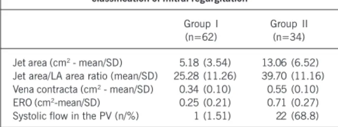
The patients with mitral regurgitation selected were classified according to at least 2 of the above-described criteria as follows: 1) group I (GI) - 62 patients with mild and moderate mitral regur-gitation; group II (G II) - 34 patients with severe mitral regurgitation. The mean values of the echocardiographic variables found in the 2 groups are shown in table I.
The left atrial and ventricular diameters were measured by using M-mode echocardiography based on the images acquired on the 2-dimensional transthoracic study 10.The results are
ex-pressed in millimeters. The calculation of the end-diastolic and end-systolic volumes of the left ventricular cavity was performed according to the Simpson technique 11. Two variables were used
for assessing the left ventricular overall systolic function: a) shor-tening fraction (delta D); and b) ejection fraction.
Patients with the following changes were considered as having severe morphological alterations consequent to mitral regurgitation: left atrial telesystolic diameter > 57 mm; left ventricular end-systolic diameter > 45 mm or index = 26 mm/m2; left ventricular
end-diastolic diameter > 70 mm or index = 40 mm/m2; left
ven-tricular end-systolic volume index > 50mL/ m2; left ventricular
end-diastolic volume index > 100 mL/m2; ejection fraction < 60% and
delta D < 32%.
The echocardiographic data of the variables described are shown in table II. The GII patients showed the greatest means of left atrial, diastolic and systolic diameters, and of end-diastolic and end-systolic volumes. The mean ejection fraction of delta D and mean thickness of the interventricular septum of the groups studied showed no significant statistical difference.
Table III shows that the rheumatic etiology predominated in both groups (GI, 85.55% and GII, 61.80%). The myxomatous degeneration occurred in 13 (38.2%) GII patients and in none of the GI patients (P = 0.008). Of the 34 GII patients, 14 were in New York Heart Association functional class (FC) II or III, and the remaining 20 in FC I. Symptoms were more frequent in GII than in GI (P < 0.001). Functional class II was identified in only 3 of the 62 GI patients.
Atrial fibrillation was present in one GI patient (1.61%) and in 4 GII patients (11.8%).
Chest teleradiography of GII patients showed a greater mean cardiothoracic index and left atrial dimensions. The alterations in pulmonary circulation analyzed through hilar enlargement, flow inversion, and presence of perivascular infiltrate were more frequent in patients with severe mitral regurgitation (GII) (tab. IV).
Blood samples for measuring BNP were obtained after venous puncture in the forearm after patients rested for 30 minutes in the supine position. Blood was placed in vacuum tubes with EDTA (ethylenediaminetetraacetic acid) anticoagulant. BNP
measure-Table I – Echocardiographic variables determining the classification of mitral regurgitation
Group I Group II (n=62) (n=34)
Jet area (cm2 - mean/SD) 5.18 (3.54) 13.06 (6.52)
Jet area/LA area ratio (mean/SD) 25.28 (11.26) 39.70 (11.16) Vena contracta (cm2 - mean/SD) 0.34 (0.10) 0.55 (0.10)
ERO (cm2-mean/SD) 0.25 (0.21) 0.71 (0.27)
Systolic flow in the PV (n/%) 1 (1.51) 22 (68.8)
n= number of individuals; SD = standard deviation; MR = mitral regurgitation; LA = left atrium; ERRO = effective regurgitant orifice; PV = pulmonary veins.
Table II – Echocardiographic variables
Group I Group II p (n = 62) (n = 34)
LA (mm-mean/SD) 40.58 (6.60) 54.21 (7.18) < 0.001 EDD (mm/SD) 53.87 (5.64) 64.91 (6.86) < 0.001 ESD (mm/SD) 33.15 (4.19) 40.00 (5.60) < 0.001 EDV (mL-mean/SD) 132.61 (36.78) 175.74 (57.83) < 0.001 ESV (mL-mean/SD) 43.20 (14.62) 58.63 (22.88) < 0.001 Delta D (mean/SD) 38.06 (2.98) 38.14 (3.96) 0.910 EF (mean/SD) 0.67 (0.04) 0.67 (0.06) 0.560 Septum (mean/SD) 7.71 (0.86) 8.09 (1.24) 0.082
MR = mitral regurgitation; n = number of individuals; SD = standard deviation; LA = left atrium; EDD = diastolic diameter; ESD = end-systolic diameter; EDV = end-diastolic volume; ESV = end-end-systolic volu-me; EF = ejection fraction.
Table III – Clinical characteristics of the patients
Group I Group II p (n = 62) (n = 34)
Etiology (n/%)
Rheumatic 53 (85.5) 21 (61.8) 0.008 Myxomatous 9 (14.5) 13 (38.2)
Clinical follow-up 91.14 (84.76) 84.16 (79.96) (months/SD)
Functional class* (n/%)
I 59 (95.2) 20 (58.8)
II 3 (4.8) 9 (26.5) < 0.001
III 0 5 (14.7)
17
ment was performed by using the fluorescent immunoassay method. The Triage Meter model (Biosite Diagnostics Incorporated, San Diego, CA, USA) device was used. The rest BNP values considered normal for this methodology are those below 100 pg/mL. The reading sensitivity is between 5 and 1300 pg/mL, according to the manufacturer 12.
The quantitative variables were compared by using the Student
t test for independent samples. For qualitative variables, the chi-square test, or Fisher exact test was used 13.
The results whose descriptive levels (P values) were lower than 0.05 were considered statistically significant.
KAPPA measurement 14, based on the number of concordant
responses, was used to assess the intensity of concordance between the 2 professionals who performed the echocardiographies. The measure of concordance with a “0” value indicates no concordan-ce, while the “1” value indicates total concordance. The analysis of concordance found in this study was 0.871 (P<0.001).
SPSS for Windows, version 11.0 was the program used for calculations.
The protocol was approved by the Committee on Ethics of the institution. All individuals, after being clearly instructed about the research and tests that would be performed, signed a written informed consent.
Results
The mean BNP levels found ranged from 0.00 to 193 pg/mL. The mean BNP levels in GI and GII patients were 18.10 ± 0.74 pg/ mL and 50.54 ± 1.46 pg/mL, respectively, which were statistically different (P=0.001).
Sensitivity and specificity for identifying patients with severe mitral regurgitation calculated for the greatest and lowest BNP levels and some intermediary cutoff values are shown in table V. Similarly, table VI shows the sensitivity and specificity for iden-tifying patients with severe mitral regurgitation and NYHA functional classes II and III.
A ROC (receiver operator characteristic) curve was drawn for assessing the discriminating capacity of BNP to detect patients with severe mitral regurgitation, and to suggest a cutoff point regarding sensitivity and specificity (fig. 1). An area of 0.76 was found under the curve, indicating an acceptable power. The most adequate cutoff point for identifying patients with severe mitral regurgitation was 15.40 pg/mL, with sensitivity of 0.73 and specificity of 0.74.
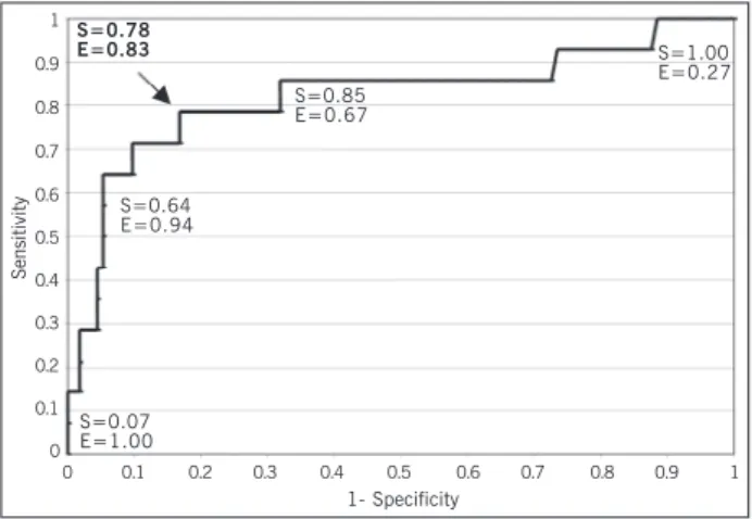
Similarly, another ROC curve was built for assessing the dis-criminating capacity of BNP to detect patients with severe mitral regurgitation and NYHA FC II and III (fig. 2). An area of 0.82 was
Table IV – Electrocardiographic and radiological alterations
Group I Group II p (n = 62) (n = 34)
Sinus rhythm (n/%) 61 (98.4) 30 (88.2) 0.052 Atrial fibrillation (n/%) 1 (1.61) 4 (11.8)
Cardiothoracic index
(mean/SD) 0.46 (0.06) 0.51 (0.06) 0.001 Left atrial dimension
(mm - mean/SD) 12.98 (7.74) 22.97 (11.60) < 0.0001 Hilar enlargement (n/%) 6 (9.7) 16 (47.1) < 0.0001 Inversion of the pulmonary 4 (6.5) 17 (50.0) < 0.0001 circulation (n/%)
Perivascular infiltrate (n/%) 5 (8.1) 17 (50.0) < 0.001
n = number of individuals; SD = standard deviation.
Table V - Sensitivity and specificity in the diagnosis of severe mitral regurgitation for the different BNP cutoff points (pg/mL)
Values of BNP cutoff points Sensitivity Specificity
0.0000 1.000 0.000 0.1000 1.000 0.032
1.1700 0.971 0.118
5.1000 0.882 0.323
5.8000 0.853 0.366
10.2000 0.765 0.602 10.5000 0.765 0.613
15.4000 0.735 0.742
15.5500 0.706 0.742 20.7000 0.618 0.828 20.9000 0.618 0.839 28.4000 0.441 0.871 40.8000 0.414 0.935 51.9000 0.353 0.946 58.4500 0.353 0.957 64.4500 0.324 0.957 68.7500 0.294 0.957 74.0000 0.265 0.957 81.8000 0.235 0.957 93.3000 0.206 0.968 103.2500 0.176 0.989 108.5000 0.147 0.989 116.0000 0.118 0.989 122.5000 0.088 0.989 147.0000 0.059 1.000 180.0000 0.029 1.000 193.0000 0.000 1.000
The first line shows the lowest cutoff value and the last line shows the greatest cutoff value. The intermediate values are in between.
Table VI - Sensitivity and specificity in the diagnosis of severe mitral regurgitation and NYHA functional classes II and III for the different
BNP cutoff points (pg/mL)
Values of BNP cutoff points Sensitivity Specificity
0.0000 1.000 0.000 0.1000 1.000 0.027
1.1500 1.000 0.071
5.1000 0.857 0.283
5.8000 0.857 0.327
10.2000 0.857 0.549 10.5000 0.857 0.558 15.4000 0.857 0.673 15.5500 0.857 0.681 20.7000 0.786 0.770 20.9000 0.786 0.779
28.4000 0.786 0.830
40.8000 0.714 0.903 51.9000 0.643 0.929 58.4500 0.643 0.938 64.4500 0.643 0.947 68.7500 0.571 0.947 74.0000 0.500 0.947 81.8000 0.429 0.947 93.3000 0.357 0.956 103.2500 0.286 0.973 108.5000 0.286 0.982 116.0000 0.214 0.982 122.5000 0.143 0.982 147.0000 0.143 1.000 180.0000 0.071 1.000 193.0000 0.000 1.000
18
radiological, and electrocardiographic criteria and adding new important variables, has contributed to the establishment of that technique as the most used for assessing patients with mitral regurgitation in the present study. Even being chosen as ideal, that technique may have limitations in defining or quantifying, or both, the severity of regurgitation 18,19. Although the severity of
mitral regurgitation in this study has been assessed by means of echocardiographic variables that measure the severity of mitral reflux, GII patients, those with severe mitral regurgitation, had the greatest repercussions of the disease evaluated through alte-rations in the electrocardiogram, in radiography, in functional class, and in left atrial and ventricular dimensions (tab. II, III, and IV). The groups differed in regard to those variables, showing that, if the clinical criteria were used for classifying the severity of mitral regurgitation, the groups would not be significantly different.
It is worth noting that the echocardiogram has interobserver variability, which could represent a bias in the present study. However, the analysis of concordance performed through the Kappa method 14 showed a high concordance, 0.871 (p<0.001), between
the 2 observers.
The mean BNP values observed in this study are lower than those observed in patients with dilated cardiomyopathy, even in the severe mitral regurgitation group, which showed mean values of 50.54 pg/mL, considered within the normal range for diagnosing heart failure. This seems to be the characteristic of patients with mitral regurgitation according to the first known publications of studies with few patients 4,5. In the study by Brookes et al 4, the
mean values observed were 20.85 ± 16.9 pmol/L in asymptomatic patients with an at least moderate mitral regurgitation on echocar-diography versus 16.9 ± 3.34 pmol/L in controls. The differences between the groups were statistically significant. Sutton et al 5
reported BNP levels of 16.9 pmol/L, 7.1 pmol/L, and 5.3 pmol/L in symptomatic and asymptomatic patients and controls, respec-tively (p<0.001). In a study of 9 symptomatic patients with mo-derate and severe mitral regurgitation, Eimer et al 20 reported
mean values of 11.8 ± 8 pg/mL versus 6 ± 4 pg/mL, in controls, a non statistically significant difference. It is worth noting that all these mean values are within the currently normal-considered range for BNP, when used for identifying individuals with and wi-thout heart failure. Thus, for using BNP measurements in managing mitral regurgitation, one cannot simply transfer the data concerning BNP application in the management of heart failure and ventricular dysfunction of other etiologies.
In the construction of a ROC curve, aiming at assessing the discriminating capacity of BNP for detecting patients with severe mitral regurgitation, a cutoff value of 15.40 pg/mL was found for a sensitivity of 0.73 and specificity of 0.74. These data confirm the previous observations that the values capable of identifying severe patients are much lower than those used for identifying heart failure. The other ROC curve constructed for assessing the discriminating capacity of BNP for detecting patients with severe mitral regurgitation and NYHA functional classes II and III had a cutoff point for BNP of 28.40 pg/mL, with sensitivity of 0.78 and specificity of 0.83. These values also suggest that one should not wait for values above 100 pg/mL to classify mitral regurgitation as severe.
All these data allow the supposition that in the natural history of mitral regurgitation, a gradual increase occurs in BNP levels according to the evolution of the disease. For decision making in found under the curve, showing a satisfactory power. The cutoff
value for identifying symptomatic patients with severe mitral regur-gitation was 28.40 pg/mL, with a sensitivity of 0.78 and a spe-cificity of 0.83.
Discussion
In recent years, the results of extensive investigations about the natriuretic peptides, especially BNP, have helped the clinical management of patients having or suspected of having heart failure, cardiomyopathies, and coronary heart disease. Thus, a study about BNP in patients with mitral regurgitation seems to be very useful in an attempt to help with the difficulties found in managing patients with that disease.
Quantifying the severity of mitral regurgitation remains a clinical challenge. Patients with mitral regurgitation can be classified as having mild, moderate, and severe lesions by using clinical history, physical examination, chest teleradiography, and electrocardio-graphy. Frequently, an intense systolic murmur 15 or the presence
of a third cardiac sound in the mitral area suggests severe mitral regurgitation 16. However, this quantification is not perfect due to
several interferences. In obese patients, those with chest defor-mities, and those with chronic pulmonary disease and severe mitral regurgitation, the systolic murmur is of low intensity or even inaudible 17. The evolution of Doppler echocardiography,
over-coming part of the subjectivity and limitations of the clinical, Fig. 1 - ROC curve for identifying severe mitral regurgitation. S = sensitivity; E = specificity.
S=0.97 E=0.11
Sensitivity
1
0.9
0.8
0.7
0.6
0.5
0.4
0.3
0.2
0.1
0
0 0.1 0.2 0.3 0.4 0.5 0.6 0.7 0.8 0.9 1
1- Specificity
S=0.73 E=0.74
S=0.08 E=0.98
S=0.44 E=0.87
S=0.85 E=0.36
Fig. 2 – ROC curve for identifying severe mitral regurgitation and NYHA functional classes II and III. S = sensitivity; E = specificity.
Sensitivity
1
0.9
0.8
0.7
0.6
0.5
0.4
0.3
0.2
0.1
0
0 0.1 0.2 0.3 0.4 0.5 0.6 0.7 0.8 0.9 1
1- Specificity
S=0.78 E=0.83
S=0.07 E=1.00
S=0.64 E=0.94
S=0.85 E=0.67
19
the management of patients, the absolute values in each evolution stage cannot be applied, because they are not known. However, this study’s findings allow supposing that sequential measurements over time may help in patient management.
The studies published 15,16 and the contribution of this study
1. de Lemos JA, McGuire DK, Drazner MH. B-type natriuretic peptide in cardiovas-cular disease. Lancet 2003; 362 (9380): 316-22.
2. Maeda K, Tsutamoto T, Wada A, Hisanga T, Kinoshita M. Plasma brain natriuretic peptide as a biochemical marker of high left ventricular end-diastolic pressure in pa-tients with symptomatic left ventricular dysfunction. Am Heart J 1998; 135:825-32. 3. Maisel AS, Krishnaswamy P, Nowak RM, et al. Rapid measurement of B-type natriuretic peptide in the emergency diagnosis of heart failure. N Engl J Med 2002; 347:161-7.
4. Brookes CIO, Kemp MW, Hooper J, Oldershaw PJ, Moat NE. Plasma brain natriu-retic peptide concentrations in patients with chronic mitral regurgitation. J Heart Valve 1997;6: 608-12.
5. Sutton TM, Stewart RAH, Gerber IL, et al. Plasma natriuretic peptide levels increase with symptms and severity of mitral regurgitation. J Am Coll Cardiol 2003; 41:2280-7.
6. Enriquez-Sarano M, Seward JB, Bailey KR, Tajik AJ. Effective regurgitant orifice area: a noninvasive Doppler development of an old hemodynamic concept. J Am Coll Cardiol 1994; 23: 443-51.
7. Bargiggia GS, Tronconi L, Sahn DJ, et al. A new method for quantification of mitral regurgitation based on color flow Doppler imaging of flow convergence proximal to the regurgitante orifice. Circulation 1991; 84:1481-9.
8. Assef JE, Barreto RBM, Barreto SNSM. Avaliação Doppler-ecocardiográfica das lesões mitrais e aórticas: a prática diária, do modo-M ao transesofágico. Rev Soc Cardiol Estado de São Paulo 1997; 7: 547-68.
9. Klein AL, Obarski TP, Stwart WJ, et al. Transesophageal Doppler echocardiogra-phy of pulmonary venous flow: a new marker of mitral regurgitation severity. J Am Coll Cardiol 1991; 18: 518-26.
10. Morcerf FR, Thevernard RS, Fuks J. Ecocardiografia: método e valores normais. Arq Bras Cardiol 1976; 29: 459-65
11. Wyatta HL, Heng M K, Meebraum S, et al. Cross-sectional echocardiography. II. Analysis of mathematic models for quantifying volume of the formalin-fixed left entricle. Circulation 1980; 61: 1119-25.
12. Triageâ BNP. Biositeâ Diagnostics, Incorporated, 1999. 16 p. ( Material Promocional).
13. Armitage P, Berry G. Statistical methods in medical research. 3. ed. Oxford: Blackwell Science, 1994.
14. Siegel S, Catellan N. Nonparametric statistics for the behavioral sciences. 2.ed. New York: Mc model. In Graw-Hill, 1988. p.284-5
15. Desjardins V, Enriquez-Sarano M, Tajik A. Intensity of murmurs correlates with severity of valvular regurgitation. Am J Med 1996; 100: 149-56.
16. Tribouilloy CM, Enriquez-Sarano M, Mohly D, et al. Pathophyfiolosic determinants of third heart sounds: a prospective clinical and Doppler echocardiographic study. Am J Med 2001; 111: 96-102.
17. Schreiber TL, Fisher J, Mangla A, Miller D. Severe “silent” mitral regurgitation: A potentially reversible cause of refratory heart failure. Chest 1989; 96: 242-6. 18. Chen C, Thomas J, Anconina J, et al. Impact of impinging wall jet on color Doppler
quantification of mitral regurgitation. Circulation 1991; 84: 712-20.
19. Kamp O, Huintik H, Van Eenige M.J, et al. Value of pulmonary venous flow charac-teristics in the assessment of severity of native mitral valve regurgitation: an angiographic correlated study. J Am Soc Echocardiogr 1992; 5: 239-6. 20. Eimer MJ, Ekery D, Rigolin V, Bonow RO. Serum B-type natriuretic peptide in
pa-tients with chronic mitral regurgitation is not elevated. [Abstract]. J Am Coll Cardiol 2003; 46 (Suppl A): 509.