A Functional Role of Fibroblast Growth Factor
Receptor 1 (FGFR1) in the Suppression of
Influenza A Virus Replication
Xin Liu1, Chengcai Lai1, Keyu Wang1, Li Xing1, Penghui Yang1,2, Qing Duan1, Xiliang Wang1*
1State Key Laboratory of Pathogen and Biosecurity, Beijing Institute of Microbiology and Epidemiology, Beijing, China,2302 Military Hospital, Beijing, China
Abstract
Influenza A virus causes annual epidemics and occasional pandemics in humans. Here, we investigated four members of the fibroblast growth factor receptor (FGFR) family; FGFR1 to 4, and examined their expression patterns in human lung epithelial cells A549 with influenza A virus infection. We identified a functional role of FGFR1 in influenza A/Puerto Rico/8/1934 (PR8) and A/Anhui/01/2005 (H5N1) virus replication. Our results showed that FGFR1 si-lencing by siRNA interference promoted influenza A/PR8 and H5N1 virus replication in A549 cells, while lentivirus-mediated exogenous FGFR1 expression significantly sup-pressed influenza A virus replication; however, FGFR4 did not have the same effects. More-over, FGFR1 phosphorylation levels were downregulated in A549 cells by influenza A virus infection, while the repression of FGFR1 kinase using PD173074, a potent and selective FGFR1 inhibitor, could enhance virus replication. Furthermore, we found that FGFR1 inhib-its influenza virus internalization, but not binding, during viral entry. These results suggested that FGFR1 specifically antagonizes influenza A virus replication, probably by blocking viral entry.
Introduction
Influenza A virus causes epidemics of respiratory diseases in humans, leading to thousands of deaths annually. This subtype of pathogen has been thoroughly studied, and many aspects in-cluding viral entry, and replication, as well as host cell-virus interactions are well-characterized. During the influenza virus life cycle, entry is the first essential process by which viral genomes are delivered from extracellular virions into the cell nucleus for viral replication [1,2], which is an ideal target to block infection. Influenza virus enters the cell by receptor-mediated endocy-tosis after binding to the sialic acid receptor [3–5]. There is increasing evidence that viral at-tachment to the cell surface by influenza hemagglutinin (HA) is not sufficient for endocytotic routes [6,7]. The adaptor protein Epsin-1 is recruited for clathrin-mediated viral entry [8]. Specific cellular signaling by receptor tyrosine kinases, including epidermal growth factor a11111
OPEN ACCESS
Citation:Liu X, Lai C, Wang K, Xing L, Yang P, Duan Q, et al. (2015) A Functional Role of Fibroblast Growth Factor Receptor 1 (FGFR1) in the Suppression of Influenza A Virus Replication. PLoS ONE 10(4): e0124651. doi:10.1371/journal. pone.0124651
Academic Editor:Ralph Tripp, University of Georgia, UNITED STATES
Received:January 14, 2015
Accepted:March 17, 2015
Published:April 24, 2015
Copyright:© 2015 Liu et al. This is an open access article distributed under the terms of theCreative
Commons Attribution License, which permits
unrestricted use, distribution, and reproduction in any medium, provided the original author and source are credited.
Data Availability Statement:All relevant data are within the paper.
Funding:This work is supported by the Ministry of Science and Technology of China, the National Basic Research Program (973, 2012CB518905,http://www.
most.gov.cn/eng/). XW received the funding. The
funders had no role in study design, data collection and analysis, decision to publish, or preparation of the manuscript.
receptor (EGFR) and c-Met receptor, is required. siRNA-induced EGFR knockdown and the inhibition of kinase activity by small molecule inhibitors leads to impaired influenza virus up-take into cells [9]. In turn, host cells express antiviral factors to defend against influenza virus entry [10]. For example, the interferon-inducible transmembrane (IFITM) proteins 1 to 3 re-strict an entry step of influenza A viral replication [11,12].
The existence of these cellular factors suggests that other receptors play a role in viral entry. Fibroblast growth factor receptor (FGFR) 1 to 4, known as the tyrosine kinase receptor super-family, regulates many biological processes including differentiation, proliferation, develop-ment, and angiogenesis [13–15]. FGFR1 is a co-receptor during adeno-associated virus 2 (AAV) infection for successful viral entry into the host cell [16]. FGFR4 knockdown reduces influenza A/WSN pseudotyped particle entry, and an FGFR4 inhibitor attenuates WSN virus replication [10]. These results increase our understanding of the role of FGFR family members in the life cycle of viral infection. Differential expression and structural complexity of the FGFR family may generate functional diversity in response to different ligands, which is crucial for regulating normal physiological processesin vivo[17]. However, it remains unclear how the expression of FGFR 1 to 4 are regulated by influenza A virus infection, and whether FGFR 1 to 4 have the same effect on influenza A (H1N1 or H5N1) virus replication requires further study.
In this study, we investigated the differential expression of four FGFR family members in A549 cells with PR8 infection and the functional roles of FGFR1 and FGFR4 on PR8 or H5N1 virus replication. We found that RNAi-induced FGFR1 gene silencing (not FGFR4) significant-ly elevated PR8 and H5N1 virus replication, while lentivirus-mediated FGFR1 overexpression reduced virus replication, which suggested that FGFR1 specifically suppressed influenza A virus replication. In addition, inhibition of FGFR1 kinase activity could enhance influenza A virus replication. During the early stages of infection, FGFR1 could inhibit viral internalization steps, which may account for the suppressive effect of FGFR1 on virus replication.
Materials and Methods
Viruses and cells
Two influenza A (H1N1) virus strains: A/Puerto Rico/8/1934 (PR8) and A/Anhui/01/2005 (H5N1) were used in this study. The strains were propagated in 9- to 11-day-old specific-pathogen-free (SPF) chicken embryos. Virus titers were determined based on the 50% tissue in-fectious dose (TCID50) assay using Madin-Darby canine kidney (MDCK) cells purchased from the ATCC according to the Reed-Muench method [18]. The human lung adenocarcinoma epi-thelial cell line A549 from the ATCC was cultured in Dulbecco’s Modified Eagle’s medium (DMEM) supplemented with 10% (v/v) fetal bovine serum (FBS) and 100 units penicillin-streptomycin ml-1at 37°C in a humidified atmosphere of 5% (v/v) CO2. All experiments with live H5N1 virus were conducted in approved bio-safety level 3 (BSL-3) laboratory facilities.
RNA isolation and real-time quantitative PCR
Total RNA was extracted from cultured cells with TRIzol reagent [19]. A total of 2μg of RNA
was treated with RNase-free DNase I (Promega) for 30 min at 37°C. Complementary DNA (cDNA) was generated by reverse transcription with 8μl of 5× PrimeScript RT Master Mix
(TaKaRa) for 15 min at 37°C. A total of 10μl of SYBR Premix Ex Taq II (TaKaRa) were mixed
with cDNA and specific primers to a total volume of 20μl. The primer pairsFGFR1,FGFR2, FGFR3,FGFR4, influenza virusM1, andGAPDH(Table 1) were designed using Primer
7300 Real-Time PCR System (Applied Biosystems). The quantification data were analyzed with ABI 7300 SDS software v.1.3.
Western blotting assay
Protein extracts from cell lysates were obtained with RIPA buffer mixed protease inhibitor cocktail (Thermo scientific). Protein extractions were boiled in 4× Protein SDS-PAGE Loading Buffer (Takara) for 5 min and resolved on a SDS-PAGE, and then transferred onto 0.45-μm
NC membrane (GE Whatman). Membranes with total FGFR proteins were blocked by 5% (w/v) non-fat dry milk (GE Healthcare), while those with phosphorylated FGFR1 protein were treated with 5% (w/v) bovine serum albumin (BSA) for 1 h at room temperature and then incu-bated overnight at 4°C with primary antibodies including anti-FGFR1 polyclonal antibody (1:500 dilution, Cell Signaling Technology), anti-phosphorylated FGFR1 polyclonal antibody (1:1000 dilution, Lianke Biotechnology Ltd.), anti-FGFR4 monoclonal antibody (1:1000 dilu-tion, Abcam; Epitomics), and anti-β-actin monoclonal antibody (1:5000 dilution, Sigma-Al-drich). After three washes with TBS containing 0.1% (v/v) Triton X-100 (TBST), they were incubated with goat anti-rabbit or anti-mouse horseradish peroxidase (HRP)-conjugated sec-ondary antibody (1:5000 dilution, Cell Signaling Technology) for 1 h at room temperature. Final detection of protein was performed using the Signal Boost Immunoreaction Enhancer Kit (Merck Millipore). Protein levels were quantified using Quantity One software (Bio-Rad).
FGFR knockdown with small interfering RNAs (siRNAs)
All siRNA transfection was performed as described previously [20]. SiRNAs against two differ-ent regions of each target genes were synthesized by RiboBio (Guangzhou, China). ForFGFR1 andFGFR4silencing, A549 cells were transfected with 20 nM siRNA using Lipofectamine RNAiMax (Invitrogen) according to the manufacturer's instructions. The medium was re-placed with fresh complete medium 6 h after transfection, and the cells were harvested 48 h after transfection for FGFR repression efficiency detection by real-time PCR and Western blotting.
Indirect immunofluorescence microscopy
After 48 h of siRNA transfection, A549 cells were infected with PR8 virus. Four hours later, A549 cells were fixed with 4% (w/v) paraformaldehyde at room temperature for 15–20 min, as described previously [21]. After two washes with PBS, cells were permeabilized with PBS con-taining 10% (v/v) FBS, 3% (w/v) BSA, and 0.5% (v/v) Triton X-100 for 15 min, after which they were incubated with an anti-influenza A virus NP primary antibody (1:1000 dilution, Merck Millipore) overnight at 4°C. After two washes with PBS containing 0.2% (w/v) BSA and 0.1% Table 1. Primer pairs used for real-time PCR.
Gene Forward Reverse
FGFR1 50- CGCCCCTGTACCTGGAGATCATCA -30 50- TTGGTACCACTCTTCATCTT -30;
FGFR2 50- GCCTGGAAGAGAAAAGGAGATTAC -30 50- GGATGACTGTTACCACCATACA -30;
FGFR3 50- CATCCGGCAGACGTACACGC -30 50- ACTGTACACCTTGCAGTGGA -30;
FGFR4 50- GTGCCCTCGGACCGCGGCACATAC -30 50- TCCGAAGCTGCTGCCGTTGATG -30;
M1 50- AAGACCAATCCTGTCACCTCTG -30 50- CAAAACGTCTACGCTGCAGTCC -30
GAPDH 50- GGTGGTCTCCTCTGACTTCAACA -30 50- GTTGCTGTAGCCAAATTCGTTGT -30 Primer pairs of humanFGFR1,FGFR2,FGFR3,FGFR4, influenza virusM1, andGAPDHwere designed using Primer 5.0 and presented inTable 1.
(v/v) Triton X-100, cells were incubated with Alexa Fluor 488-labeled secondary antibodies (Invitrogen) at room temperature for 1 h. Cell nuclei were stained with Hoechst 33342 (Sigma) for 10 min followed by three PBS washes. Images were captured using a Nikon Eclipse
TE2000-U inverted microscope. Analysis was performed with Image Pro Plus software (Media Cybernetics). Number of total cells and virus-infected positive cells in each of three areas (× 250 field) was counted, and then the average count of the three fields was recorded.
Lentivirus-mediated FGFR overexpression in A549 cells
DNA fragments corresponding to the coding sequence of humanFGFR1andFGFR4genes were obtained from A549 cDNA by PCR amplification and subcloned into plasmid pWPXL between restriction sitesPacI andNdeI orEcoRI. The primer sequences used were as follows: FGFR1forward, 5’- CGGTTAATTAACCATGTGGAGCTGGAAGTGCC -3’and reverse, 5’
-CTGCATATGTCAGCGGCGTTTGAGTCC -3’;FGFR4forward, 5’- GCTTAATTAACCAT GCGGCTGCTGCTGGCCCTGT -3’and reverse, 5’- TTGGAATTCCTGTCTGCACCCCAG ACCCGAAG -3’. The 293T cells were transfected with pWPXL-R1 or pWPXL-R4 and two other helper plasmids. After 48 h transfection, recombinant lentivirus was obtained from 293T cells and filtered through 0.45-μm filters. Empty lentivirus vector was used as a control. For
lentivirus infection, A549 cells were incubated with diluted virus supernatant (>95% infection efficiency) supplemented with 6μg polybrene (Sigma-Aldrich) ml-1for 8 h. Medium was then
replaced with fresh complete medium and cultured for a further 40 h. Cells were harvested for FGFR ectopic expression efficiency determination by real-time PCR and Western blotting assay.
Virus binding and internalization assay
For influenza virus binding, A549 cells with FGFR1, FGFR4, or GFP overexpression were pretreated with or without 0.01 units of sialidase (Sigma-Aldrich) ml-1at 37°C for 8 h [9], and then pre-cooled for 10 min, incubated with PR8 or H5N1 virus at a multiplicity of infec-tion (MOI) of 1 at 4°C for 1 h, and finally washed twice with ice-cold PBS [1]. Cell lysates were prepared at 4°C with ice-cold RIPA buffer containing protease inhibitor cocktail. For virus internalization, cells with prebound virus were warmed to 37°C for 30 min and then washed with acidic cold PBS-HCl (pH 1.3) for detection of internalized virus particles, but not attached particles [9]. Cell lysates were obtained to determine influenza virus NP levels by Western blotting.
Statistical analyses
Statistical analysis of the results was performed in GraphPad Prism software 4.0 (GraphPad Soft-ware, CA). Data was shown as means ± s.e.m. Statistical significance was assessed by one-way analysis of variance (ANOVA). Statistical significances are indicated asP<0.05;P<0.01;
P<0.001.
Results
Expression patterns of FGFR family members in A549 cells with
influenza A (H1N1) virus infection
while FGFR4 was intermediate and FGFR2 and FGFR3 were lowest. The results showed that the FGFR1 and FGFR4 mRNA expression decreased sharply at 12 hpi, and then stabilized. PR8 virus infection also decreased FGFR2 and FGFR3 mRNA expression at 12 hpi, which increased subsequently. We next detected the protein levels of four FGFR members by Western blotting using specific antibodies. The results showed that the FGFR1 and FGFR4 protein levels in A549 cells significantly decreased at 48 hpi (Fig1Eand1F), followed by mRNA expression lev-els (Fig1Aand1D). However, FGFR2 and FGFR3 protein expression could hardly be detected within the sensitivity of our experiment, which was most likely due to low levels of mRNA ex-pression in A549 cells.
The expression repression of FGFR1 by siRNA interference increased
influenza A virus replication
According to expression patterns of FGFR family members in A549 cells, we investigated the effect of FGFR1 and FGFR4 on influenza A/PR8 and H5N1 virus infection. A549 cells were transiently transfected with siRNAs for specific genetic interference, and then infected with Fig 1. Time-course analysis of FGFR family member expression in A549 cells infected with influenza A/PR/8/34 virus.(A-D) A549 cells were infected with PR8 virus at an MOI of 1 for the indicated times. FGFR mRNA levels were detected using real-time PCR analysis. The mRNA expression of FGFR1 (A), FGFR2 (B), FGFR3 (C), and FGFR4 (D) relative to the reference gene GAPDH was calculated. (E,F) The lysates of A549 cells were obtained at the indicated times post-PR8 infection. Protein levels of FGFR1 (E) and FGFR4 (F) were determined by Western blotting using specific antibodies. Densitometric analysis relative toβ-actin levels was expressed as fold change. All graphs present the means±s.e.m. (n = 3). Values of P<0.001***were considered
statistically highly significant.
PR8 or H5N1 virus. The mRNA levels of influenza virus M1 were detected using real-time PCR. The results showed that the M1 mRNA levels in PR8-infected A549 cells were markedly elevated by FGFR1 siRNA#1 and siRNA#2, but not by FGFR4 siRNA#1, siRNA#2, or control siRNA (Fig 2A). In addition, we determined progeny virus titers using MDCK cells with the TCID50assay. Consistent with virus M1 mRNA expression, the viral titers were also increased in FGFR1 silenced cells (Fig 2C). Similar results were observed in H5N1-infected A549 cells (Fig2Band2D). The mRNA expression levels of FGFR1 and FGFR4 were analyzed using real-time PCR after 48 h transfection, and showed up to 60% knockdown by RNAi (Fig 2E). The ef-fective repression of two proteins was also determined by Western blotting (Fig 2F). These Fig 2. FGFR1 silencing by RNAi increased influenza A/PR8 and H5N1 virus replication.(A-D) A549 cells were transiently transfected with specific siRNA targeting FGFR1, FGFR4, or negative control siRNA. Forty-eight hours later, A549 cells were infected with PR8 virus at an MOI of 1. The cell culture supernatants and cell lysates were obtained at 24 hpi. Influenza virus M1 mRNA expression in A549 cells with PR8 (A) or H5N1 (B) infection was detected using real-time PCR. Progeny virus titers of PR8 (C) or H5N1 (D) were determined using MDCK cells with the TCID50assay. (E) The knockdown efficiencies of FGFR1 and FGFR4 by target siRNA were tested using real-time PCR. (F) Protein expressions of FGFR1 and FGFR4 were detected using specific antibodies by Western blotting assay. All graphs represent the means±s.e.m. (n = 3). Values of P<0.01**and P<0.001***were considered statistically
highly significant.
results suggested that FGFR1 silencing promoted PR8 and H5N1 virus replication in A549 cells, while FGFR4 had no effect.
FGFR1 silencing resulted in a significant elevation of influenza A virus
infection efficiency
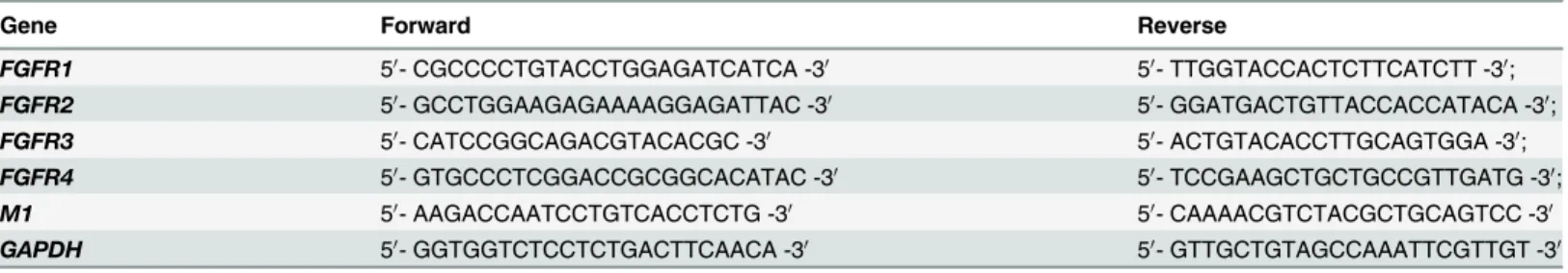
Incoming vRNPs, containing nucleoprotein (NP)-encapsidated viral genomic RNAs with asso-ciated viral polymerase proteins (PA, PB1, and PB2), are mostly located in the nuclei at 4 hpi; after 7 hours, newly synthesized vRNPs are predominantly in the cytoplasm [22]. To investi-gate the effect of siRNA-mediated FGFR1 silencing on early stage of influenza virus infection, incoming vRNPs were detected at 4 h after PR8 virus infection using indirect immunofluores-cence assays with anti-influenza A virus NP antibodies (Fig 3A–3C) [22,23]. The results showed that the incoming vRNPs were mainly located in the nuclei of A549 cells at 4 hpi, while they could hardly be detected in the cytoplasm. The percentage of NP-positive cells to the total number of cells was then calculated (Fig 3D). Cells exposed to FGFR1 siRNA interference showed the highest percentage of NP-positive cells compared with FGFR4-repressed cells or the negative control. These results indicated that FGFR1 silencing significantly increased PR8 entry efficiency at an early stage of the viral life cycle.
Fig 3. Specific siRNA target FGFR1 markedly increased PR8 virus entry at an early stage of the viral life cycle.(A-C) A549 cells were transfected with FGFR1 siRNA#1, FGFR4 siRNA#1, and negative control siRNA. After 48 h of transduction, A549 cells were incubated with PR8 virus at an MOI of 0.01 for 4 h, followed by indirect immunofluorescence assays. A549 cells were stained with anti-influenza A virus NP antibodies (green) and Hoechst 33342 (nucleus, blue). (D) The data of PR8-infected cells were presented as the percentages of NP-positive cells to the total number of cells. The bars represent the
means±s.e.m. (n = 3). P<0.001***were considered statistically highly significant.
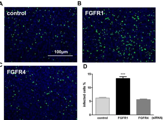
Influenza A virus replication could be reduced by lentivirus-mediated
FGFR1 overexpression
To confirm the effect of FGFR1 on influenza A virus infection, we explored whether overex-pression of FGFR1 in A549 cells downregulated influenza A virus replication. Thus, recombi-nant lentivirus vectors for efficient delivery of exogenous FGFR1, FGFR4, or GFP to A549 cells were constructed. A549 cells with lentivirus-mediated FGFR1 or FGFR4 overexpression were infected with PR8 or H5N1 virus. The virus M1 mRNA levels and viral titers were then de-tected as described previously. As expected, FGFR1 significantly decreased virus M1 mRNA levels in PR8-infected A549 cells, but not FGFR4 or GFP (Fig 4A). Viral titers were also re-duced in FGFR1-overexpressing cells, but not in the other two groups (Fig 4C). Similar results were observed in H5N1-infected A549 cells (Fig4Band4D). There was more than a 2.5-fold
Fig 4. Lentivirus-mediated FGFR1 overexpression in A549 cells significantly decreased influenza A virus replication.A549 cells were infected with recombinant lentivirus expressing FGFR1, FGFR4, or GFP (as a control). After 48 h, A549 cells were infected with PR8 virus (MOI = 1). The supernatants and lysates of A549 cells were harvested after 24 h virus infection. (A,B) Influenza M1 mRNA levels in A549 cells with PR8 and H5N1 were detected using real-time PCR. (C,D) Progeny virus titers of PR8 and H5N1 were determined as described previously. (E,F) FGFR1 and FGFR4 expression efficiencies were detected using real-time PCR and Western blotting. All graphs present the means±s.e.m. (n = 3). Values of P<0.05*were considered statistically
significant and P<0.001***statistically highly significant.
increase in mRNA expression of FGFR1 or FGFR4 in A549 cells (Fig 4E). The effective exoge-nous expression of FGFR1 or FGFR4 was also confirmed by Western blotting (Fig 4F). These results indicated that FGFR1 as a cellular antiviral factor could inhibit influenza A virus infec-tion; meanwhile, influenza A virus may evade the inhibitory effect by downregulating FGFR1 protein levels.
Treatment of PD173074, a FGFR kinase activity inhibitor, enhanced
influenza A/PR8 virus replication
FGFR1 is a receptor tyrosine kinase. Tyrosine autophosphorylation of FGFR1 can be induced by extracellular stimuli [24]. Our previous results suggested that influenza A (H1N1) virus in-fection downregulated FGFR1 expression (Fig1Aand1E), but whether influenza virus infec-tion stimulated FGFR1 phosphorylainfec-tion remains unknown. FGFR1 could inhibit influenza virus replication, but whether FGFR1 kinase activation had an effect on virus replication has not yet been explored. Next, the effect of influenza infection in A549 cells on FGFR1 phosphor-ylation for the indicated times was investigated (Fig 5A). The results showed that FGFR1 phos-phorylation was obviously reduced by PR8 infection over time, which suggested that influenza A (H1N1) virus infection downregulated FGFR1 phosphorylation in A549 cells. To address the second question, PD173074 as a potent and highly selective inhibitor of the FGFR family that can inhibit autophosphorylation of FGFR1 in a dose-dependent manner with an IC50 in the range 1–5 nM [25] was used to verify the effect of FGFR1 kinase activity on virus replication. A549 cells were pretreated with PD173074 and then infected with PR8 virus. The results showed that FGFR1 phosphorylation in PR8-infected A549 cells was significantly suppressed by PD173074 in a dose-dependent manner (Fig 5B). There was little difference in M1 expres-sion in A549 cells with 5 nM of PD173074 treatment; however, when the dose of PD173074 in-creased to 10 and 20 nM, virus M1 expression in A549 cells remarkably inin-creased (Fig 5C). For cytotoxicity analysis, the viability of A549 cells with varying amounts of PD173074 with or without PR8 infection was measured using the MTT assay. The results showed that the range 5–20 nM of PD173074 did not induce significant cytotoxicity (Fig 5D). These results suggested that the repression of FGFR1 phosphorylation in A549 cells with PD173074 treatment could enhance influenza virus replication.
FGFR1 overexpression inhibited influenza virus internalization, but not
binding, during viral entry
Discussion
In this study, we found that FGFR1 silencing by RNAi remarkably increased IAV replication, while lentivirus-mediated FGFR1 overexpression effectively reduced IAV replication. Differ-ences caused by FGFR1 silencing or overexpression were almost two-fold, which indicated that FGFR1 played a suppressive role in influenza A virus (IAV) PR8 and H5N1 replication. It is known that influenza virus attaches to the cell surface by binding of the HA protein, and then enters the cell through receptor-mediated endocytosis. Several studies have shown that influen-za virus HA binding to sialic acid receptor is not sufficient to initiate virus entry. Host factors including dynamin, Epsin-1, EGFR, and PLC-γ1 etc. are known to be involved in this process Fig 5. Repression of FGFR1 phosphorylation by the PD173074 inhibitor enhanced influenza A/PR8virus replication.(A) The lysates of A549 cells with PR8 virus infection were harvested for the indicated times. FGFR1 phosphorylation levels were determined by Western blotting with phospho-specific antibodies. Densitometric analysis of the phosphorylated/total FGFR1 ratio was shown as fold change. (B-D) A549 cells were pretreated with PD173074 (FGFR1 kinase inhibitor) at indicated concentrations for 30 min, and then infected with PR8 (indicated-dose inhibitor was also added to virus diluent). After 1 h, the medium was replaced with fresh medium supplemented with PD173074. (B) The cell lysates were collected at 12 hpi and then subjected to Western blotting for FGFR1 phosphorylation detection. The phosphorylated/total FGFR1 ratio indicated the fold change of FGFR1 phosphorylation. (C) Influenza M1 mRNA in PR8 infected-A549 cells with PD173074 treatment as indicated was detected by real-time PCR. (D) MTT assay of A549 cells with PD173074 treatment as indicated with or without PR8 infection. The results were expressed as means±s.e.m. (n = 3). Values of P<0.05*were considered statistically significant and P<0.001***considered statistically highly significant.
and facilitate viral internalization [8,9,26,27]. Our results suggested that FGFR1 knockdown notably elevated virus entry efficiency during the early stages of IAV infection; furthermore, FGFR1 could inhibit virus internalization, which indicated that host cells may have evolved a specific strategy of utilizing plasma membrane as a barrier to block influenza virus entry.
Four FGFR members have distinct spatial patterns of distribution in normal human adult tissues [28]. Our results showed that abundant FGFR1 and FGFR4 mRNA transcripts and pro-tein expressions were observed in A549 cells, while FGFR2 and FGFR3 with lower mRNA lev-els could not be detected at the protein level, which was indicative of the differential roles of FGFR family members in A549 cells. A previous study using genome-wide siRNA screening as-says showed that FGFR4 knockdown reduced influenza A/WSN pseudotyped particle entry in A549 cells, and also that the FGFR4 inhibitor attenuated WSN virus replication [10]. In our study, FGFR4 knockdown had no significant effect on inhibiting PR8 and H5N1 virus replica-tion; in contrast, we demonstrated that FGFR1 not FGFR4 remarkably suppressed PR8 and H5N1 virus replication. Since FGFR2 and FGFR3 were not further investigated in our study be-cause of their undetectable protein expression in A549 cells, it remains possible that they play a role in influenza virus replication.
Fig 6. Lentivirus-delivered FGFR1 overexpression affected influenza A virus internalization, but not binding, during virus entry.A549 cells with FGFR1, FGFR4, or GFP overexpression were pretreated with or without sialidase, and then infected with influenza A/PR8 (A) or H5N1 (B). The procedures were described in detail in the Methods, virus binding and internalization assay. Influenza virus NP was detected by Western blotting using anti-influenza NP antibodies.β-actin protein was used as an internal control. The ratio of NP/β -actin was determined based on densitometric analysis. The results were expressed as means±s.e.m.
(n = 3). Values of P<0.05*were considered statistically significant and P<0.01**highly significant.
FGFR signaling pathways play critical roles in many aspects of biological processes. In this study, we found that both FGFR1 protein expression and kinase phosphorylation levels were downregulated by influenza A/PR8 infection, which may be related to pathogenesis of influen-za virus-induced disease. FGFR contains a highly conserved region between the first and sec-ond IgG-like domains, which exhibit extreme sequence similarity to the amino termini of the HA1 chains of influenza A virus HA [29]. Deletion of this HA-containing region completely abolished FGFR function of responding to basic FGF (FGFR ligand), which suggested that the HA-containing region plays a crucial role in regulating ligand binding or receptor oligomeriza-tion. In addition, FGFR dimerization is required for receptor tyrosine kinase activation [24, 30]; thus, an interaction may exist between influenza virions and the HA-containing region of FGFR1, by which receptor dimerization was blocked and kinase activity was inhibited.
Receptor tyrosine kinase signaling is known to play important roles in influenza A virus (IAV) replication [22]. The inhibition of tyrosine kinase activity with broad-range tyrosine ki-nase inhibitor genistein results in impaired IAV uptake into cells; EGFR kiki-nase activation is re-quired for efficient viral internalization. Besides, two other inhibitors, AG879 (known to inhibit the nerve growth factor receptor and epidermal growth factor receptor) and tyrphostin A9 (a selective inhibitor of the platelet-derived growth factor receptor) block multiple steps of IAV replication [9]. In our study, the inhibition of FGFR1 phosphorylation with a selective in-hibitor PD173074 enhanced IAV replication.
Our results indicated that FGFR1 suppressed influenza A virus replication, probably by in-hibiting viral internalization, but it remains unclear whether FGFR1 was implicated in other steps of the IAV life cycle. The mechanism of FGFR1-induced inhibition of virus internaliza-tion has not been fully explored. In this study, a funcinternaliza-tional role of FGFR1 on PR8 and H5N1 infection was found in A549 cells, but whether FGFR1 has a universal effect in other human cell lines requires further study.
Author Contributions
Conceived and designed the experiments: XL XW. Performed the experiments: XL CL KW. Analyzed the data: XL LX PY QD. Contributed reagents/materials/analysis tools: CL KW. Wrote the paper: XL XW.
References
1. Matlin KS, Reggio H, Helenius A and Simons K (1981) Infectious entry pathway of influenza virus in a canine kidney cell line. J Cell Biol 91: 601–613. PMID:7328111
2. Sun X and Whittaker GR (2013) Entry of influenza virus. Adv Exp Med Biol 790: 72–82. doi:10.1007/
978-1-4614-7651-1_4PMID:23884586
3. Patterson S, Oxford JS and Dourmashkin RR (1979) Studies on the mechanism of influenza virus entry into cells. J Gen Virol 43: 223–229. PMID:479843
4. Yoshimura A, Kuroda K, Kawasaki K, Yamashina S, Maeda T and Ohnishi S (1982) Infectious cell entry mechanism of influenza virus. J Virol 43: 284–293. PMID:7109028
5. Lakadamyali M, Rust MJ and Zhuang X (2004) Endocytosis of influenza viruses. Microbes Infect 6: 929–936. PMID:15310470
6. Rust MJ, Lakadamyali M, Zhang F and Zhuang X (2004) Assembly of endocytic machinery around indi-vidual influenza viruses during viral entry. Nat Struct Mol Biol 11: 567–573. PMID:15122347
7. Chu VC and Whittaker GR (2004) Influenza virus entry and infection require host cell N-linked glycopro-tein. Proc Natl Acad Sci U S A 101: 18153–18158. PMID:15601777
8. Chen C and Zhuang X (2008) Epsin 1 is a cargo-specific adaptor for the clathrin-mediated endocytosis of the influenza virus. Proc Natl Acad Sci U S A 105: 11790–11795. doi:10.1073/pnas.0803711105
9. Eierhoff T, Hrincius ER, Rescher U, Ludwig S and Ehrhardt C (2010) The epidermal growth factor re-ceptor (EGFR) promotes uptake of influenza A viruses (IAV) into host cells. PLoS Pathog 6: e1001099. doi:10.1371/journal.ppat.1001099PMID:20844577
10. König R, Stertz S, Zhou Y, Inoue A, Hoffmann H-H, Bhattacharyya S, et al. (2010) Human host factors required for influenza virus replication. Nature 463: 813–817. doi:10.1038/nature08699PMID:
20027183
11. Brass AL, Huang IC, Benita Y, John SP, Krishnan MN, Feeley EM, et al. (2009) The IFITM proteins me-diate cellular resistance to influenza A H1N1 virus, West Nile virus, and dengue virus. Cell 139: 1243–1254. doi:10.1016/j.cell.2009.12.017PMID:20064371
12. Feeley EM, Sims JS, John SP, Chin CR, Pertel T, Chen LM, et al. (2011) IFITM3 inhibits influenza A virus infection by preventing cytosolic entry. PLoS Pathog 7: e1002337. doi:10.1371/journal.ppat. 1002337PMID:22046135
13. Mima T, Ueno H, Fischman DA, Williams LT and Mikawa T (1995) Fibroblast growth factor receptor is required for in vivo cardiac myocyte proliferation at early embryonic stages of heart development. Proc Natl Acad Sci U S A 92: 467–471. PMID:7831312
14. Presta M, Dell'Era P, Mitola S, Moroni E, Ronca R and Rusnati M (2005) Fibroblast growth factor/fibro-blast growth factor receptor system in angiogenesis. Cytokine Growth Factor Rev 16: 159–178. PMID:
15863032
15. Turner N and Grose R (2010) Fibroblast growth factor signalling: from development to cancer. Nat Rev Cancer 10: 116–129. doi:10.1038/nrc2780PMID:20094046
16. Qing K, Mah C, Hansen J, Zhou S, Dwarki V and Srivastava A (1999) Human fibroblast growth factor re-ceptor 1 is a co-rere-ceptor for infection by adeno-associated virus 2. Nat Med 5: 71–77. PMID:9883842 17. Givol D and Yayon A (1992) Complexity of FGF receptors: genetic basis for structural diversity and
functional specificity. The FASEB Journal 6: 3362–3369. PMID:1464370
18. Reed LJ and Muench H (1938) A simple method of estimating fifty per cent endpoints. American journal of epidemiology 27: 493–497.
19. Liu X, Yang N, Tang J, Liu S, Luo D, Duan Q, et al. (2014) Downregulation of angiotensin-converting enzyme 2 by the neuraminidase protein of influenza A (H1N1) virus. Virus Res 185: 64–71. doi:10.
1016/j.virusres.2014.03.010PMID:24662240
20. Yang N, Ma P, Lang J, Zhang Y, Deng J, Ju X, et al. (2012) Phosphatidylinositol 4-kinase IIIbeta is re-quired for severe acute respiratory syndrome coronavirus spike-mediated cell entry. J Biol Chem 287: 8457–8467. doi:10.1074/jbc.M111.312561PMID:22253445
21. Yang N, Hong X, Yang P, Ju X, Wang Y, Tang J, et al. (2011) The 2009 pandemic A/Wenshan/01/2009 H1N1 induces apoptotic cell death in human airway epithelial cells. J Mol Cell Biol 3: 221–229. doi:10.
1093/jmcb/mjr017PMID:21816972
22. Kumar N, Liang Y and Parslow TG (2011) Receptor tyrosine kinase inhibitors block multiple steps of in-fluenza a virus replication. J Virol 85: 2818–2827. doi:10.1128/JVI.01969-10PMID:21209112 23. Pleschka S, Wolff T, Ehrhardt C, Hobom G, Planz O, Rapp UR, et al. (2001) Influenza virus propagation
is impaired by inhibition of the Raf/MEK/ERK signalling cascade. Nat Cell Biol 3: 301–305. PMID:
11231581
24. Eswarakumar VP, Lax I and Schlessinger J (2005) Cellular signaling by fibroblast growth factor recep-tors. Cytokine Growth Factor Rev 16: 139–149. PMID:15863030
25. Mohammadi M, Froum S, Hamby JM, Schroeder MC, Panek RL, Lu GH, et al. (1998) Crystal structure of an angiogenesis inhibitor bound to the FGF receptor tyrosine kinase domain. EMBO J 17: 5896–5904. PMID:9774334
26. Zhu L, Ly H and Liang Y (2014) PLC-γ1 Signaling Plays a Subtype-Specific Role in Postbinding Cell Entry of Influenza A Virus. Journal of virology 88: 417–424. doi:10.1128/JVI.02591-13PMID:
24155396
27. Roy AM, Parker JS, Parrish CR and Whittaker GR (2000) Early stages of influenza virus entry into Mv-1 lung cells: involvement of dynamin. Virology 267: 17–28. PMID:10648179
28. Hughes SE (1997) Differential expression of the fibroblast growth factor receptor (FGFR) multigene family in normal human adult tissues. Journal of Histochemistry & Cytochemistry 45: 1005–1019. 29. Byers S, Amaya E, Munro S and Blaschuk O (1992) Fibroblast growth factor receptors contain a
con-served HAV region common to cadherins and influenza strain A hemagglutinins: a role in protein-protein interactions? Developmental biology 152: 411–414. PMID:1322849