HLA Typing for the Next Generation
Neema P. Mayor1,2, James Robinson1,2, Alasdair J. M. McWhinnie1, Swati Ranade3, Kevin Eng3, William Midwinter1, Will P. Bultitude1, Chen-Shan Chin3, Brett Bowman3, Patrick Marks3, Henny Braund1, J. Alejandro Madrigal1,2, Katy Latham1, Steven G. E. Marsh1,2*
1Anthony Nolan Research Institute, Royal Free Hospital, London, United Kingdom,2UCL Cancer Institute, Royal Free Campus, London, United Kingdom,3Pacific Biosciences, Menlo Park, California, United States of America
Abstract
Allele-level resolution data at primary HLA typing is the ideal for most histocompatibility test-ing laboratories. Many high-throughput molecular HLA typtest-ing approaches are unable to de-termine the phase of observed DNA sequence polymorphisms, leading to ambiguous results. The use of higher resolution methods is often restricted due to cost and time limita-tions. Here we report on the feasibility of using Pacific Biosciences’Single Molecule Real-Time (SMRT) DNA sequencing technology for high-resolution and high-throughput HLA typing. Seven DNA samples were typed for HLA-A, -B and -C. The results showed that SMRT DNA sequencing technology was able to generate sequences that spanned entire HLA Class I genes that allowed for accurate allele calling. Eight novel genomic HLA class I sequences were identified, four were novel alleles, three were confirmed as genomic se-quence extensions and one corrected an existing genomic reference sese-quence. This meth-od has the potential to revolutionize the field of HLA typing. The clinical impact of achieving this level of resolution HLA typing data is likely to considerable, particularly in applications such as organ and blood stem cell transplantation where matching donors and recipients for their HLA is of utmost importance.
Introduction
The HLA genes are located within one of the most gene rich regions of the human genome, the Major Histocompatibility Complex (MHC), on the short arm of chromosome 6 (6p21.3). Many of these genes, including HLA, encode proteins that have a critical role in immune re-sponses[1,2]. The MHC is divided into three distinct regions referred to as class I, II and III, with the HLA genes being located within the class I and class II regions. The HLA genes are known to be the most polymorphic genes of the human genome[1,3]. This polymorphism is predominantly found within the six classical HLA genes: the class I genes HLA-A,-B and-C and the class II genes HLA-DRB1,-DQB1 and -DPB1. Over 12,200 HLA alleles have been iden-tified to date (December 2014), with in excess of 9,200 being variants of the HLA class I genes alone [www.ebi.ac.uk/imgt/hla] [4,5].
OPEN ACCESS
Citation:Mayor NP, Robinson J, McWhinnie AJM, Ranade S, Eng K, Midwinter W, et al. (2015) HLA Typing for the Next Generation. PLoS ONE 10(5): e0127153. doi:10.1371/journal.pone.0127153
Received:January 7, 2015
Accepted:April 12, 2015
Published:May 27, 2015
Copyright:© 2015 Mayor et al. This is an open access article distributed under the terms of the
Creative Commons Attribution License, which permits unrestricted use, distribution, and reproduction in any medium, provided the original author and source are credited.
Data Availability Statement:All relevant data are within the paper and supporting information files. In addition, the genomic sequences described have been submitted to both the EMBL and IMGT/HLA Databases. Accession numbers for all sequences are provided in the manuscript.
Funding:Pacific Biosciences provided support in the form of salaries for authors SR, KE, C-SC, BB and PM. The authors affiliated with Pacific Biosciences were involved in the study design, data collection and analysis, and have reviewed, commented on and approved the content of the manuscript. The authors have reviewed the authors' roles in the online form and confirm they are correct.
HLA proteins function as antigen presentation molecules presenting self and non-self pep-tides to T-cells, a fundamental step in the initiation of certain adaptive immune responses. Much of the described polymorphism within the HLA class I genes is located within the exons that encode the peptide binding groove and the points at which T-cells interact with the mole-cule itself. This diversity has evolved as a mechanism to ensure on-going pathogen recognition and eradication by increasing the repertoire of peptide motifs that can be bound and presented to T cells [6,7]. Over-dominant selection is also thought to have driven the extent of polymor-phism. HLA heterozygosity is favoured in a population because it increases the number of pep-tide motifs that can be presented by the co-dominantly expressed HLA molecules[7]. This strong heterozygote advantage is of particular importance in the event of infection by a patho-gen that is specifically able to evade presentation by a particular HLA allele by ensuring that an individual is capable of initiating immune responses against the pathogen by presentation of the peptide by the second allele.
As the majority of described polymorphisms are located within the peptide binding groove that is encoded by exons 2 and 3 of the HLA class I genes, and that these differences have such an important functional relevance, many of the routinely used high-throughput HLA typing methods are focused on identifying variation within this limited region. A common problem encountered is the inability to determine the phase of polymorphisms identified in a single in-dividual, a problem that is exacerbated by the extensive genetic diversity seen in HLA genes [8, 9]. The result of this is ambiguous HLA types and the reporting of HLA typing strings. The high workload, cost and time required to generate true allele-level HLA typing using current methods makes it preclusive for most histocompatibility laboratories.
The recent development of second-generation sequencing methods has been of great inter-est to the HLA typing community due to the possibility of sequencing a single DNA strand in isolation. These techniques provide an opportunity for single allele definition at primary HLA typing as opposed to cross-referencing results from different molecular techniques and sero-logical testing. Previously, sequencing an entire HLA gene in isolation was achieved through the use of PCR-cloning processes, which are lengthy and often problematic. Second-generation sequencing methods have the potential to negate the use of such challenging laboratory prac-tices. These technologies offered the first realistic solution to the problem of phasing polymor-phisms throughout the HLA gene, enabling definitive allele typing. Consequently, many second-generation technologies have now been optimised for use by the HLA typing market and use of Sequence-Based Typing (SBT) protocols are common [10–14]. A current limitation of these methods are the read lengths that can be generated, resulting in the need for multiple over-lapping sequences to achieve full gene and even partial gene sequencing. A common con-cern with these methodologies is that incorrectly aligned fragments could result in HLA typing errors. It is possible that in a system as polymorphic as the HLA genes, incorrect phasing of SNPs that are distant to each other across the gene but otherwise show complete homology could result in an incorrect allele being assigned. Additionally rare or novel allele formed by a recombination event may be missed if the consensus sequence analysis tools are biased towards the more common alleles.
The ideal solution to resolve both HLA ambiguity and the potential problems caused by phasing multiple fragments would be to produce multiple long sequence reads encompassing whole gene PCR amplicons, in isolation. The development of Pacific Biosciences’Single Mole-cule Real Time (SMRT) DNA sequencing technology offers the first realistic option to achieve this goal [15]. The SMRT sequencing method is able to generate exceptionally long read lengths that would allow coverage of the 3 kb or more of a HLA class I gene sequence, and thus deter-mine the phase of the resolving polymorphisms seen. In addition, the technology has the po-tential to sequence read length in excess of 20 kb that could allow for entire HLA class II gene declare. SR, KE, C-SC, and BB are or were
sequencing, which at over 10 kb for some genes, are substantially longer in length than the HLA class I genes.
SMRT DNA sequencing makes use of SMRTbell templates, single stranded hairpin adaptors that can be ligated on to the ends of PCR products. The function of these adaptors is to turn an essentially linear PCR amplicon into a circular molecule. The advantage of generating a circular molecule is that the enzyme added to facilitate the reaction is capable of processively generating sequence from both strands of the PCR amplicon until either the enzyme expires or the end of the run-time is achieved. Under optimal experimental conditions, the result of the continuous sequencing process is the generation of a Continuous Long Read (CLR); one exceptionally long read which contains multiple regions of sequence specific to the PCR amplicon (known as sub-reads) interspersed with the sequence of the SMRTbell adaptors (Fig 1). This novel method of generating DNA sequence means that it is possible to interrogate the same DNA strand multi-ple times within a single experiment, achieving exceptionally high depth of sequence coverage. Here we describe the results of a study to determine whether the SMRT DNA sequencing methodology could be adapted for use in the Anthony Nolan Histocompatibility Laboratory to facilitate stem cell donor registry typing. The aims of this study were fourfold; i) to determine whether the methods were suitable for adaptation with Anthony Nolan DNA samples and PCR amplicons; ii) to determine if basic levels of multiplexing were possible; iii) to see if geno-mic HLA class I sequences could be generated; and iv) to determine the accuracy and specifici-ty of the sequences generated.
Material and Methods
Seven DNA samples were selected for HLA class I genotyping using Pacific Biosciences’SMRT sequencing methodology. In recent years, Anthony Nolan has changed from blood to Oragene saliva (DNA Genotek, Ottawa, Canada) as their primary source of DNA when recruiting do-nors to our stem cell donor register and thus DNA from this source makes up a large part of our workload. In accordance with our in-house protocol, blood samples are still requested from donors who are short-listed as potential matches for confirmatory HLA typing, virology and other associated tests. To ensure that DNA from both starting materials were suitable for use with SMRT sequencing methods, two samples were selected from each category. Written consent for HLA typing was obtained from all blood and saliva sample donors at the point of collection for the purposes of transplantation. These consent forms are in accordance with the Human Tissue Authority (HTA) UK, European Federation for Immunogenetics (EFI), and Clinical Pathology Accreditation (CPA) regulatory body guidelines. Anthony Nolan’s Medical Advisory committee has also reviewed and approved the consent form. Specific approval from the local ethics committee was not sought, as the purpose of the study is to assess the method of HLA typing in comparison with existing HLA typing techniques. No new genetic informa-tion outside of the HLA genes that would affect the donors of the material used in this study has been gained.
Three B-Lymphoblastoid Cell Lines (B-LCLs) were also selected from a well-characterized panel that have been extensively analysed for their HLA genes. HLA class I genotyping was un-dertaken for all donor-derived DNA samples using Luminex LABType SSO typing kits (One Lambda, CA, USA). HLA genotype information for the B-LCLs was obtained from the IMGT/ HLA Database website [www.ebi.ac.uk/ipd/imgt/hla/][8].
where possible; c) alleles that have known indels; and d) one DNA sample that is well charac-terized and known to be homozygous for HLA-A,-B and-C. The homozygous cell line chosen was the B-LCL COX (sample AN5) and was selected for HLA typing using the SMRT sequenc-ing method as it is known to be consanguineous and has previously undergone in-depth se-quence analysis of the entire Major Histocompatibility Complex (MHC) region on
chromosome 6, which includes the HLA gene family [16,17]. This previous in-depth analysis ensured full-length HLA class I gene sequences were available for the constituent alleles. In ad-dition, the HLA haplotype observed in this cell line has remained evolutionarily well-conserved and is one of the most frequently observed haplotypes in our tested population [18].
HLA class I amplicons were generated using primers as described for the 13th International Histocompatibility workshop [19]. This protocol enables amplification of the entire HLA Class I gene from 5´ to 3´ UTR. Fragment sizes were estimated to be 3500 bp, 3400 bp and 3450 bp for HLA-A,-B and-C respectively. The amplification method used TaKaRa LA DNA polymer-ase (Takara Bio Europe SAS, Saint-Germain-en-Laye, France). Agarose gel electrophoresis was used to confirm amplification and correct fragment size, as well as to check for non-specific product contamination. A 10 KB sizing marker was included to confirm size specificity (HyperLadder I; Bioline Reagents LTD, London, UK).
The HLA class I amplicons were sequenced according to Pacific Biosciences’standard pro-tocol for PCR amplicons greater than 3 KB in length. Briefly, HLA class I amplicons underwent quality and quantity confirmation using a Bioanalyser instrument and the Agilent DNA 12000 kit (Agilent Technologies, Santa Clara, CA, USA). As we aimed to test basic multiplexing capa-bilities of the SMRT DNA sequencing system, a pool of each of the HLA class I amplicons for a single sample were pooled at equimolar concentrations. After performing DNA damage and Fig 1. Basic stages of the Single Molecule Real-Time (SMRT) DNA sequencing method.SMRTbell adaptors are ligated onto the ends of a blunt-ended PCR amplicon to facilitate continuous sequencing of both strands of the amplicon. The entire sequence generated may include multiple copies of the sense and anti-sense strands of the PCR amplicon in a single read known as the Continuous Long Read (CLR). The post-sequencing bioinformatic post-processes are able to break down the CLR into shorter sub-reads, which encompass the sequence of one strand of the amplicon. These sub-reads can then be compared and used to create a consensus sequence.
end repair, the SMRTbell adaptors were blunt-end ligated onto the PCR amplicons in the pool. Following the ligation of the adaptors, an adaptor-specific sequencing primer and enzyme were bound to the templates. For this study, sequencing was enabled by the use of the P4 enzyme and C2 chemistry. Finally, the SMRTbell templates were loaded on to MagBeads, magnetic beads that facilitate even sample loading into the SMRT Cell. DNA samples were sequenced on the PacBio RS II SMRT DNA Sequencing System with a movie capture time of 120 minutes. All stages of the sequencing process, including library preparation, SMRT Cell loading and data collection were achieved within three working days.
The DNA sequences derived from Pacific Biosciences’SMRT sequencing technologies un-derwent post-processing using the SMRT analysis tool v2.1, and were assigned HLA types using Anthony Nolan in-house Bioinformatics methods. The PacBio methodology provides a number of sequences for analysis for each sample. The optimal consensus sequences for each run were selected by Anthony Nolan and Pacific Biosciences’researchers and analysis was per-formed. HLA types were assigned based on identity to known sequences within the IMGT/ HLA Database. Where novel sequences were reported, assignment of a HLA type was based on aligning the novel consensus sequence at both the cDNA, gDNA and protein level, to identify the nearest known HLA allele.
Sanger sequencing was used to determine the accuracy of regions of DNA sequence ob-tained from SMRT sequencing that either differed to the existing genomic sequences for the ex-pected allele, or if no genomic sequence were available, differed to that seen in the closest matching allele. SBT was enabled using BigDye Terminator sequencing kit V3.1 (Applied Bio-systems, Foster City, California, USA) and utilised primers designed in-house. Fragments were sequenced on an ABI 3730XL Genetic Analyser (Applied Biosystems, Foster City, California, USA). As the majority of the tested samples were heterozygous for each of the HLA class I loci, generic PCR and SBT was not sufficient in some cases to enable confirmation of discrepancies. For these samples, cloning of full-length HLA gene PCR amplicons was used to allow separa-tion of the two alleles. HLA class I PCR products were cloned using the Zero Blunt TOPO clon-ing kit (Life Technologies, Paisley, UK) before targeted sequencclon-ing as previously described.
Results
Seven DNA samples were selected for SMRT DNA sequencing based on a set of defined inclu-sion criteria which included different starting material from which the DNA was extracted and the inclusion of as many commonly seen HLA class I alleles as was feasible. Each of the seven samples tested were able to generate sufficient quality sequence data to create a consensus se-quence for all of the alleles expected. Variation was seen in the number of sub-reads achieved for each allele due to allelic imbalance that occurs during PCR amplification that is not routine-ly detected with HLA typing strategies that do not allow sequencing of single gene sequences in isolation. Despite these imbalances, the minimum depth of coverage was still in excess of 150x (median 462.5; range 154–2931), that is there were in excess of 150 sub-reads of sufficient qual-ity for each allele once subjected to the qualqual-ity checks in the post-processing stage of SMRT data analysis, that could be used to generate a consensus sequence (Table 1). 100% of the total number of consensus sequences generated achieved a mean Quality Value (QV) of over 70 (mean QV 74.079, range 71.937–80).
HLA-A03:01:01:01 in samples AN1 and AN4) were considered as different consensus se-quences. Therefore, a total of 38 possible consensus sequences were expected.
Thirty of these 38 possible HLA consensus sequences immediately showed complete identi-ty with reference sequences available in the IMGT/HLA Database (Table 2). Both alleles for each of the three HLA class I loci were accurately called in three samples, AN1, AN5 and AN7. SMRT sequencing of sample AN5 correctly identified this sample to be homozygous for all three HLA class I loci tested. Unexpectedly, an additional HLA gene sequence was also Table 1. Depth of coverage achieved using SMRT sequencing.
ID HLA-A HLA-B HLA-C
Allele Number of sub-reads Allele Number of sub-reads Allele Number of sub-reads
AN1 A*03:01 303 B*07:02 841 C*05:01 569
A*11:01 385 B*44:02 780 C*07:02 726
AN2 A*25:01/02 353 B*15:01 817 C*03:03 514
A*68:01:02 184 B*18:01 583 C*12:03 422
AN3 A*26:01 238 B*14:01 1263 C*02:02 282
A*31:01:02 371 B*27:05:02 498 C*08:02 162
AN4 A*03:01 300 B*27:05:18 197 C*01:02 213
A*32:01 799 B*35:01 836 C*04:01 584
AN5 A*01:01 1477 B*08:01 2134 C*07:01 2931
AN6 A*02:01 516 B*52:01 247 C*07:01 278
B*73:01 427 C*15:05 156
AN7 A*23:01 349 B*42:01 327 C*06:02 1080
A*24:02 313 B*50:01 1390 C*17:01 1840
doi:10.1371/journal.pone.0127153.t001
Table 2. A comparison of expected HLA types, as typed by Anthony Nolan, with those generated by the Single Molecule Real-Time (SMRT) DNA Sequencing method.
ID DNA source Results group* HLA-A allele 1 HLA-A allele 2 HLA-B allele 1 HLA-B allele 2 HLA-C allele 1 HLA-C allele 2
AN1 Saliva AN *03 *11 *07 *44 *05 *07
SMRT *03:01:01:01 *11:01:01 *07:02:01 *44:02:01:01 *05:01:01:02 *07:02:01:03
AN2 Blood AN *25:01/02 *68 *15 *18 *03:03 *12:03
SMRT *25:01:01 *68:01:02:02 *15:01:01:01 *18:01:01:02 *03:03:01 *12:03:01:01
AN3 Saliva AN *26:01:01 *31:01:02 *14:01 *27:05/13 *02 *08
SMRT *26:01:01 *31:01:02 *14:01:01 *27:05:02 *02:02:02:02 *08:02:01:02
AN4 Blood AN *03 *32:01 *27:05:18 *35:01 *01 *04
SMRT *03:01:01:01 *32:01:01 *27:05:18 *35:01:01:02 *01:02:01 *04:01:01:01
AN5 B-LCL AN *01:01:01:01 *01:01:01:01 *08:01:01 *08:01:01 *07:01:01:01 *07:01:01:01
SMRT *01:01:01:01 *01:01:01:01 *08:01:01 *08:01:01 *07:01:01:01 *07:01:01:01
AN6 B-LCL AN *02:01 *02:01 *52:01:01 *73:01 *07:01 *15:05
SMRT *02:01:01:01 *02:01:01:01 *52:01:01:03 *73:01 *07:01:01:01 *15:05:01
AN7 B-LCL AN *23:01 *24:02:01:01 *50:01 *42:01 *06:02 *17:01
SMRT *23:01:01 *24:02:01:01 *50:01:01 *42:01:01 *06:02:01:02 *17:01:01:02
HLA alleles in bold highlight novel alleles, genomic sequence corrections or genomic sequence extensions.
*AN—Anthony Nolan typing data as generated by Luminex LABType SSO typing kits (One Lambda, CA, USA), Sequencing-based typing and/or PCR using Sequence specific primers (PCR-SSP).
SMRT—Single Molecule Real-Time DNA sequencing method from Pacific Biosciences
identified in this sample. Sequences corresponding to the HLA pseudogene allele
HLA-H02:01:01:01 were identified, although the numbers of reads were low (n = 12). Despite the suboptimal number of reads, the HLA-H sequences were correctly called for this sample. The reason for this non-specific product is due to the co-amplification of HLA-H in the reac-tions for another class I product, presumably HLA-A due to sequence similarities between the two genes. Samples AN6 and AN7 were included in this test cohort as they contain alleles that have notable non-coding deletions in their genomic DNA (gDNA) sequences (B73:01 and C17:01). SMRT sequencing methods were able to generate consensus sequences that accurate-ly identified these two alleles.
Four consensus sequences generated with SMRT DNA sequencing methods matched alleles that only had either partial gene or Coding DNA Sequences (CDS) available in the IMGT/HLA Database. As described previously, HLA types were assigned to the consensus sequences by comparison to CDS sequences available on all HLA alleles as well as with genomic sequences of closest related alleles (Table 3). Data for sample AN6 (HLA-C15:05:01 expected) was iden-tical to the reference genomic sequence used as a comparison (HLA-C15:05:02) except for the single nucleotide difference in exon 1 that differentiates the two alleles (gDNA 24T>C). Sam-ples AN3 (HLA-B14:01:01 expected) and AN4 (HLA-B27:05:18 expected) showed sequence variation in intron sequences in addition to those that define the differences between the ob-served allele and that to which it was being compared. Sanger sequencing was used to confirm allele identity (AN6) or to confirm the existence of novel non-coding variants (AN3 and AN4). All positions tested matched those generated with SMRT sequencing technology confirming the accuracy of the method. Data for the second HLA-B allele in sample AN3 initially sug-gested an intron 5 variant of HLA-B27:05:02 (gDNA 2086C>T). Sanger sequencing of the re-gion of interest confirmed the nucleotide substitution when compared to the existing genomic reference sequence. An analysis of all available intron 5 sequences for HLA-B27 alleles showed all other alleles had the variant base at the queried position (2086T), possibly suggesting that there was an error in the original sequence submitted to the IMGT/HLA Database. The original DNA source used to generate the HLA-B27:05:02 genomic sequence was identified and re-se-quenced. The data confirmed that there was an error in the original genomic sequence and that the consensus sequence generated by SMRT DNA sequencing was correct. These novel geno-mic HLA sequences have been submitted to the IMGT/HLA Database as extensions or correc-tions to existing alleles (Table 3).
Four of the 38 tested alleles showed novel genomic HLA sequences when compared to the expected sequences (Table 4); AN2 (HLA-A68 variant), AN6 (HLA-B52 variant) and AN3 (two HLA-C variants, C02 and C08). Sanger SBT of the regions of interest for each of the four alleles confirmed the variant bases, and thus the novel alleles identified using SMRT se-quencing. These novel genomic sequences have been submitted to the IMGT/HLA Database and have been officially named according to the WHO Nomenclature Committee for Factors of the HLA System (Table 4) [3].
When Sanger sequencing confirmations of novel genomic sequences were included, the final analysis showed absolute concordance between the consensus sequences generated with Pacific Biosciences’SMRT DNA sequencing method and the expected/observed alleles.
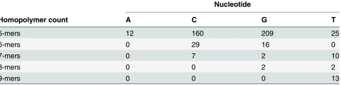
The total number of bases sequenced was 130117 bp within which 487 homopolymer regions were identified. The most frequently observed nucleotide repeat regions were 5-mers, which occurred multiple times for each of the four nucleotides (range 12–209 times). The longest ho-mopolymer region found in the tested alleles was a 9-mer; this occurred 13 times but only for the T nucleotide. Noticeably fewer homopolymer regions were observed for the A, C and G nu-cleotides, particularly in 7-, 8- and 9-mers. In all cases, SMRT DNA sequencing methodology accurately determined the correct number of nucleotides present in each allele. Additionally, 99.354% of the homopolymer regions had a mean QV of 70 or more across the homopolymer region (mean QV 74.084; range 64.286–80). Details on individual QV data for each of the 38 HLA sequences generated can be found in the supplementary information (S1 Table).
Discussion
Next generation sequencing technologies have offered the first feasible laboratory-based solu-tion to the problem of phasing the complex polymorphisms seen in the HLA gene family. Limi-tations in read length have meant that a shotgun approach has to be applied, with multiple fragments covering an entire region of interest being necessary [10,12,20–24]. The SMRT DNA sequencing method from Pacific Biosciences has overcome the need to sequence multiple overlapping fragments allowing sequencing of a single fragment in excess of 20 kb in one se-quencing reaction. The implications of this technology in the field of HLA typing could be enormous, allowing for true allelic HLA typing in a single experimental set-up and making re-dundant the need for multiple experiments on different typing platforms, cross-referencing of results and/or the need for re-sequencing using an allele specific protocol. We have described here the results of a feasibility study which shows that whole HLA class I gene sequencing is possible using the SMRT DNA sequencing platform. The sequence data generated was high quality and allowed for accurate allele calling. In addition, all stages of the experimental set-up were completed within three working days and sequence data were captured over 120 minutes. In combination, these factors make the SMRT DNA sequencing method amenable for use in a high-throughput HLA typing laboratory.
The primary aim in testing this methodology was to determine whether accurate genomic consensus sequences could be generated using our current in-house protocol for full gene HLA class I amplification and with our DNA samples using SMRT sequencing technology. Our Table 3. Extensions and corrections to known HLA alleles identified in PacBio results.
Sample Expected Allele
Allele used for genomic sequence comparisons
Novel/ differentiating variants
Sequence type
EMBL Accession numbers
AN3 B*14:01:01 B*14:02:01 Intron 2 G>T gDNA 665 Extension HG794368
AN3 B*27:05:02 B*27:05:02 Intron 5 C>T gDNA 2086 Correction HG794364
AN4 B*27:05:18 B*27:05:02 Exon 2 C>T gDNA 269; Extension HG530757
AN6 C*15:05:01 C*15:05:02 None Extension HG794367
doi:10.1371/journal.pone.0127153.t003
Table 4. Anomalies observed in the PacBio SMRT consensus sequences as compared to the expected allele.
Sample Expected Allele Variants Sequence Confirmed New allele name EMBL Accession number
AN2 A*68:01:02 Intron 7 G>A gDNA 2770 Confirmed,new variant A*68:01:02:02 HG794362
AN3 C*02:02:02 Intron 5 T>C gDNA 2487 Confirmed,new variant C*02:02:02:02 HG794365 AN3 C*08:02:01 Intron 3 A>G gDNA 1338 Confirmed new variant C*08:02:01:02 HG794366 AN6 B*52:01:01:02 5´ UTR C>A gDNA -180 Confirmed new variant B*52:01:01:03 HG794363
findings have confirmed that our blood and saliva specimens and subsequent DNA extraction procedures are suitable for the isolation of high molecular weight genomic DNA, an essential prerequisite for the PCR amplification of HLA whole gene amplicons. The PCR primers and amplification conditions led to specific amplification of the genes of interest, namely HLA-A,-B and-C. There was minimal co-amplification of HLA-H, most likely with HLA-A primers, but this did not have a detrimental effect on allele calling. Some allelic imbalance was observed in the data generated for HLA-B. The most likely explanations for this observation are either that SMRT DNA sequencing is a more sensitive methodology and is therefore more likely to identify imbalance in the PCR which is not seen in SBT, or that possible nucleotide differences between the primer and allele sequences caused inefficient or inhibited binding.
Differences between the numbers of reads seen for each locus of a single sample were also observed. A potential reason for these differences is that there was imbalance during the equi-molar pooling stages, with some loci being over or under represented. Additionally, the kit used to quantify the PCR amplicons prior to equimolar pooling is limited to quantifying sam-ples within a range of 0.5–50 ng/μl. As all amplicons in this experiment were of concentrations
towards the upper limits of the kit, it is possible that the sizing and quantification values were affected, which consequently affected the volumes required for equimolar pooling and causing the imbalance between loci. The use of single molecule sequencing methodologies is challeng-ing our previous perceptions of what constitutes‘good’or‘successful’PCR amplifications, with significantly lower quantities of amplicons required for most processes.
Despite imbalance issues, significant depth of coverage was achieved for all alleles that were sequenced and allowed for accurate HLA allele assignment. Future experiments where the ex-tent of multiplexing different DNA samples or HLA loci is tested should consider the affect of allelic imbalance on the depth of coverage achievable, although these issues should be easily rectified with additional amplification optimisation. Additionally, future experiments should either take final concentrations of samples and quantification kit limitations into consideration before proceeding with the sequencing experiment, or alternatively, PCR conditions altered to allow for lower quantities of amplicon to be generated.
The concentration of amplicons for all three class I loci was sufficient for pooling at equimo-lar levels prior to library preparation. The multiplexing of the three amplicons from a single sample in a designated SMRT cell allowed for 150x read depth for all alleles present with the re-sultant sequence reads being successfully aligned and assigned to the relevant HLA class I genes with analysis software. The amplicon lengths were similar enough to negate the potential problem of loading bias towards smaller PCR products in a pool when dispensed into SMRT Cells. The generated sequence exhibited complete coverage from the sites of the PCR primers. Depending on the loci and alleles present, this was inclusive of the terminal 300 bp in the 5 ´UTR, exons, introns and the leading 200 bp in the 3´UTR.
Table 5. Homopolymer count in 38 PacBio sequences (total length: 130117 bp).
Nucleotide
Homopolymer count A C G T
5-mers 12 160 209 25
6-mers 0 29 16 0
7-mers 0 7 2 10
8-mers 0 0 2 2
9-mers 0 0 0 13
The quality of the HLA class I genomic sequences generated can partly be confirmed by the high percentage of those reaching QV70, in some cases higher, but also by the accurate assign-ment of HLA types to these sequences. Of particular interest was the accuracy of the data pro-duced for the homopolymer regions present in the different alleles due to the known cross-platform problem of enzymes incurring slippage when sequencing through long stretches of a single continuous base. In all cases, SMRT DNA sequencing technology was able to call the cor-rect number of bases for each allele. The longest homopolymer region sequenced here was a 9-mer and although this was seen multiple times, only 9-mers of the T nucleotide were ob-served. Thus it remains to be seen whether the technology can adequately sequence through longer homopolymer regions and whether different bases introduce other problems.
The accuracy of the methodology for sequencing the tested samples was substantiated by the correct identification of novel HLA class I alleles, each of which was separately confirmed using Sanger-based sequencing methods. The high number of novel alleles found in this small test cohort (4/38 sequences; 10.5%) highlights the extensive polymorphism seen in the HLA genes outside of the routinely typed exons, much of which may as yet be unknown. As previ-ously stated, most histocompatibility laboratories would like to be able to generate allele-level resolution for all samples processed, but this is often unattainable due to financial, time and ex-perimental constraints. SMRT DNA sequencing technology could offer a resolution to these is-sues, providing sequences for ultra-high resolution HLA typing in a single sequencing reaction and being achievable in less time than it would take using current methodologies.
The down-stream uses of HLA typing data are varied and include assessing compatibility between donors and recipients prior to transplantation, drug hypersensitivity and disease asso-ciations. The potential impact of using SMRT DNA sequencing in the future to generate such high-resolution HLA typing on many of these areas of medicine are likely to be considerable. For example, high resolution HLA typing has been shown to significantly improve outcome when stem cell transplant recipients and their unrelated donors are matched for both alleles at five of the classical HLA loci (HLA-A,-B,-C,-DRB1 and-DQB1, a 10/10 match) [25–28], as it is thought that disparity at these important compatibility loci can contribute to complications such as graft-versus-host disease and consequently, to mortality. SMRT DNA sequencing has the potential to detect previously unidentified polymorphisms in regions of the HLA genes that could be significantly contributing to these complications. This could ultimately result in con-siderable improvement in survival rates post transplant.
HLA class I genes is possible and produces accurate typing results, suggesting that this technol-ogy is viable for use in a high-throughput clinical laboratory (unpublished data).
The number of DNA samples tested here were low although multiple genes were sequenced for each sample. It is important that larger and more diverse cohorts of DNA samples are se-quenced using SMRT DNA technology to confirm suitability for HLA typing. Future studies should also test the maximum multiplexing capabilities of the SMRT sequencing system, both with increased numbers of samples and the number of HLA loci included per SMRT Cell. It also remains to be seen whether accurate and high-quality HLA class II consensus sequences can be generated on this platform, which would be necessary for clinical use of this technology.
This method offers a realistic solution to the issues encountered in clinical HLA typing and has the potential to significantly improve clinical prognoses.
Supporting Information
S1 Table. Accession numbers and QV values of all HLA genomic sequences generated using SMRT DNA sequencing method and submitted to EMBL.
(DOCX)
Author Contributions
Conceived and designed the experiments: NPM JR AJMM SR KE C-SC HB JAM KL SGEM. Performed the experiments: NPM JR AJMM KE WM WPB C-SC BB PM. Analyzed the data: NPM JR AJMM SR KE WM WPB C-SC BB PM HB JAM KL SGEM. Contributed reagents/ma-terials/analysis tools: SR C-SC HB JAM KL SGEM. Wrote the paper: NPM JR AJMM SR KE WM WPB C-SC BB PM HB JAM KL SGEM.
References
1. Horton R, Wilming L, Rand V, Lovering RC, Bruford EA, Khodiyar VK, et al. Gene map of the extended human MHC. Nat Rev Genet. 2004; 5(12):889–99. PubMed PMID:15573121.
2. Consortium TMs. Complete sequence and gene map of a human major histocompatibility complex. The MHC sequencing consortium. Nature. 1999; 401(6756):921–3. PubMed PMID:10553908.
3. Marsh SGE, Albert ED, Bodmer WF, Bontrop RE, Dupont B, Erlich HA, et al. Nomenclature for factors of the HLA system, 2010. Tissue antigens. 2010; 75(4):291–455. Epub 2010/04/02. doi:10.1111/j. 1399-0039.2010.01466.xPubMed PMID:20356336; PubMed Central PMCID: PMC2848993.
4. Robinson J, Waller MJ, Parham P, Bodmer JG, Marsh SGE. IMGT/HLA Database—a sequence data-base for the human major histocompatibility complex. Nucl Acids Res. 2001; 29(1):210–3. doi:10. 1093/nar/29.1.210PMID:11125094
5. Robinson J, Halliwell JA, Hayhurst JD, Flicek P, Parham P, Marsh SG. The IPD and IMGT/HLA data-base: allele variant databases. Nucleic Acids Res. 2014. Epub 2014/11/22. doi:10.1093/nar/gku1161 PubMed PMID:25414341.
6. Parham P, Ohta T. Population biology of antigen presentation by MHC class I molecules. Science. 1996; 272(5258):67–74. PubMed PMID:8600539.
7. Hughes AL, Nei M. Pattern of nucleotide substitution at major histocompatibility complex class I loci re-veals overdominant selection. Nature. 1988; 335(6186):167–70. PubMed PMID:3412472.
8. Robinson J, Halliwell JA, McWilliam H, Lopez R, Parham P, Marsh SG. The IMGT/HLA database. Nu-cleic acids research. 2013; 41(Database issue):D1222–7. Epub 2012/10/20. doi:10.1093/nar/gks949 PubMed PMID:23080122; PubMed Central PMCID: PMC3531221.
9. Tu B, Cha N, Yang R, Ng J, Hurley CK. A one-step DNA sequencing strategy to HLA type hematopoiet-ic stem cell donors at recruitment—rethinking typing strategies. Tissue Antigens. 2013; 81(3):150–60. doi:10.1111/tan.12072PubMed PMID:23398508.
11. Wang C, Krishnakumar S, Wilhelmy J, Babrzadeh F, Stepanyan L, Su LF, et al. High-throughput, high-fidelity HLA genotyping with deep sequencing. Proc Natl Acad Sci U S A. 2012; 109(22):8676–81. doi: 10.1073/pnas.1206614109PubMed PMID:22589303; PubMed Central PMCID: PMCPMC3365218.
12. Shiina T, Suzuki S, Ozaki Y, Taira H, Kikkawa E, Shigenari A, et al. Super high resolution for single mol-ecule-sequence-based typing of classical HLA loci at the 8-digit level using next generation sequenc-ers. Tissue Antigens. 2012; 80(4):305–16. doi:10.1111/j.1399-0039.2012.01941.xPubMed PMID: 22861646.
13. De Santis D, Dinauer D, Duke J, Erlich HA, Holcomb CL, Lind C, et al. 16(th) IHIW: Review of HLA typ-ing by NGS. International journal of immunogenetics. 2013; 40(1):72–6. doi:10.1111/iji.12024PubMed PMID:23302098.
14. Rozemuller EH, Chadwick B, Charron D, Baxter-Lowe LA, Eliaou JF, Johnston-Dow L, et al. Seque-nase sequence profiles used for HLA-DPB1 sequencing-based typing. Tissue Antigens. 1996; 47 (1):72–9. Epub 1996/01/01. PubMed PMID:8929715.
15. Eid J, Fehr A, Gray J, Luong K, Lyle J, Otto G, et al. Real-time DNA sequencing from single polymerase molecules. Science. 2009; 323(5910):133–8. Epub 2008/11/22. doi:10.1126/science.1162986 PubMed PMID:19023044.
16. Traherne J, Horton R, Roberts A, Miretti M, Hurles M, Stewart C, et al. Genetic analysis of completely sequenced disease-associated MHC haplotypes identifies shuffling of segments in recent human histo-ry. PLoS genetics. 2006; 2(1):e9. PubMed PMID:16440057.
17. Stewart CA, Horton R, Allcock RJN, Ashurst JL, Atrazhev AM, Coggill P, et al. Complete MHC haplo-type sequencing for common disease gene mapping. Genome Res. 2004; 14(6):1176–87. doi:10. 1101/gr.2188104PubMed PMID:15140828.
18. Degli-Esposti M, Leaver A, Christiansen F, Witt C, Abraham L, Dawkins R. Ancestral haplotypes: con-served population MHC haplotypes. Human immunology. 1992; 34(4):242–52. PubMed PMID: 1464552.
19. Tilanus MGJ. 13th IHWS Technology Joint Report. In: Hansen J, editor. Immunobiology of the human MHC Proceedings of the 13th International Histocompatability Workshop and Conference. 12006. p. 304-.
20. Danzer M, Niklas N, Stabentheiner S, Hofer K, Pröll J, Stückler C, et al. Rapid, scalable and highly auto-mated HLA genotyping using next-generation sequencing: a transition from research to diagnostics. BMC genomics. 2013; 14(1):221. doi:10.1186/1471-2164-14-221PubMed PMID:23557197.
21. Moonsamy PV, Williams T, Bonella P, Holcomb CL, Höglund BN, Hillman G, et al. High throughput HLA genotyping using 454 sequencing and the Fluidigm Access Array system for simplified amplicon li-brary preparation. Tissue Antigens. 2013; 81(3):141–9. doi:10.1111/tan.12071PubMed PMID: 23398507.
22. Lind C, Ferriola D, Mackiewicz K, Heron S, Rogers M, Slavich L, et al. Next-generation sequencing: the solution for high-resolution, unambiguous human leukocyte antigen typing. Human immunology. 2010; 71(10):1033–42. doi:10.1016/j.humimm.2010.06.016PubMed PMID:20603174.
23. Lind C, Ferriola D, Mackiewicz K, Sasson A, Monos D. Filling the gaps—The generation of full genomic sequences for 15 common and well-documented HLA class I alleles using next-generation sequencing technology. Human immunology. 2012. doi:10.1016/j.humimm.2012.12.007PubMed PMID:
23246585.
24. Hosomichi K, Jinam TA, Mitsunaga S, Nakaoka H, Inoue I. Phase-defined complete sequencing of the HLA genes by next-generation sequencing. BMC genomics. 2013; 14:355. doi: 10.1186/1471-2164-14-355PubMed PMID:23714642; PubMed Central PMCID: PMCPMC3671147.
25. Lee S, Klein J, Haagenson M, Baxter-Lowe L, Confer D, Eapen M, et al. High-resolution donor-recipient HLA matching contributes to the success of unrelated donor marrow transplantation. Blood. 2007; 110 (13):4576–83. PubMed PMID:17785583.
26. Spellman SR, Eapen M, Logan BR, Mueller C, Rubinstein P, Setterholm MI, et al. A perspective on the selection of unrelated donors and cord blood units for transplantation. Blood. 2012; 120(2):259–65. doi: 10.1182/blood-2012-03-379032PubMed PMID:22596257.
27. Shaw BE, Mayor NP, Russell NH, Apperley JF, Clark RE, Cornish J, et al. Diverging effects of HLA-DPB1 matching status on outcome following unrelated donor transplantation depending on disease stage and the degree of matching for other HLA alleles. Leukemia: official journal of the Leukemia Soci-ety of America, Leukemia Research Fund, UK. 2010; 24(1):58–65. doi:10.1038/leu.2009.239PubMed PMID:19924143.