Received: 29 Aug 2012, Accepted: 2 Jun 2013
* Corresponding Address: P.O. Box: 16635-148, Department of Endocrinology and Female Infertility at Reproductive Biomedicine Research Center, Royan Institute for Reproductive Biomedicine, ACECR, Tehran, Iran
The Role of Toll Like Receptors in Pregnancy
Elham Amirchaghmaghi, M.D.1, Seyed Abdolvahab Taghavi, M.Sc.2, Farnaz Shapouri, M.Sc.3, Shaghayegh Saeidi, M.Sc.3, Abbas Rezaei, Ph.D.1, Reza Alatoonian, M.D., Ph.D.2,3*
1. Department of Immunology, Faculty of Medicine, Isfahan University of Medical Sciences, Isfahan, Iran 2. Department of Anatomy, School of Medicine, Iran University of Medical Sciences, Tehran, Iran 3. Department of Endocrinology and Female Infertility at Reproductive Biomedicine Research Center,
Royan Institute for Reproductive Biomedicine, ACECR, Tehran, Iran
Abstract
For many years, the innate immunity was of less interest than the adaptive immunity because it was perceived to have secondary importance in the functionality of the immune system. During the past decades, with the advancement of knowledge about innate immune system, interest in innate immunity has grown dramatically and thus its function has been extensively studied. Innate immunity plays fundamental roles in the initiation and induction of adaptive immune responses. It consists of several cells and receptors including natural killer (NK) cells, macrophages (MQs), den-dritic cells (DCs) and pattern recognition receptors (PRRs). Two decades ago, Toll like receptors (TLRs) family was known as one of the important PRRs with unique functions especially in protection against invading pathogens. Since the female re-productive tract has access to the outside environment and has a unique interaction with different pathogens whether invading microorganisms or normal flora, allo-genic sperm and semi alloallo-genic fetus, it has an essential need for effective immune responses. It has therefore been suggested that TLRs may play important roles in the immune regulation of the female reproductive tract. In addition, it has been demonstrated that immune disturbance may be responsible for some adverse preg-nancy outcomes such as preeclampsia (PE), recurrent spontaneous abortion (RSA) and intrauterine growth restriction (IUGR). Our focus in this review is to show the importance of TLRs in pregnancy with emphasis on the expression of these recep-tors in different tissues related to pregnancy.
Keywords: Innate Immunity, TLRs, Pregnancy, PRRs
Citation: Amirchaghmaghi E, Taghavi SA, Shapouri F, Saeidi Sh, Rezaei A, Alatoonian R. The role of Toll like
receptors in pregnancy. Int J Fertil Steril. 2013; 7(3): 147-154.
Royan Institute
International Journal of Fertility and Sterility
Introduction
All organisms are in a continuous challenge with the surrounding environment during their life. The defense mechanism of every organism which is called the immune system has two main branches in vertebrates. Innate (natural)
immu-nity, which is the irst branch of immune sys -tem, is the ancient form of host defense against infection and plays a critical role in activation of adaptive (acquired) immunity, the another branch of the immune system. The adaptive
im-mune system responds to speciic 'non-self '
anti-gens and generates immunologic memory. Innate immunity comprises different cells and receptors
which provides irst line of defense against in -vading microorganisms (1). Pathogens that in-vade a vertebrate host are initially recognized by the innate immune system via a limited number of germline-encoded receptors called pattern-recognition receptors (PRRs) (1, 2).
mo-lecular patterns (PAMPs). PAMPs are essential for the survival of the microorganism and therefore it could not alter them without threatening its life. 2. Their expression is constitutive in the host and thus could detect the pathogens during their life time. 3. PRRs are germline encoded and nonclonal which
are expressed on all cells of a speciic type (2, 3).
PRRs not only recognize exogenous components derived from both pathogenic and non pathogenic mi-croorganisms known as PAMPs, but also respond to endogenous molecules released from dying host cells upon cellular stress or tissue injury known as damage associated molecular patterns (DAMPs) (4, 5).
PRRs exist in every compartment of the body. Some of them are humoral proteins circulating in the plasma while endocytic receptors expressed on the cell surface and signaling receptors can be expressed either on the cell surface or intracellularly (3). The PRRs come under two types of Toll like receptors (TLRs) and nod-like receptors (NLRs) as membra-nous and intracellular receptors, respectively (6).
Toll like receptors
One of the main subgroups of PRRs which are conserved during evolution is TLRs. They are type I transmembrane glycoproteins which consist of ex-tracellular domains containing varying numbers of leucine-rich-repeat (LRR) motifs, a trans membrane portion and a cytoplasmic signaling domain homol-ogous to that of the interleukin-1 receptor (IL-1R), termed the Toll/ IL-1R homology (TIR) domain (7).
To date, different TLRs have been identiied in
different species. TLRs 1-9 are conserved between human and mouse. Although TLR10 is not func-tional in mice, they express TLR11, TLR12 and TLR13 which are not expressed in humans (8).
In a host, TLRs are expressed in various cells. They are not only expressed in immune cells such as macrophages (MQ), dendritic cells (DCs), B
lymphocytes and speciic types of T cells but are also expressed in non-immune cells including i -broblasts and epithelial cells. In addition, expres-sion of TLRs is dynamic and rapidly changes in response to pathogens, a variety of cytokines and environmental stresses (2).
History of TLRs
TLRs are named because of their similarity to a
molecule identiied in the fruit ly, Drosophila melanogaster called 'Toll'. Toll was irst iden
-tiied by Anderson et al. in 1985 as a gene in
Drosophila which its protein product plays an important role during its embryogenesis in de-velopment of dorsal-ventral axis (9). In 1996, Lemaitre et al. (10) revealed that Toll protein had an important role in the immune response
of the ly against fungal infection by inducing
antifungal peptide expression.
Later, receptors were identiied which were
similar to Toll so were named "Toll like
recep-tors". The irst human TLR was reported by
Nomura et al. in 1994 (11) and was mapped on chromosome 4 by Taguchi et al. in 1996 (12). At that time, it was assumed that TLRs are im-portant in the developmental process. In 1997, Charles Janeway and Ruslan Medzhitov showed that activation of a TLR could induce the acti-vation of certain genes necessary for initiating an adaptive immune response. They cloned and characterized a human homologue of the Dros-ophila Toll protein and showed that like Toll in Drosophila, human Toll is a type I transmem-brane protein with extracellular and cytoplasmic domains. They showed that activation of this protein can induce the activation of the tran-scription factor, nuclear factor -kappa B (NF-kB). Subsequently, it induces the expression of
NF-kB-controlled genes including the inlam -matory cytokines IL-1, IL-6 and IL-8 as well as the expression of B7.1. As a co-stimulatory molecule, B7.1 is required for the activation of
naive T cells (1, 3). Of note, the irst identiied
human TLR is now known as TLR4.
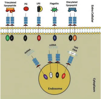
TLRs classiication
Fig 1: Distribution and dimerisation of TLRs in different cellular compartments (2, 8).
PG; Peptidoglycan, LPS; Lipopolysaccharide, dsRNA; Dou
-ble stranded RNA and ssRNA; Single stranded RNA.
TLRs signaling
Although extracellular domain of each TLR (corre-sponding to ligand recognition) is different from oth-ers, all of them have a great similarity in their intracel-lular domain and respective transduction pathways.
After ligand binding, the intracellular cascades start. As mentioned previously, all TLRs have cyto-plasmic signaling domain homologous to that of IL-1 receptor, known as TIR domain. Several adaptor mol-ecules containing TIR domain interact with the TIR domain of TLRs. These adaptors include myeloid differentiation primary response gene -88 (MyD88), TIR domain containing adaptor protein/ MyD88 adapter-like protein (TIRAP/Mal),
TIR-domain-containing adapter-inducing interferon-β (TRIF)
and TRIF-related adaptor molecule (TRAM) (Fig 2). Overall, the downstream signaling pathways of TLRs can be divided into two main groups: MyD88 dependent and MyD88 independent (TRIF depend-ent) pathways. All TLRs except TLR3 use MyD88 dependent pathway whereas TLR4 could utilize both pathways (2). In addition, TLR4 is the only TLR which recruits all 4 adaptor molecules (MyD88, TI-RAP, TRIF and TRAM) for signaling (13). In brief, TLRs 5, 7, 8 and 9, in contrast to TLR3 which uses TRIF, use only MyD88 while TLR2 in dimer form
with TLR1 or TLR6 recruits both MyD88 and TI-RAP (13). TLRs activation could result in:
i. production of inlammatory cytokines and
chemokines.
ii. induction of anti viral response by production of type 1 interferons.
iii. maturation of dendritic cells by upregulation of costimulatory molecules (13).
Fig 2: Adaptor molecules involved in different TLRs signal
-ing (2, 13).
MyD88 dependent pathway
Following the engagement of adaptor molecules, different molecules are recruited including several IL-1 receptor associated kinases (IRAKs) and TNF receptor associated factors (TRAFs) and mitogen activated protein kinases (MAPKs). Afterwards, inhibitory kappa B kinase (IKK) is engaged and modulates the activation and translocation of the
transcription factor NF-κB. Another transcription
factor activated by MAPKs is activating protein-1 (AP-1) (13). In case of TLRs 7, 8 and 9, interferon response factor 7 (IRF7), a transcription factor, is activated and translocated to the nucleus. Finally,
this pathway leads to the production of inlamma -tory cytokines and/or type 1 interferons (IFN I) (2) depending on which TLR was activated.
MyD88 independent (TRIF dependent) pathway
Both TLR3 and TLR4 use this pathway which leads to the recruitment of TRAFs and IRF3 and production
of both inlammatory cytokines and IFN I (2) (Fig 3).
TLRs ligands
Each TLR has its own distinct PAMP ligands. The cell surface TLRs recognize different ligands including components of gram positive and gram negative bacteria, fungi and parasites such as popolysaccharide (LPS), peptidoglycan (PG),
li-poteichoic acid (LT), lagellin, mannan, zymosan
and glycoinositolphospholipids. On the other hand, intracellular TLRs are usually stimulated by nucle-ic acids of viruses and bacteria including ssRNA, dsRNA and CpG unmethylated DNA (Table 1) (2, 14-18). Recently, some host derived molecules are
identiied as endogenous ligands for TLRs such as
some heat shock proteins (HSP) 60 and 70 (19), neutrophil elastase (20), fatty acid, heme (21),
beta defensin (22), reactive oxygen species (ROS)
(23), ibronectin (24), oligosaccharides of hyalu -ronic acid, heparan sulfate (25) and chromatin-IgG complexes (26).
As mentioned above, despite relatively limited types of TLRs known in human (TLR1-10), they can react with a wide spectrum of PAMPs. This could be explained by a special characteristic of TLRs. TLRs usually form dimers (homo or hetero dimers) which increases the diversity of ligands recognized by them, for example, TLR2 usually forms heterodimers with TLR1 or TLR6 with each
dimer having different ligand speciicity (27, 28).
The varieties of ligands recognized by TLRs un-derline the importance of the TLRs in the host in-nate immune response.
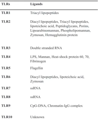
Table 1: Human TLRs and their respective ligands (2, 14-19, 26)
Ligands TLRs
Triacyl lipopeptides TLR1
Diacyl lipopeptides, Triacyl lipopeptides, lipoteichoic acid, Peptidoglycans, Porins, Lipoarabinomannan, Phospholipomannan, Zymosan, Hemagglutinin protein TLR2
Double stranded RNA TLR3
LPS, Mannan, Heat-shock protein 60, 70, Fibrinogen
TLR4
Flagellin TLR5
Diacyl lipopeptides, lipoteichoic acid, Zymosan
TLR6
ssRNA TLR7
ssRNA TLR8
CpG-DNA, Chromatin-IgG complex TLR9
Unknown TLR10
TLRs and female reproductive tract
Several reports have revealed that TLRs are ex-pressed throughout different parts of the female re-productive tract (29, 30). In addition, it was shown that their expression is altered during different phases of the menstrual cycle (31), suggesting that alterations during the menstrual cycle may be un-der the control of sex hormones including estro-gen and progesterone (31, 32). During pregnancy, special hormonal changes and immunologic chal-lenges occur. With regard to the immune system, normal pregnancy consists of three different im-munologic phases:
1. Pro-inlammatory environment during embryo im -plantation, placentation and early stage of pregnancy.
2. Anti-inlammatory milieu during mid-pregnancy 3. Pro-inlammatory environment at third trimester
and end of pregnancy (33).
During normal pregnancy, different parts of fe-male reproductive tract including endometrium, myometrium, cervix and vagina undergo
histo-logical and functional changes while speciic preg -nancy related tissues such as amnion, chorion and placenta are created.
In the following, we will overview the research-es done on the exprresearch-ession of different TLRs and their function during pregnancy in different tissues of the female reproductive tract which are closely in relation to embryo.
Placenta
The placenta serves as an active barrier between the embryo and the surrounding environment. Dif-ferent PRRs are considered to play roles in this in-teraction including TLRs and NLRs (34).
Various studies have evaluated the presence and function of TLRs or their related molecules in placental tissues. By using immunohistochemis-try, Kumazaki et al. investigated the expression of TLR4 protein in human placentas obtained from normal and complicated pregnancies delivered in the second and third trimesters. They showed that TLR4 was found on the extravillous trophoblasts, intermediate trophoblasts/X cells in the degenera-tive villi, and villous Hofbauer cells of both
pre-term and pre-term placentas and on the inlammatory
cells in placentas with chorioamnionitis (CAM). TLR4 immunoreactivity was increased in the vil-lous Hofbauer cells of preterm CAM placentas compared with those of preterm placentas without CAM or those of term placentas with or without CAM (35). Later, Abrahams et al. studied the
ex-pression and function of TLR2 and TLR4 in irst
trimester trophoblast cells. They found that acti-vation of TLR4 induces cytokine production by trophoblast cells, but TLR2 activation induced apoptosis using Fas associated death domain, the inactivation of the X-linked inhibitor of apopto-sis, and the activation of caspases 3, 8 and 9. They suggested a pathogenic role for TLR2 during some intrauterine infections (36).
Klaffenbach et al. studied the chorioncarcimoma cell lines by using different techniques including real time polymerase chain reaction (real time
PCR), luorescence-activated cell sorter analysis
dur-ing elective caesarean sections (ECS) with those of normal vaginal delivery (NVD) to explore the effect of completion of labor on TLR expression
proile. They showed that both groups of placentas
expressed the TLR1-TLR10 and revealed for the
irst time that human term placenta can respond to
TLR3, TLR5 and TLR7/8 agonists (39).
Activation of TLRs on trophoblast has various consequences during pregnancy including immune cell recruitment, cytokine secretion and protective responses to invading pathogens (40). Recently, Aboussahoud et al. used an in vitro model of hu-man embryo implantation and showed that activa-tion of TLR5 decreases the attachment of human trophoblast cells to endometrial cell line (41). In addition, it has been suggested that pregnancy complications associated with placental dysfunc-tion, such as preterm labor, may be a result of TLR activation (40). It seems that proper interaction between TLRs and respective ligands on placenta plays important roles during different phases of pregnancy including implantation and labor.
Chorion and amnion
The expression of TLRs in fetal membrane such as amnion has been studied but not as extensive as placenta. Kim et al. showed that the expression of TLR2 and TLR4 is increased in chorioamnion membrane at time of labor and in presence of cho-rioamnionitis. They also showed that TLR2 was polarized to the basal surface of amniotic epithelial
cells in women without inlammation but this dis -tribution was lost in the presence of chorioamnio-nitis (42). Recently, Choi et al. studied the immu-nohistochemical expression of TLR4 in different histological layers and anatomical regions of hu-man fetal membranes. They showed that chorion
expressed signiicantly higher levels of TLR4 than
the amnion and this expression did not differ with regard to anatomical regions (uterine fundus vs. uterine low segment). Furthermore, histological presence of chorioamnionitis did not alter TLR4 expression while the progression of gestation
sig-niicantly decreased the TLR4 expression (43). Myometrium
Little data exists in regard to TLRs expression in human myometrium. Youssef et al. demonstrated
that expression of TLRs 2 and 4 was signiicantly
higher in pregnant myometrium at term in compar-ison with preterm. In addition, they showed that
the level of TLR2 protein signiicantly increased
during labor. The authors suggest that these TLRs may be important in labor and their function could be suppressed by progesterone (44).
Endometrium
For the irst time, Young et al. showed that TLRs
1-6 and 9 are expressed in both whole endome-trium and separated endometrial epithelial cells using reverse transcriptase-PCR (RT-PCR). They also showed that Ishikawa cells expressed TLRs 2 and 5 while RL95-2 cells expressed TLRs 3, 5 and 9 (29). Subsequently, several studies were un-dertaken in this regard using different techniques. Pioli et al. reported the expression of TLRs 1 to 6, MyD88 and CD14 in different parts of the female reproductive tract including uterine endometrium (45). Fazeli et al. (30) showed that TLRs 1, 2, 3, 5 and 6 proteins were present in different parts of female reproductive tract. Also they found that TLR4 was only present in upper parts of the re-productive tract including the endocervix, endo-metrium and fallopian tubes and absent in vagina and ectocervix.
Alatoonian et al. studied endometrium obtained
from normal women at different phases of men-strual cycle (menstruation, proliferative and secre-tory) and detected TLRs 7-10 proteins in both en-dometrial epithelium and stroma. The authors also demonstrated that all ten TLRs were expressed in human endometrial tissue and most of them had
signiicantly higher expression during the secre -tory phase in comparison with other phases of the
menstrual cycle (31). Their indings support that
TLRs expression may be under the control of fe-male sex hormones (estrogen and progesterone). It seems that estrogen has an inhibitory effect on TLRs expression while progesterone may have stimulating effects because TLRs expression was at its highest in secretory phase (31, 32).
of human endometrium (46).
Decidua
Decidua is deined as the transformed endome -trium during pregnancy, which forms the maternal
part of the placenta. For the irst time , Krikun et al.
studied the decidual tissues and cells obtained from
women undergoing irst trimester elective termina -tions or repeat cesarean sec-tions and showed that human decidua differentially express TLRs (47). Canavan and Simhan showed that decidual cells from term unlabored pregnancies express TLRs 1, 2, 4 and 6 which respond to LPS and PG stimula-tion (48). Recently, Hayati et al. studied TLRs 2 and 4 expression in decidua and amniotic cells of
non inlamed placenta and placenta with infection.
They reported higher expression of TLR2 in the
amniotic and decidua cells of inlamed placenta than non-inlamed placenta, supporting the po -tential role of TLR2 in defense against infection (49). In addition, Schatz et al. studied the immu-nostaining of TLR4 protein in decidual cells com-pared with trophoblasts in different trimesters and showed that TLR4 expression is higher in decidual cells than trophoblasts. They suggested that de-cidual cells are primary targets for gram negative bacterial infection (50).
Conclusion
Pregnancy is a fundamental stage in life of every woman in reproductive age. The immunologic fea-tures of normal pregnancy are unique because the mother tolerates the semi allogenic embryo. The immunologic changes during pregnancy are very important not only in normal tissues of the female reproductive tract but also in embryo-related tis-sues created during pregnancy such as fetal
mem-brane and placenta. According to different indings
obtained using several study models including in vitro models, animal models or studies done on tissues obtained during normal or complicated human pregnancies, it seems that the TLRs fam-ily as one of the main regulators of the innate immunity, is not only involved in protecting the female reproductive tract against invading patho-gens, but also is a key regulator in immunologic events during stages of normal pregnancy such as implantation or labor. One of the issues that make researches about TLRs so interesting is the accessibility to the agonists and antagonists of
TLRs and the possibility to stimulate or suppress TLRs function. Although researches in regard to roles of TLRs in pregnancy are in progression, more studies are needed especially in cases of pregnancy complications such as preeclampsia, abortion and preterm labor.
References
1. Janeway CA Jr, Medzhitov R. Innate immune recognition. Annu Rev Immunol. 2002; 20: 197-216.
2. Akira S, Uematsu S, Takeuchi O. Pathogen recognition and innate immunity. Cell. 2006; 124 (4): 783-801. 3. Medzhitov R, Janeway CA Jr. Innate immunity: the virtues
of a nonclonal system of recognition. Cell. 1997; 91(3): 295-298.
4. Essakalli M, Atouf O, Bennani N, Benseffaj N, Ouadghiri S, Brick C. Toll-like receptors. Pathol Biol (Paris). 2009; 57(5): 430-438.
5. Fischer M, Ehlers M. Toll-like receptors in autoimmunity. Ann N Y Acad Sci. 2008; 1143: 21-34.
6. Martinon F, Tschopp J. NLRs join TLRs as innate sensors of pathogens. Trends Immunol. 2005; 26 (8): 447-454. 7. Bowie A, O'Neill LA. The interleukin-1 receptor/Toll-like
re-ceptor superfamily: signal generators for pro-inlammatory
interleukins and microbial products. J Leukoc Biol. 2000; 67(4): 508-514.
8. Kawai T, Akira S. The role of pattern-recognition receptors in innate immunity: update on Toll-like receptors. Nat Im-munol. 2010; 11(5): 373-384.
9. Anderson KV, Bokla L, Nüsslein-Volhard C. Establishment of dorsal-ventral polarity in the Drosophila embryo: the in-duction of polarity by the Toll gene product. Cell. 1985 ; 42 (3): 791-798.
10. Lemaitre B, Nicolas E, Michaut L, Reichhart JM, Hoffmann JA. The dorsoventral regulatory gene cassette spatzle/ Toll/cactus controls the potent antifungal response in Drosophila adults. Cell. 1996; 86(6): 973-983.
11. Nomura N, Miyajima N, Sazuka T, Tanaka A, Kawarabaya-si Y, Sato S, et al. Prediction of the coding sequences of
unidentiied human genes. I. The coding sequences of 40
new genes (KIAA0001-KIAA0040) deduced by analysis of randomly sampled cDNA clones from human immature myeloid cell line KG-1. DNA Res. 1994; 1(1): 27-35. 12. Taguchi T, Mitcham JL, Dower SK, Sims JE, Testa JR.
Chromosomal localization of TIL, a gene encoding a protein related to the Drosophila transmembrane recep-tor Toll, to human chromosome 4p14. Genomics. 1996; 32(3): 486-488.
13. Kawai T, Akira S. TLR signaling. Cell Death Differ. 2006; 13(5): 816-825.
14. Ahmad-Nejad P, Hacker H, Rutz M, Bauer S, Vabulas RM, Wagner H. Bacterial CpG-DNA and lipopolysaccharides activate Toll-like receptors at distinct cellular compart-ments. Eur J Immunol. 2002; 32(7): 1958-1968.
15. Alexopoulou L, Holt AC, Medzhitov R, Flavell RA. Rec-ognition of double-stranded RNA and activation of NF-kappaB by Toll-like receptor 3. Nature. 2001; 413(6857): 732-738.
16. Andersen-Nissen E, Smith KD, Strobe KL, Barrett SL, Cookson BT, Logan SM, et al. Evasion of Toll-like receptor
5 by lagellated bacteria. Proc Natl Acad Sci USA. 2005;
102 (26): 9247-9252.
303(5663): 1529-1531.
18. Hemmi H, Takeuchi O, Kawai T, Kaisho T, Sato S, Sanjo H, et al. A Toll-like receptor recognizes bacterial DNA. Na-ture. 2000; 408(6813): 740-745.
19. Wan T, Zhou X, Chen G, An H, Chen T, Zhang W, et al. Novel heat shock protein Hsp70L1 activates dendritic cells and acts as a Th1 polarizing adjuvant. Blood. 2004; 103(5): 1747-1754.
20. Devaney JM, Greene CM, Taggart CC, Carroll TP, O’Neill SJ, McElvaney NG. Neutrophil elastase up-regulates in-terleukin-8 via toll-like receptor 4. FEBS Lett. 2003; 544(1-3): 129-132.
21. Yu L, Wang L, Chen S. Endogenous toll-like receptor
li-gands and their biological signiicance. J Cell Mol Med.
2010; 14(11): 2592-2603.
22. Biragyn A, Rufini PA, Leifer CA, Klyushnenkova E, Shak -hov A, Chertov O, et al. Toll-like receptor 4-dependent activation of dendritic cells by beta-defensin 2. Science. 2002; 298 (5595): 1025-1029.
23. Frantz S, Kelly RA, Bourcier T. Role of TLR-2 in the ac-tivation of nuclear factor kappa B by oxidative stress in cardiac myocytes. J Biol Chem. 2001; 276: 5197-5203. 24. Okamura Y, Watari M, Jerud ES, Young DW, Ishizaka ST,
Rose J, et al. The extra domain A of ibronectin activates
Toll-like receptor 4. J Biol Chem. 2001; 276: 10229-10233. 25. Taylor KR, Trowbridge JM, Rudisill JA, Termeer CC, Si-mon JC, Gallo RL. Hyaluronan fragments stimulate en-dothelial recognition of injury through TLR4. J Biol Chem. 2004; 279: 17079-17084.
26. Leadbetter EA, Rifkin IR, Hohlbaum AM, Beaudette BC, Shlomchik MJ, Marshak-Rothstein A. Chromatin-IgG complexes activate B cells by dual engagement of IgM and Toll-like receptors. Nature. 2002; 416(6881): 603-607. 27. Jin MS, Kim SE, Heo JY, Lee ME, Kim HM, Paik SG, et al.
Crystal structure of the TLR1-TLR2 heterodimer induced by binding of a tri-acylated lipopeptide. Cell. 2007; 130(6): 1071-1082.
28. Kang JY, Nan X, Jin MS, Youn SJ, Ryu YH, Mah S, et al. Recognition of lipopeptide patterns by Toll-like receptor 2-Toll-like receptor 6 heterodimer. Immunity. 2009; 31(6): 873-884.
29. Young SL, Lyddon TD, Jorgenson RL, Misfeldt ML. Ex-pression of Toll-like receptors in human endometrial epi-thelial cells and cell lines. Am J Reprod Immunol. 2004; 52(1): 67-73.
30. Fazeli A, Bruce C, Anumba DO. Characterization of Toll-like receptors in the female reproductive tract in humans. Hum Reprod. 2005; 20(5): 1372-1378.
31. Alatoonian R, Tuckerman E, Elliott SL, Bruce C, Alatoo -nian A, Li TC, et al. Menstrual cycle-dependent changes of Toll-like receptors in endometrium. Hum Reprod. 2007; 22(2): 586-593.
32. Alatoonian R, Fazeli A. Toll-like receptors in female repro -ductive tract and their menstrual cycle dependent expres-sion. J Reprod Immunol. 2008; 77 (1): 7-13.
33. Mor G. Inlammation and pregnancy: the role of toll-like re -ceptors in trophoblast-immune interaction. Ann N Y Acad Sci. 2008; 1127: 121-128.
34. Abrahams VM. The role of the Nod-like receptor family in trophoblast innate immune responses. J Reprod Immunol. 2011; 88(2): 112-117.
35. Kumazaki K, Nakayama M, Yanagihara I, Suehara N,
Wada Y. Immunohistochemical distribution of Toll-like re-ceptor 4 in term and preterm human placentas from nor-mal and complicated pregnancy including chorioamnioni-tis. Hum Pathol. 2004; 35(1): 47-54.
36. Abrahams VM, Bole-Aldo P, Kim YM, Straszewski-Chavez SL, Chaiworapongsa T, Romero R, et al. Divergent troph-oblast responses to bacterial products mediated by TLRs. J Immunol. 2004; 173(7): 4286-4296.
37. Klaffenbach D, Rascher W, Röllinghoff M, Dötsch J, Meissner U, Schnare M. Regulation and signal transduc-tion of toll-like receptors in human chorioncarcinoma cell lines. Am J Reprod Immunol. 2005; 53(2): 77-84. 38. Nishimura M, Naito S. Tissue-speciic mRNA expression
proiles of human toll-like receptors and related genes.
Biol Pharm Bull. 2005; 28(5): 886-892.
39. Patni S, Wynen LP, Seager AL, Morgan G, White JO, Thornton CA. Expression and activity of Toll-like receptors 1-9 in the human term placenta and changes associated with labor at term. Biol Reprod. 2009; 80(2): 243-248. 40. Riley J K, Nelson DM. Toll-like receptors in pregnancy
disorders and placental dysfunction. Clinic Rev Allerg Im-munol. 2010; 39(3): 185-193.
41. Aboussahoud W, Bruce C, Elliott S, Fazeli A. Activation of Toll-like receptor 5 decreases the attachment of hu-man trophoblast cells to endometrial cells in vitro. Hum Reprod. 2010; 25(9): 2217-2228.
42. Kim YM, Romero R, Chaiworapongsa T, Kim GJ, Kim MR, Kuivaniemi H, et al. Toll-like receptor -2 and -4 in the cho-rioamniotic membranes in spontaneous labor at term and in preterm parturition that are associated with chorioam-nionitis. Am J Obstet Gynecol. 2004; 191(4): 1346-1355. 43. Choi SJ, Jung SH, Eom M, Han KH, Chung IB, Kim SK.
Immunohistochemical distribution of toll-like receptor 4 in preterm human fetal membrane. J Obstet Gynaecol Res. 2012; 38(1): 108-112.
44. Youssef RE, Ledingham MA, Bollapragada SS, O'Gorman N, Jordan F, Young A, et al. The role of toll-like receptors (TLR-2 and -4) and triggering receptor expressed on my-eloid cells 1 (TREM-1) in human term and preterm labor. Reprod Sci. 2009; 16(9): 843-856.
45. Pioli PA, Amiel E, Schaefer TM, Connolly JE, Wira CR, Guyre PM. Differential expression of Toll-like receptors 2 and 4 in tissues of the human female reproductive tract. Infect Immun. 2004; 72 (10): 5799-5806.
46. Aboussahoud W, Alatoonian R, Bruce C, Elliott S, Ward J, Newton S, et al. Expression and function of Toll-like recep-tors in human endometrial epithelial cell lines. J Reprod Immunol. 2010; 84(1): 41-51.
47. Krikun G, Lockwood CJ, Abrahams VM, Mor G, Paidas M, Guller S. Expression of Toll-like receptors in the human decidua. Histol Histopathol. 2007; 22(8): 847-854. 48. Canavan TP, Simhan HN. Innate immune function of the
human decidual cell at the maternal-fetal interface. J Re-prod Immunol. 2007; 74(1-2): 46-52.
49. Hayati AR, Mohamed AE, Tan GC. An immunohistochemi-cal study of Toll-like receptors 2 and 4 in placenta with and without infection. Malays J Pathol. 2010; 32(1): 13-19. 50. Schatz F, Kayisli UA, Vatandaslar E, Ocak N, Guller S,