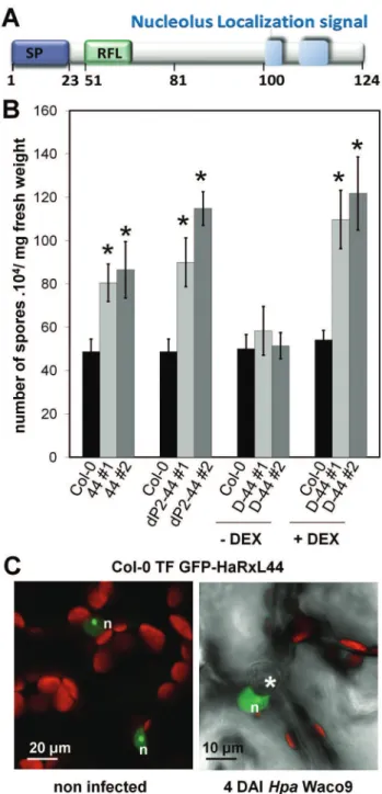
A downy mildew effector attenuates salicylic acid-triggered immunity in Arabidopsis by interacting with the host mediator complex.
Texto
Imagem


Documentos relacionados
Transposon insertion mutants defective in heme uptake or intracellular heme transport would show both cytochrome bd and catalase deficiency when grown in the presence of hemin..
Despite important progress in the un- derstanding of leprosy immunity, much of the health staff in many countries is unacquainted with leprosy epidemiology or with
Estimation and partitioning of actual daily evapotranspiration at an intensive olive grove using the STSEB model based on remote
citri effector that plays an essential role in citrus canker, while limiting the host range of CC strains to citrus because it triggers immunity in all other tested plant species
To better compare salicylic acid degradation by different cell concentrations, the specific degradation rate of salicylic acid (quantity of salicylic acid removed per quantity per
Cette promesse réalise donc cet événement de la Pentecôte car tous furent remplis de l’Esprit Saint (Cfr. Evénement de taille et fondateur par lequel naît l’Eglise.
citri effector that plays an essential role in citrus canker, while limiting the host range of CC strains to citrus because it triggers immunity in all other tested plant species
161 virulence genes essentially associated with the invasion of the host-cell. We also observed that chromosomal genes known to be regulated by the plasmid were
