faecalis
Michael Baureder*, Lars Hederstedt
Microbiology Group, Department of Biology, Lund University, Lund, Sweden
Abstract
Little in general is known about how heme proteins are assembled from their constituents in cells. The Gram-positive bacteriumEnterococcus faecaliscannot synthesize heme and does not depend on it for growth. However, when supplied with heme in the growth medium the cells can synthesize two heme proteins; catalase (KatA) and cytochromebd(CydAB). To identify novel factors important for catalase biogenesis libraries ofE. faecalisgene insertion mutants were generated using two different types of transposons. The libraries of mutants were screened for clones deficient in catalase activity using a colony zymogram staining procedure. Analysis of obtained clones identified, in addition tokatA (encoding the catalase enzyme protein), nine genes distributed over five different chromosomal loci. No factors with a dedicated essential role in catalase biogenesis or heme trafficking were revealed, but the results indicate the RNA degradosome (srmB,rnjA), an ABC-type oligopeptide transporter (oppBC), a two-component signal transducer (etaR), and NADH peroxidase (npr) as being important for expression of catalase activity in E. faecalis. It is demonstrated that catalase biogenesis in E. faecalis is independent of the CydABCD proteins and that a conserved proline residue in the N-terminal region of KatA is important for catalase assembly.
Citation: Baureder M, Hederstedt L (2012) Genes Important for Catalase Activity in Enterococcus faecalis. PLoS ONE 7(5): e36725. doi:10.1371/ journal.pone.0036725
Editor:Riccardo Manganelli, University of Padova, Italy
ReceivedMarch 9, 2012;AcceptedApril 11, 2012;PublishedMay 10, 2012
Copyright:ß2012 Baureder, Hederstedt. This is an open-access article distributed under the terms of the Creative Commons Attribution License, which permits unrestricted use, distribution, and reproduction in any medium, provided the original author and source are credited.
Funding:The work was supported by grant 621-2010-5672 from the Swedish Research Council (http://www.vr.se/). The funder had no role in study design, data collection and analysis, decision to publish, or preparation of the manuscript.
Competing Interests:The authors have declared that no competing interests exist.
* E-mail: [email protected]
Introduction
Catalase is a well-known enzyme found in all three domains of life. It plays a role in oxidative stress defense by degrading hydrogen peroxide to molecular oxygen and water [1].
Enterococcus faecalisis one of the few species of lactic acid bacteria that exhibit catalase activity, but only when grown in the presence of heme [2]. E. faecalis catalase (KatA) belongs to the group of heme-containing mono-functional catalases (EC 1.11.1.6). It is a homo-tetrameric protein containing one heme group (protoheme IX) per subunit and its crystal structure has been determined [3]. Catalase protectsE. faecalisagainst hydrogen peroxide stress but is not the only enzyme with this function. The bacterium can produce a NADH peroxidase (Npr) that degrades hydrogen peroxide to water and which seems sufficient for protection against high concentrations of hydrogen peroxide when heme is not available [4].E. faecaliscells cannot synthesize heme and thus have to take it up from the environment in order to form active catalase. Despite the fact that catalase has been studied intensively for many years biogenesis of this enzyme is not understood. The chronological order of assembly events such as KatA polypeptide folding, tetramer formation and heme insertion into the apopro-tein is not known. The complex structure of the catalase tetramer as well as the hydrophobic nature of heme and its potential in generating reactive oxygen species indicate the need for tightly controlled and assisted assembly of catalase. No catalase biogenesis factors have been identified except in the bacteriumCampylobacter jejuni which was recently reported to contain a protein (Cj1386)
involved in heme trafficking to catalase [5]. E. faecalis does not encode any apparent homolog of this protein.
In this work we usedE. faecalis as an experimental system to search for genes encoding factors necessary for biogenesis of functional catalase, i.e. genes for proteins that function in heme uptake, heme trafficking or catalase assembly. We generated libraries of transposon insertion mutants in E. faecalis strain OG1RF which were screened for clones deficient in catalase activity. Identification of the chromosomal transposon insertion site in those mutants revealed a set of genes in addition tokatA.
Results
Colony assay for detection of catalase activity
The classical method for detection of catalase activity in bacteria grown on agar plates is the observation of effervescence after flushing colonies on the plate with dilute hydrogen peroxide solution. With this procedure it is difficult to find rare catalase-deficient colonies among a large number of catalase-positive colonies. We therefore made use of a zymogram staining reagent, modified from that described by Hanker and Rabin [6], which leads to the formation of a purple color around colonies due to catalase activity.
EMB1 remained unstained. As expected, colonies of these strains did not stain after growth on media without hemin (Figure 1).
Transposon mutagenesis and screening of libraries Two different transposon mutagenesis systems, pG+host8:ISS1 [7] and EfaMarTn [8], were used to generate libraries of insertion mutants in E. faecalis OG1RF. The colony staining method described above was used to screen the libraries for mutants lacking catalase activity when grown on agar plates containing hemin. In total 8,180 ISS1 and 7,994 EfaMarTn transposon insertion mutants were tested for catalase activity resulting in the isolation of 11 ISS1and 32 EfaMarTn mutants with no or reduced catalase activity stain. The transposon insertion site in the chromosome of 42 of these mutants was identified by inverse PCR combined with DNA sequence analysis. The insertion site in one strain, EMB5, could not be identified. All isolated catalase-deficient clones seemingly contained a single transposon insertion, i.e. amplification of Tn insertion sites by inverse PCR yielded only one product per clone. The insertion sites were found distributed over 23 different loci on the OG1RF chromosome (Figure 2, Tables 1 and S2). For six loci two or more independent transposon insertion mutants were obtained (Table 1 and Figure 3). One of them was the expected OG1RF_11314 (katA) gene which validated the used mutagenesis and screening procedure for isolation of catalase-deficient mutants. The other five loci were OG1RF_10576 (srmB), OG1RF_10635-37 (opp), OG1RF_10782-83 (gnd and etaR), OG1RF_10983 (npr), and OG1RF_12223-24 (rnjA).
The isolation of several independent insertions in some genes indicated that the screen was near saturation and strongly suggested linkage between genetic locus and selected phenotype. Transposon insertions often generate polar effects on downstream genes. For this reason we cannot conclude whether the respective observed phenotype is caused by the gene disruption or a polar effect or a combination of the two. Insertions in three genes (katA, npr, and etaR) were obtained with both transposon mutagenesis systems.
Catalase activity and KatA polypeptide in cell lysates The catalase-deficient phenotype of the mutants found in the screen was first confirmed by catalase activity determination and immunoblot analysis for KatA polypeptide in the soluble (cytoplasmic) fraction of cell extracts. Subsequently unfractionated cell lysates of the mutants were similarly analyzed for catalase. Figure 1. Colony catalase activity assay.E. faecalisOG1RF (wild-type) and EMB1 (katA::ISS1) colonies grown on TH agar plates with and without 8mM hemin added and zymogram stained for catalase activity. doi:10.1371/journal.pone.0036725.g001
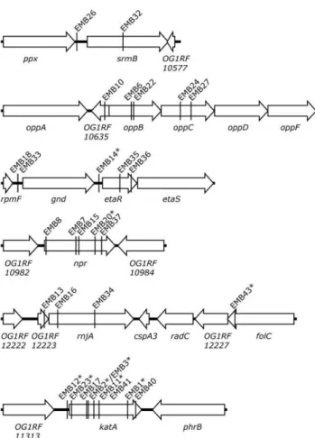
Figure 2. Distribution of transposon insertion sites.Transposon insertion sites in the OG1RF genome of 42 catalase-deficient mutants isolated in this work are shown. The strain names (EMBx) are indicated and ISS1-derived mutants are marked with an asterisk.
doi:10.1371/journal.pone.0036725.g002
Figure 3. Genomic context of loci for which two or more independent catalase-deficient transposon insertion mutants were obtained.Insertion sites in theE. faecalisOG1RF chromosome are marked with the designation of the respective isolate (EMBx) and ISS1-derived mutants are marked with an asterisk. See Table 1. doi:10.1371/journal.pone.0036725.g003
E. faecalisCatalase Defective Mutants
Locus tag(s)a Gene Annotated function Strain Tn insertion position in genomea Catalase activityb KatA protein
Locus 1 OG1RF_10576 srmB ATP-dependent RNA helicase DeaD EMB26 604,149 0.10 nd
OG1RF_10576 EMB32 605,068 0.33 0.37
Locus 2 OG1RF_10635 ---- Hypothetical protein EMB10 674,142 0.11 0.37 OG1RF_10636 oppB Oligopeptide ABC superfamily ATP binding cassette
transporter, membrane protein
EMB6 674,670 0.12 0.30
OG1RF_10636 EMB22 674,715 0.29 0.32
OG1RF_10637 oppC Oligopeptide ABC superfamily ATP binding cassette transporter, membrane protein
EMB24 675,641 0.42 0.35
OG1RF_10637 EMB27 675,858 0.54 0.44
Locus 3 OG1RF_10782 gnd Phosphogluconate dehydrogenase (decarboxylating) EMB18 814,391 0.11 0.23
OG1RF_10782 EMB33 814,391 0.25 0.24
OG1RF_10783 etaR Response regulator EMB14c 815,997 0.19 0.26
OG1RF_10783 EMB35 816,411 0.26 0.28
OG1RF_10783 EMB36 816,640 0.29 0.34
Locus 4 OG1RF_10983 npr NADH peroxidase EMB8 1,022,177 0.08 0.90
OG1RF_10983 EMB7 1,022,772 0.08 0.68
OG1RF_10983 EMB15 1,022,828 0.15 1.00
OG1RF_10983 EMB20c 1,023,154 0.05 0.96
OG1RF_10983 EMB37 1,023,286 0.12 1.19
Locus 5 OG1RF_12223 ---- Hypothetical protein EMB16 2,343,191 0.42 0.83
OG1RF_12224 rnjA Ribonuclease J1 EMB13 2,343,456 0.61 0.94
OG1RF_12224 EMB34 2,344,190 0.98 1.12
Locus 6 OG1RF_11314 katA Catalase EMB12c 1,371,597 na na
OG1RF_11314 EMB23c 1,371,681
OG1RF_11314 EMB17 1,371,981
OG1RF_11314 EMB2c 1,372,016
OG1RF_11314 EMB3c 1,372,016
OG1RF_11314 EMB11c 1,372,126
OG1RF_11314 EMB41 1,372,226
OG1RF_11314 EMB1c 1,372,790
OG1RF_11314 EMB40 1,372,951
In the screen for catalase deficiency two or more independent transposon insertions were obtained in six loci. Catalase activity and KatA content in cell extracts are expressed as fraction relative to those of the parental strain OG1RF.
nd, not done;na, not applicable. aGenBank: CP002621.1.
bCatalase activity of strain OG1RF was 14 U/mg of protein. c
E.
faecalis
Catalase
Defective
Mutants
ONE
|
www.plos
one.org
3
May
2012
|
Volume
7
|
Issue
5
|
Extracts ofE. faecalisOG1RF served as reference in the analyses. The results are presented in Tables 1 and S2.
Catalase activity in the extracts correlated with the KatA polypeptide concentration except for the fivenprinsertion mutants which showed wild-type levels of the enzyme protein but only low activity. Notably, rnjA insertion mutants uniquely showed 5–10 fold decreased catalase activity when the cells were grown in BHI supplemented with hemin compared to TSBG supplemented with hemin. Irrespective of growth medium the colony size of these mutants was reduced compared to the wild-type and other catalase-deficient mutants.
Identification of acydC katAdouble mutant
To exclude the possibility of fortuitous mutations in katA in transposon insertion mutants thekatAgene of mutants showing no catalase activity stain was sequenced. A single strain, EMB4 with cydC(the OG1RF_11664 gene) inactivated by transposon insertion (Tables 2 and S2), was found to carry a point mutation (C82A) in katAwhich results in a threonine instead of a proline residue at position 28 in KatA. Full-length KatA polypeptide was found in the particulate cell fraction of strain EMB4. This is in contrast to the parental strain OG1RF and all other KatA-containing mutants which had the polypeptide in the soluble fraction. Cell lysates, as well as the particulate and soluble fractions, of EMB4 lacked catalase activity (,5% activity compared to OG1RF).
Cytochromebdis not important for catalase biogenesis Cytochromebdand catalase are the only known heme proteins inE. faecalis[9]. Transposon insertion mutants defective in heme uptake or intracellular heme transport would show both cytochromebdand catalase deficiency when grown in the presence of hemin. Light absorption redox spectroscopy of membranes isolated from cells grown in the presence of hemin demonstrated the presence of cytochrome bd in all isolated catalase-deficient mutants except for EMB4 (Figure 4). This property of strain EMB4 was expected sincecydCandcydDof thecydABCDoperon are in other bacteria known to be essential for the synthesis of cytochrome bd [10,11,12]. To analyze whether any of the CydABCD proteins play a role in heme trafficking or maturation of catalase thecydABCDoperon was deleted in OG1RF, yielding strain EMB44. When grown in the presence of hemin this mutant contained normal amounts of both KatA polypeptide and catalase activity, and, as expected, lacked cytochrome bd. From these results we conclude that CydC is important for cytochrome bd biogenesis also inE. faecalisand that the CydABCD proteins are not important for biogenesis of catalase. The results furthermore
show that thekatA82point mutation alone is responsible for the catalase-negative phenotype of strain EMB4.
Discussion
Biogenesis of catalase requires insertion of one heme group per KatA monomer and assembly of the homotetramer. How and in what order these events occur are not known. To identify genes important for biogenesis of a functional catalase enzyme and for heme trafficking, libraries ofE. faecalismutants were constructed using two different transposon systems and screened for clones defective in catalase activity.
The multiple obtainedkatA-deficient mutants validated the used mutagenesis and screening procedure for isolation of catalase-deficient mutants. AkatApoint mutation (katA82) was detected in strain EMB4 carrying ISS1inserted incydC. It is remarkable that EMB4 is defective in both of the only two known heme proteins of E. faecalis. Our analysis of mutants defective in catalase or cytochromebddid not reveal any interdependence of biogenesis or activity of the two enzymes. KatA82 polypeptide, with Pro28 replaced by Thr, was found inactive in the particulate fraction of cell extracts. The mutated Pro residue is conserved in
mono-Table 2.Bacterial strains.
Strain Characteristics Reference/source
Enterococcus faecalis OG1RF Rifampicin and fusidic acid resistant Laboratory stock
OG1RF/pCJK55 EfaMarTn recipient [8]
CK111/pCF10-101/pCJK72 EfaMarTn donor [8]
OG1RF/pG+
host8:ISS1 ISS1system [7] & this work
EMB4 cydC::ISS1,katA82 This work
EMB44 DcydABCD::tetL This work
Escherichia coli TOP10 Cloning host Invitrogen
TOP10/pLUMB25 cydA’-tetL-cydD’in pCR-Blunt II TOPO This work
TOP10/pLUMB28 cydA’-tetL-cydD’in pJRS233 This work
doi:10.1371/journal.pone.0036725.t002
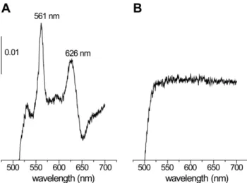
Figure 4. Light absorption spectra.Difference (dithionite-reduced minus air-oxidized) absorption spectra of cytoplasmic membranes isolated from strain OG1RF (A) and EMB4 (B) grown in the presence of hemin. The spectrum of strain EMB44 was similar to that of EMB4. The absorbance scale is indicated by the vertical bar.
doi:10.1371/journal.pone.0036725.g004
E. faecalisCatalase Defective Mutants
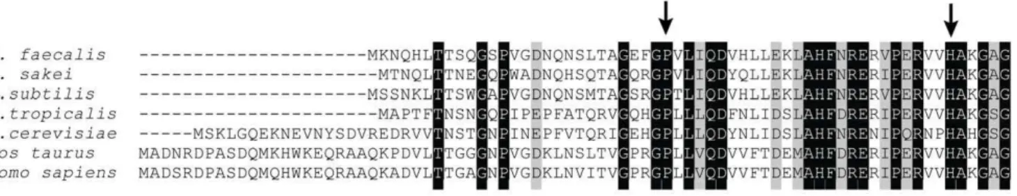
functional catalases and present in the N-terminal arm which interconnects with a neighboring subunit in the fully assembled tetrameric catalase (Figure 5) [3]. Apparently residue Pro28 is important for assembly of functional catalase. We conclude that the mutant KatA protein aggregates and therefore is found in the particulate cell fraction.
In addition to katA we identified five chromosomal loci seemingly important for expression of catalase activity inE. faecalis cells. The proteins encoded by those genes have diverse functions such as global regulation of RNA turnover (rnjA, srmB), NADH oxidation and hydrogen peroxide detoxification (npr), stress response (etaR), and membrane transport (oppBC). The connection between these physiological functions and expression of catalase activity in colonies remains to be elucidated but are probably indirect as discussed here below.
Five catalase-deficient insertion mutants had the gene for NADH peroxidase (Npr) inactivated. Similar to catalase Npr degrades hydrogen peroxide but in contrast it oxidizes NADH and generates water (instead of molecular oxygen) [13]. Interestingly, the amount of KatA polypeptide in Npr-negative mutants was found to be normal but the catalase activity was low. Since catalase polypeptide is unstable without the heme cofactor inserted [2] the low activity seems not explained by lack of heme in assembled catalase. The connection between Npr-deficiency and low catalase activity remains unexplained.
Several mutants isolated in the screen for catalase deficiency carried the transposon insertion in a gene related to RNA degradation. Two mutants were found defective in ribonuclease J1 (rnjA) and one in a hypothetical protein encoded by a gene upstream ofrnjA(Figure 3). Ribonuclease J1 plays a central role in mRNA turnover thereby indirectly influencing the expression of a multitude of genes (reviewed in [14]). It forms a complex with ribonuclease J2 (rnjB) for which a single mutant (EMB29; Table S2) was obtained in our screen. RNases J1/J2 are essential for growth of group A streptococci [15] and RNase J1 is essential in Bacillus subtilis[16,17]. Our results indicate that neither J1 nor J2 is essential in E. faecalis. Recently, it was reported that J2 is not essential for E. faecalis and involved in regulation of Ebp pili expression [18]. The DEAD-box RNA helicase SrmB, for which two insertion mutants were found in our screen, is thought to be a part of the RNA degradosome. InS. aureus, the homologous RNA helicase, CshA, interacts with RNase J1 [19]. These findings together suggest that normal catalase synthesis depends on a fully functional RNA degradosome complex.
Three transposon insertion mutants were obtained for etaR encoding the response regulator of the EtaRS two-component system. This system shows high similarity to LisRK of Listeria
monocytogenes[20] and CsrRS ofStreptococcus pyogenes[21]. EtaRS is involved in stress response and virulence [22,23]. Catalase deficiency in EtaRS-negative strains has not been reported, but this might have passed undetected in previous studies if hemin was not included in the growth media. The gene upstream of etaR encodes a putative 6-phoshogluconate dehydrogenase (Gnd). Two mutants with the same transposon insertion site in the gnd promoter region were obtained in our screen. Except for colocalization of the genes no connection between Gnd and EtaRS is apparent. ThelisRKandgndgenes inL. monocytogenes, but not inS. pyogenes, have the same organization as inE. faecalis.
It is not known how heme is transported intoE. faecalis. Five transposon insertions were found inoppB,oppCand a putative gene upstream of oppB (Figure 3) encoding an oligopeptide ABC transporter (OppBCDF). For unknown reasons no insertion was found in oppD or oppF. In Escherichia coli, the housekeeping dipeptide permease (DppBCDF) functions as a heme transporter [24] and heme is delivered to the permease by the periplasmic peptide-binding proteins DppA and MppA. It has been suggested that DppB and DppC bind heme and thus contribute to substrate specificity of the transporter. Two homologs ofE. coliDppBCDF can be identified by a BLAST search ofE. faecalisstrain OG1RF; OppBCDF and OG1RF_12367-70. This could suggest that E. faecalisOppBCDF plays a role in heme uptake. To test this we grew strain EMB6, and the wild-type strain as a reference, in the presence of 0.1mM hemin, which is a limiting concentration with respect to assembly of active catalase. No difference in amount of catalase between the strains was observed. This result suggests that E. faecalisOppBCDF does not transport heme or that it is a low affinity heme transporter present alongside other transporters with similar or higher affinity for heme.
The presence of cytochrome bd in all the isolated catalase-deficient mutants, except for EMB4 with thecydCgene inactivated, indicates that mutants with a general block in heme uptake or heme protein assembly cannot be found by the experimental approach taken in this work. Reasons for this can be (i) presence of functionally redundant proteins, (ii) bias in insertion sites for the transposons used for mutagenesis such that certain genes will not be mutated or be overrepresented, or (iii) that the sought function is essential for growth. There is also the possibility that catalase biogenesis inE. faecalisis not assisted by any specific protein. It is not known if the heme groups for catalase in the cytoplasm and cytochromebdin the membrane are taken up and delivered by the same cellular route. ThecydCDgenes encode an ABC transporter which is essential for biosynthesis of cytochromebdand might play a role in heme insertion into the oxidase (CydAB) specifically [10,11]. ThecydABCDoperon is however not required for catalase
Figure 5. Sequence alignment of catalase polypeptides.Multiple amino acid sequence alignment of six catalases showing conserved residues in the N-terminal arm. Human and bovine catalase enzymes have an extended N-terminal region containing an additionala-helix. Residues Pro28 (mutated to a Thr in strain EMB4) and the essential His54 (proximal to heme iron) inE. faecalisKatA are indicated by arrows.E. faecalis(UniProt F2MTL2),B. subtilis(P26901),Bos taurus(P00432),Homo sapiens(P04040),Lactobacillus sakei(P30265),Saccharomyces cerevisiae(P15202),Candida tropicalis(P07820).
biogenesis in E. faecalis, as demonstrated by the properties of a deletion mutant EMB44.
In Streptococcus agalactiae, alkyl hydroperoxide reductase C (AhpC) was shown to bind intracellular heme and thereby protects it from degradation [25]. In addition, it was recently found that intracellular heme homeostasis is controlled by heme sensing and efflux systems in Lactococcus lactis [26] and S. agalactiae [27]. Disruption of the two S. agalactiae efflux systems, PefAB and PefCD, led to accumulation of heme in the cell. In our screen for catalase-deficient mutants we did not reveal any putative heme efflux mutants, i.e., all,16,000 transposon insertion mutants grew in the presence of 8mM hemin.
Materials and Methods
Bacterial strains and culture media
E. faecalisstrains were grown in Todd-Hewitt (TH), Brain Heart Infusion (BHI) or Tryptic Soy Broth (TSB). The latter was supplemented with 1% glucose (TSBG). When indicated 8mM hemin (Sigma-Aldrich) was added to the media from a 10 mM stock solution in dimethyl sulfoxide (DMSO).
Colony catalase activity assay
The staining reagent [6] was prepared by mixing one part of 80 mg/mL dopamine (Fluka) in 0.2 M potassium phosphate buffer pH 8.0, one part of 40 mg/mLp-phenylenediamine (ICN Pharmaceuticals) in buffer, one part 12% hydrogen peroxide, and two parts of DMSO. Two mL of the reagent were mixed with 4 mL molten soft agar (0.7%) in water and subsequently poured on bacterial growth on an agar plate (d = 8.5 cm). Catalase activity caused within minutes the appearance of a purple color around bacterial colonies.
Transposon mutagenesis – ISS1 system
E. faecalisOG1RF was transformed with plasmid pG+
host8:ISS1 by electroporation as described in [28], but cells were incubated for 2 hours at 28uC after electroshock. Transformants were selected on TH plates containing 10mg/mL tetracycline incubat-ed at 28uC and the presence of the intact plasmid was checked by restriction enzyme digestion of isolated plasmid DNA with EcoRI and HindIII (New England Biolabs). Generation of random transposon mutants was done as described by Maguinet al.[7]; TH broth was inoculated with cells of strain OG1RF/pG+
hos-t8:ISS1to an OD600of 0.05 and incubated for 2.5 hours at 28uC
and 200 rpm. Then the culture was shifted to 37.5uC and incubated for a further 2.5 hours. Finally cells were diluted 1000-fold in 2 mM EDTA pH 7.5 and plated on TH agar containing 10mg/mL tetracycline. After overnight incubation at 37uC transposants were picked and streaked on both TH containing 10mg/mL tetracycline and TH containing additionally 8mM hemin (100 mutants per plate). Colonies on the latter plate were stained for catalase activity.
Transposon mutagenesis – EfaMarTn system
Generation of random transposon mutants was done as described by Kristich et al. [8]. In short; strains E. faecalis CK111/pCF10-101/pCJK72 (donor) and E. faecalis OG1RF/ pCJK55 (recipient) were grown in BHI broth containing 10mg/ mL erythromycin. After overnight incubation the cultures were diluted 20-fold in BHI containing 1mg/mL erythromycin. Nisin (Sigma-Aldrich) was added to the recipient culture to a final concentration of 10, 20, 30 or 40 ng/mL and the cultures were incubated at 30uC for 105 minutes. Donor and recipient were then mixed, plated on BHI containing the same concentration of
nisin, and incubated at 30uC for 21 hours. All cells on the plate were suspended in BHI containing 2 mM EDTA pH 7.5 and dilutions (50- and 100-fold) were spread on BHI agar containing 10mg/mL chloramphenicol, 250mg/mL 5-bromo-4-chloro-3-indolyl-b-D-galactopyranoside (X-gal), and 25mg/mL fusidic acid. Plates were incubated at 37uC overnight. ‘‘White’’ colonies were picked and streaked on both BHI containing 10mg/mL chlor-amphenicol and BHI containing additionally 8mM hemin (100 mutants per plate). Colonies on the latter plate were stained for catalase activity.
Inverse PCR
Chromosomal DNA was isolated as described by Marmur [29]. Twomg of the DNA were digested with HindIII or HincII (New England Biolabs) in a 50mL reaction. Ligation, to obtain circularized DNA, was done after heat inactivation of the restriction enzyme in a total volume of 750mL containing 2.5mL T4 DNA ligase (1000 NEB units, 15 Weiss units) and 25mL (1mg) digested DNA. The reaction was incubated for 16 hours at 15uC. The DNA was purified and concentrated using the Montage PCR cleanup kit (Millipore). PCR was done using Phusion DNA polymerase (Finnzymes) and primers invCATR2 and invGFPR1 (Table S1) for the EfaMarTn system or primers invISS1fwd and invISS1rev for the ISS1 system. PCR products were sequenced using primers invISS1fwd or invGFPR1.
Construction of strain EMB44
A gene deletion cassette consisting of the tetracycline resistance gene tetLfrom pDG1515 [30] flanked by segments of cydAand cydDfromE. faecalisOG1RF was constructed usingE. coliTOP10 as a cloning host (Table 2). The segments were amplified by PCR using primer pair tetL01/tetL02, cydA03/cydA04, and cydD03/ cydD04, respectively (Table S1). Vector pLUMB25 was con-structed by first cloning thetetLPCR product into plasmid pCR-Blunt II TOPO (Invitrogen) and subsequently adding thecydAand cydDsegments via restriction sites introduced by the primers. The cydA’-tetL-cydD’cassette was transferred from pLUMB25 into the E. coli/E. faecalis shuttle vector pJRS233 [31] via XhoI and HindIII restriction sites resulting in vector pLUMB28.E. faecalis OG1RF was transformed with pLUMB28 by electroporation [28] and transformants were selected at 30uC on TH containing 10mg/mL erythromycin. Chromosomal integration of the plasmid was selected by incubation at 42uC in the presence of erythromy-cin and 10mg/mL tetracycline. Excision of the plasmid by homologous recombination was accomplished by incubation at 30uC in the absence of antibiotics. Clones containing the cassette were selected on tetracycline-containing TH plates incubated at 37uC. The presence of the cassette and the absence of the vector backbone were confirmed by streaking colonies on TH containing tetracycline and erythromycin, respectively. Colonies that were Tetrand Ermswere checked for the loss of thecydlocus by colony PCR using primers cyd01 and cyd02 (Table S1).
katAsequence analysis
ThekatAgene in isolated chromosomal DNA was amplified by PCR using primers KatA03 and KatA04 (Table S1). Purified PCR products were sequenced using primers KatA03, KatA04 and KatAR01.
Large scale cell extracts
Bacteria from a starter culture in hemin supplemented medium (TH or BHI) were used to inoculate 600 mL of the same medium. Cultures were incubated at 37uC and 200 rpm overnight E. faecalisCatalase Defective Mutants
protected from light. Cells were harvested by centrifugation for 20 min at 5,0006gand 4uC. The cell pellet was washed once in 50 mM potassium phosphate buffer pH 8.0 and stored at220uC. For cell lysis the pellet was thawed and suspended in buffer containing 1 mM MgSO4, 1 mg/mL lysozyme and some grains of
DNase. The cell suspension was incubated for 1 hour at 37uC and subsequently passed two times through a pre-cooled French Press cell operated at 16,000 psi. Unbroken cells and debris were then removed by centrifugation for 20 minutes at 5,0006gand 4uC. After centrifugation for 90 min at 200,0006 g and 4uC the supernatant was saved as cytoplasmic fraction and the pellet (particulate fraction) was washed once in buffer using a homogenizer. The respective fraction contained 5–20 mg/mL protein and were stored at280uC until analysis.
Small scale lysates
Fifty mL of TSBG supplemented with hemin were inoculated with bacteria to an OD600 of 0.05. The culture was grown for
18 hours at 37uC and 200 rpm. Cells were harvested by centrifugation for 10 min at 8,0006gand 4uC. The pellet was washed once in 50 mM potassium phosphate buffer pH 8.0 and was subsequently stored at220uC. Cells were thawed, suspended in buffer and lysis was done in a FastPrep instrument (MP Biomedicals) at 6 m/s for 3620 seconds with 0.1 mm zirconia/ silica beads. Cell debris and unbroken cells were removed by centrifugation for 30 min at 5,0006gand 4uC. Lysates contained about 5 mg/mL protein and were stored at220uC until analysis.
SDS-PAGE and Western Blot
SDS-PAGE was done using the NuPAGE system (Invitrogen) with precast 10% Bis-Tris gels and MOPS SDS running buffer. Proteins were then transferred by electroblot onto a PVDF membrane (Millipore). KatA antigen was detected using rabbit KatA antiserum [2] and a HRP-coupled anti-rabbit secondary antibody (GE Healthcare). For detection of bound antibodies the Super Signal West pico kit (Pierce Chem. Co.) and a Kodak Imager station were used.
Catalase activity
Cell extract (10–50mL,<200mg total protein) was added to a cuvette containing 0.1% hydrogen peroxide in 3 mL 50 mM potassium phosphate buffer pH 7.0. The rate of hydrogen peroxide decomposition was recorded as the change in absorption
at 240 nm. The values were normalized for protein concentration determined using the BCA protein assay (Pierce Chem. Co.).
Absorption spectroscopy
The presence of cytochromebdwas detected by light absorption difference (dithionite-reduced minus air-oxidized) spectroscopy of isolated membranes (2.5–5 mg protein/mL) in 50 mM potassium phosphate buffer pH 8.0. Spectra were recorded at room temperature on a Shimadzu UV 3000 spectrophotometer using a 1 nm slit and 1 mL cuvettes.
Heme saturation ofoppB-defective mutant
Strains EMB6 and OG1RF were grown in TSBG until OD600
reached 0.2. Then 0.1mM hemin was added to the cells and incubation continued. Cells were harvested at early stationary phase and KatA polypeptide was detected in cell lysates by immunoblot.
Supporting Information Table S1 List of primers. (PDF)
Table S2 Loci for which a single transposon insertion was found.
Colonies of all strains showed decreased catalase activity on hemin-supplemented plates. Catalase activity and KatA content in cell extracts are expressed as fraction relative to those of the parental strain OG1RF.
(PDF)
Acknowledgments
The authors thank Farshid Jalalvand, Carole Darmanin, Ilker Murat Bildircin, Asisa Jana Saile, Sofie Nilsson, Sofia Hossini, Anna O’Halloran, Emma Mattsson, Ineˆs Fragoso Grilo, Jesper Sohlmer, Ali Nasser Radhy, Stina Linde´n, Marie Uksa and Emelie Holmqvist for their help in screening for mutants. We thank Prof. Gary M. Dunny for generously providing the EfaMarTn system and Elisabeth Barane for expert technical assistance.
Author Contributions
Conceived and designed the experiments: MB LH. Performed the experiments: MB. Analyzed the data: MB LH. Contributed reagents/ materials/analysis tools: LH. Wrote the paper: MB LH.
References
1. Zamocky M, Furtmuller PG, Obinger C (2008) Evolution of catalases from bacteria to humans. Antioxid Redox Signal 10: 1527–1548.
2. Frankenberg L, Brugna M, Hederstedt L (2002) Enterococcus faecalis heme-dependent catalase. J Bacteriol 184: 6351–6356.
3. Hakansson KO, Brugna M, Tasse L (2004) The three-dimensional structure of catalase from Enterococcus faecalis. Acta Crystallogr D Biol Crystallogr 60: 1374–1380.
4. La Carbona S, Sauvageot N, Giard JC, Benachour A, Posteraro B, et al. (2007) Comparative study of the physiological roles of three peroxidases (NADH peroxidase, Alkyl hydroperoxide reductase and Thiol peroxidase) in oxidative stress response, survival inside macrophages and virulence ofEnterococcus faecalis. Mol Microbiol 66: 1148–1163.
5. Flint A, Sun YQ, Stintzi A (2012) Cj1386 Is an Ankyrin-Containing Protein Involved in Heme Trafficking to Catalase inCampylobacter jejuni. J Bacteriol 194: 334–345.
6. Hanker JS, Rabin AN (1975) Color reaction streak test for catalase-positive microorganisms. J Clin Microbiol 2: 463–464.
7. Maguin E, Prevost H, Ehrlich SD, Gruss A (1996) Efficient insertional mutagenesis in lactococci and other gram-positive bacteria. J Bacteriol 178: 931–935.
8. Kristich CJ, Nguyen VT, Le T, Barnes AM, Grindle S, et al. (2008) Development and use of an efficient system for randommariner transposon mutagenesis to identify novel genetic determinants of biofilm formation in the coreEnterococcus faecalisgenome. Appl Environ Microbiol 74: 3377–3386.
9. Winstedt L, Frankenberg L, Hederstedt L, von Wachenfeldt C (2000)Enterococcus faecalisV583 contains a cytochromebd-type respiratory oxidase. J Bacteriol 182: 3863–3866.
10. Georgiou CD, Fang H, Gennis RB (1987) Identification of the cydClocus required for expression of the functional form of the cytochromedterminal oxidase complex inEscherichia coli. J Bacteriol 169: 2107–2112.
11. Poole RK, Hatch L, Cleeter MW, Gibson F, Cox GB, et al. (1993) Cytochrome
bdbiosynthesis inEscherichia coli: the sequences of thecydCandcydDgenes suggest that they encode the components of an ABC membrane transporter. Mol Microbiol 10: 421–430.
12. Winstedt L, Yoshida K, Fujita Y, von Wachenfeldt C (1998) Cytochromebd
biosynthesis in Bacillus subtilis: characterization of the cydABCD operon. J Bacteriol 180: 6571–6580.
13. Ross RP, Claiborne A (1991) Cloning, sequence and overexpression of NADH peroxidase from Streptococcus faecalis 10C1. Structural relationship with the flavoprotein disulfide reductases. J Mol Biol 221: 857–871.
14. Condon C (2010) What is the role of RNase J in mRNA turnover? RNA Biol 7: 316–321.
15. Bugrysheva JV, Scott JR (2010) The ribonucleases J1 and J2 are essential for growth and have independent roles in mRNA decay inStreptococcus pyogenes. Mol Microbiol 75: 731–743.
17. Mader U, Zig L, Kretschmer J, Homuth G, Putzer H (2008) mRNA processing by RNases J1 and J2 affectsBacillus subtilisgene expression on a global scale. Mol Microbiol 70: 183–196.
18. Gao P, Pinkston KL, Nallapareddy SR, van Hoof A, Murray BE, et al. (2010)
Enterococcus faecalis rnjB is required for pilin gene expression and biofilm formation. J Bacteriol 192: 5489–5498.
19. Roux CM, DeMuth JP, Dunman PM (2011) Characterization of components of the Staphylococcus aureus mRNA degradosome holoenzyme-like complex. J Bacteriol 193: 5520–5526.
20. Cotter PD, Emerson N, Gahan CG, Hill C (1999) Identification and disruption oflisRK, a genetic locus encoding a two-component signal transduction system involved in stress tolerance and virulence inListeria monocytogenes. J Bacteriol 181: 6840–6843.
21. Levin JC, Wessels MR (1998) Identification ofcsrR/csrS, a genetic locus that regulates hyaluronic acid capsule synthesis in group A Streptococcus. Mol Microbiol 30: 209–219.
22. Teng F, Wang L, Singh KV, Murray BE, Weinstock GM (2002) Involvement of PhoP-PhoS homologs in Enterococcus faecalis virulence. Infect Immun 70: 1991–1996.
23. Le Breton Y, Boel G, Benachour A, Prevost H, Auffray Y, et al. (2003) Molecular characterization ofEnterococcus faecalistwo-component signal trans-duction pathways related to environmental stresses. Environ Microbiol 5: 329–337.
24. Letoffe S, Delepelaire P, Wandersman C (2006) The housekeeping dipeptide permease is theEscherichia coliheme transporter and functions with two optional peptide binding proteins. Proc Natl Acad Sci U S A 103: 12891–12896. 25. Lechardeur D, Fernandez A, Robert B, Gaudu P, Trieu-Cuot P, et al. (2010)
The 2-Cys peroxiredoxin alkyl hydroperoxide reductase C binds heme and participates in its intracellular availability inStreptococcus agalactiae. J Biol Chem 285: 16032–16041.
26. Lechardeur D, Cesselin B, Liebl U, Vos MH, Fernandez A, et al. (2012) Discovery of an intracellular binding protein, HrtR, that controls heme-efflux by the conserved HrtB HrtA transporter inLactococcus lactis. J Biol Chem 287: 4752–4758.
27. Fernandez A, Lechardeur D, Derre-Bobillot A, Couve E, Gaudu P, et al. (2010) Two coregulated efflux transporters modulate intracellular heme and protopor-phyrin IX availability inStreptococcus agalactiae. PLoS Pathog 6: e1000860. 28. Dunny GM, Lee LN, LeBlanc DJ (1991) Improved electroporation and cloning
vector system for gram-positive bacteria. Appl Environ Microbiol 57: 1194–1201.
29. Marmur J (1961) A Procedure for the Isolation of Deoxyribonucleic Acid from Micro-organisms. J Mol Biol 3: 208–218.
30. Guerout-Fleury AM, Shazand K, Frandsen N, Stragier P (1995) Antibiotic-resistance cassettes forBacillus subtilis. Gene 167: 335–336.
31. Perez-Casal J, Price JA, Maguin E, Scott JR (1993) An M protein with a single C repeat prevents phagocytosis of Streptococcus pyogenes: use of a temperature-sensitive shuttle vector to deliver homologous sequences to the chromosome ofS. pyogenes. Mol Microbiol 8: 809–819.
E. faecalisCatalase Defective Mutants