Blockade of the Programmed Death-1 (PD1) Pathway
Undermines Potent Genetic Protection from Type 1
Diabetes
Nora M. Kochupurakkal1, Annie J. Kruger2, Sudipta Tripathi1, Bing Zhu3, La Tonya Adams1, Daniel B. Rainbow4, Aldo Rossini2, Dale L. Greiner2, Mohamed H. Sayegh1, Linda S. Wicker4, Indira Guleria1*
1Transplantation Research Center, Brigham and Women’s Hospital and Children’s Hospital Boston, Harvard Medical School Renal Division, Boston, Massachusetts, United States of America,2Program in Molecular Medicine, University of Massachusetts Medical School, Worcester, Massachusetts, United States of America,3Center for Neurologic Diseases, Brigham and Women’s Hospital, Boston, Massachusetts, United States of America, 4Cambridge Institute for Medical Research, University of Cambridge, Cambridge, United Kingdom
Abstract
Aims/Hypothesis: Inhibition of PD1-PDL1 signaling in NOD mice accelerates onset of type 1 diabetes implicating this pathway in suppressing the emergence of pancreatic beta cell reactive T-cells. However, the molecular mechanism by which PD1 signaling protects from type 1 diabetes is not clear. We hypothesized that differential susceptibility ofIddmouse strains to type 1 diabetes when challenged with anti PDL1 will identify genomic loci that collaborate with PD1 signaling in suppressing type 1 diabetes.
Methods:Anti PDL1 was administered to NOD and variousIddmouse strains at 10 weeks of age and onset of disease was monitored by measuring blood glucose levels. Additionally, histological evaluation of the pancreas was performed to determine degree of insulitis. Statistical analysis of the data was performed using Log-Rank and Student’s t-test.
Results:Blockade of PDL1 rapidly precipitated type 1 diabetes in nearly all NODIddcongenic strains tested, despite the fact that all are moderately (Idd5, Idd3andIdd10/18) or highly (Idd3/10/18andIdd9) protected from spontaneous type 1 diabetes by virtue of their protectiveIddgenes. Only theIdd3/5strain, which is nearly 100% protected from spontaneous disease, remained normoglycemic following PDL1 blockade.
Conclusions:These results indicate that multipleIddloci collaborate with PD1 signaling. Anti PDL1 treatment undermines a large portion of the genetic protection mediated by Idd genes in the NOD model of type 1 diabetes. Basal insulitis correlated with higher susceptibility to type 1 diabetes. These findings have important implications since the PD1 pathway is a target for immunotherapy.
Citation:Kochupurakkal NM, Kruger AJ, Tripathi S, Zhu B, Adams LT, et al. (2014) Blockade of the Programmed Death-1 (PD1) Pathway Undermines Potent Genetic Protection from Type 1 Diabetes. PLoS ONE 9(2): e89561. doi:10.1371/journal.pone.0089561
Editor:Rasheed Ahmad, Dasman Diabetes Institute, Kuwait
ReceivedSeptember 18, 2013;AcceptedJanuary 22, 2014;PublishedFebruary 28, 2014
Copyright:ß2014 Kochupurakkal et al. This is an open-access article distributed under the terms of the Creative Commons Attribution License, which permits unrestricted use, distribution, and reproduction in any medium, provided the original author and source are credited.
Funding:This work was supported by JDRF Grant#1-2007-756 (I. Guleria). LSW is supported by Wellcome Trust Grant 096388 and Juvenile Diabetes Research Foundation International Grant 9-2011-253. This work was also supported by National Institute of Allergy and Infectious Disease (NIAID) grants AI 070351, P01AI039671 (LSW), and AI 46629. The Cambridge Institute for Medical Research is the recipient of a Wellcome Trust Strategic Award (100140). The funders had no role in study design, data collection and analysis, decision to publish, or preparation of the manuscript.
Competing Interests:The authors have declared that no competing interests exist.
* E-mail: [email protected]
Introduction
Type 1 diabetes is a multi-factorial autoimmune disease resulting from the destruction of pancreatic beta cells by autoreactive T cells. Both environmental factors and variations in multiple genetic loci have been implicated in the etiology of type 1 diabetes. The NOD mouse recapitulates many features of human type 1 diabetes and is used extensively as an experimental model.
Programmed death-1 (PD1) and its ligand PDL1 have been shown to play an important role in regulating T cell activation and peripheral tolerance. The PD1- PDL1 pathway is being explored for developing therapies against recurrent solid tumors and
infectious diseases (such as HIV), since blocking the pathway results in an increased immune response against tumors and infections [1–3].
We and others have shown that PD1-PDL1 interaction is critical for the regulation of CD4 and CD8 autoreactive T cells involved in the development of type 1 diabetes [4,5]. Further, while PD1 deficiency resulted in lupus-like symptoms in C57BL6 or BALB/c mice, it led to accelerated onset and frequency of type 1 diabetes in NOD mice [6].
susceptibility to autoimmune diseases, particularly in individuals harboring susceptibility alleles. To date, numerous MHC-linked and non-MHC-linked genes and genetic regions influencing the susceptibility to autoimmune diseases have been identified in humans, rats and mice. In insulin dependent type 1 diabetes, many genes implicated in the control of glycemia have also been described in the NODIddcongenic mouse strains. Congenic NOD strains have genetic loci from diabetes resistant parental strains inserted (introgressed) into their genome (reviewed in [7]).
In recent years, NOD H2-Ag7 and H2-Enull MHC class II genes have been unequivocally identified as susceptibility genes within Idd1 [8]. Additionally, accumulated data support the existence of particular susceptibility genes within otherIddregions.
Idd3is the most well studiedIddregion [9–11]. Protective alleles inIdd3reduce type 1 diabetes frequency andIl2andIl21are the prime candidate genes. The protective effects ofIdd3are evident in multiple cell types including antigen-presenting cells, effector T cells and regulatory (FoxP3+
) T cells which are critical for maintaining immune cell homeostasis [12,13].
The prime gene candidate forIdd10isCd101whose expression on regulatory T cells and dendritic cells is affected in NOD/B6 polymorphisms [14]. Vav3, which encodes a guanine nucleotide exchange factor important for signaling in immune cells, is the only complete gene present in the 604 kb Idd18.1 region on Chromosome 3. Gene expression evidence indicates that alter-ation ofVav3expression is an etiological factor in the development of autoimmune beta-cell destruction in NOD mice, making it the most likely candidate [15]. TheIdd5region is composed of at least 5 sub-regions. Idd5 contributes to islet-specific CD8 T cell tolerance and to loss of CD4 tolerance through both lymphocytic and non-lymphocytic compartments [9,16,17]. Candidate genes forIdd5sub regions includeCtla4forIdd5.1[18],Slc11a1forIdd5.2
[19] andAcadlforIdd5.3[20]. TheIdd9region on chromosome 4 is composed of at least three separate intervals,Idd9.1,Idd9.2, and
Idd9.3and numerous candidate genes are present. Fine mapping of type 1 diabetes regions Idd9.1 and Idd9.2 revealed further genetic complexity [21]. TheIdd9.1sub-region has been shown to influence regulatory T cells and iNKT cells [22,23].Idd9.2 and
Idd9.3 have been linked to limit the expansion of islet specific autoreactive CD8 T cells [24]. TheIdd9.3candidate gene encodes 4-1bb, which is important for CD4 and CD8 T cell activation [25]. TheIdd9locus has also been previously described to play a role in homing of islet-specific T cells [26]. Overall, Idd9 mice display profound resistance to diabetes even though nearly all develop insulitis.
In this study, we made use of four loci on Chromosome 3, four on Chromosome 1, and three on Chromosome 4 to determine whichIddregions conferring resistance to type 1 diabetes remain so in the presence of anti PDL1 negative co-stimulatory blockade. We show that blockade of the PD1-PDL1 interaction results in accelerated onset of type 1 diabetes in all the NOD Iddstrains except NODIdd3/5. Additionally, basal insulitis levels correlated with higher susceptibility to type 1 diabetes induction by anti PDL1 treatment.
Methods
Mice
Female NOD mice were obtained from Taconic (Germantown, NY, USA). NOD congenics were obtained through the Taconic Emerging Models program; NOD.B10-Idd9.1/9.2/9.3 (line 905) [27], NOD.B10-Idd9.1 (line 1565) [22], NOD.B10-Idd9.2 (line 1566) [22], NOD.B10-Idd9.3(line 1106) [22], NOD.B6-Idd10/18
(line 7754) [14,15,27–30], NOD.B10-Idd5.1 Idd5.2 Idd5.3 (line
1094) [31], NOD.B10-Idd5.1 (line 2193) [31], NOD.B10-Idd5.2
(line 6146) [31], NOD.B10-Idd5.3(line 6360) [32], NOD.B6-Idd3/ 10/18 (line 1538) [15,27], NOD.B6-Idd3 (line 1098) [12,27], NOD.B6-Idd3B10-Idd5(line 6109) [27], NOD.B10-Idd5.2 Idd5.3
(line 1595) [32] and NOD.B10-Idd5.2 Idd5.3Idd3 (lines 7380 and 9245, data combined in this study)[33]. The NOD congenic strains will be referred to by theirIdd numbers without adding NOD before the designated Idd region. When referring to congenic mice containing two or moreIdd loci, the loci will be separated by slashes. For example,Idd9.1 Idd9.2 Idd9.3(line 905) mice will be referred to as Idd9.1/9.2/9.3 for simplicity. Spontaneous development of diabetes in females from these strains of mice has been published (references noted above). BDC2.5 TCR Tg mice were a gift of Drs. Diane Mathis and Christophe Benoist [34]. NY8.3 mice were obtained from JDRF’s Resource Sharing Program. All mouse experiments were approved by the Institutional Animal Care and Use Committee of Children’s Hospital Boston and University of Massachusetts Medical School. All mice were cared for in accordance with Boston Children’s Hospital and the University of Massachusetts Medical School institutional guidelines.
Antibodies and Treatment Protocol
Anti mouse PDL1 mAb (MIH6, rat IgG2a) was generated as previously described, [35] and was manufactured by BioXCell (West Lebanon, NH, USA). Rat IgG (Sigma-Aldrich, St.Louis, MO, USA) served as a control. Anti PDL1 was injected in PBS i.p.; 500mg on day 0, followed by 250mg on days 2, 4, 6, 8, and 10 unless indicated otherwise. Mice were 10 weeks of age at the start of treatment.
Monitoring for Diabetes
The onset of type 1 diabetes was defined as a random blood glucose reading of 250 mg/dl or greater for three consecutive days. Blood glucose levels were measured daily for the first two weeks followed by 2–3 times per week by One Touch Ultra meter and One Touch Ultra test strips (LifeScan, Milipitas, CA, USA).
Histology
Pancreases were fixed in 10% neutral buffered formalin in PBS for 16 h and transferred to 70% ethanol before being embedded in paraffin. Tissue sections were stained with H&E (Dana Farber Cancer Institute’s Research Pathology Core, Boston, MA, USA) and insulitis was graded by scoring a minimum of 10 islets per mouse. Each mouse received a score from the average of graded islets. Scores were defined as: 0 -no insulitis, 1 –peri-insulitis, 2, 50% insulitis, 3.50% insulitis, 4 -100% insulitis.
Adoptive transfer of BDC2.5 TCR-transgenic cells
Anti-CD25 mAb (clone 7D4, ATCC, Manassas, VA, USA) and rabbit complement (Cedarlane, Burlington, NC, USA) were incubated with splenocytes of BDC2.5 TCR Tg mice at 37uC for 45 min to remove CD25+
cells (technique described in [36]. Remaining cells were labeled with 7.5mM CFSE (Invitrogen, Carlsbad, CA, USA) according to the manufacturer’s instructions. The percentage of CD4+
T cells in the splenocyte suspension was determined by flow cytometry to calculate the volume needed for injection of 0.56106 BDC2.5tg CD4+T cells. Splenocytes were labeled with CD3, CD4 and Vb4 antibodies and analyzed by flow cytometry. Half a million CD4+
on days 2 and 4. Pancreatic LN and spleens were harvested on day 6 and cells were stained for CD4, Vb4 (KT4, BD Biosciences, USA), labeled with CFSE and analyzed by flow cytometry.
Adoptive transfer of NY8.3 TCR transgenic cells
Splenocytes from NY8.3-NOD TCR Tg mice were used for adoptive transfer studies. Splenocytes were enriched for CD8+
T cells using the CD8+
T cell untouched isolation kit II (Miltenyi, Auburn, CA, USA). One million CD8+
T cells were injected i.v. into the tail veins of pre-diabetic 8–10 week old female NOD and age matchedIdd3/10/18mice. The recipients received 500mg of either anti PDL1 mAb or IgG Ab one day before transfer (day 0), and 250mg on days 2 and 4. The pancreatic lymph node and spleen were harvested on day 6 and the cells were acquired by flow cytometry for CFSE labeling.
RNA extraction and Real time PCR of pancreas tissue
Pancreas tissue fromIdd9mice was stored in RNAlater solution (Ambion, Austin, TX, USA) and total RNA was extracted using the RNAeasy Mini kit (Qiagen, Gaithersburg, MD, USA). RNA was redissolved in RNAse-free water and the yield quantified by spectrophotometry. Equal amounts of RNA were used for quantitative real time PCR. First strand cDNA synthesis was performed using Superscript III (Invitrogen, Grand Island, NY, USA). All reactions were run in triplicates in an ABI Prism 7300 (Applied Biosystems, Foster City, CA, USA) and normalized to GAPDH. For a list of primers used, see Electronic Supplemental Material.
Statistical Analysis
Kaplan-Meier survival analysis was performed to compare the frequency of diabetes in sub-congenic strains using the Log-Rank test. Differences in insulitis between congenic strains were analyzed by unpaired two-tailed Student’s t test. A p value of, 0.05 was considered significant.
Results
Diabetes-resistant NOD Idd strains develop diabetes upon anti PDL1 treatment
In order to determine if blocking the PD1-PDL1 pathway would induce autoimmune diabetes in mice genetically protected from developing the disease, strains of mice protected from type 1 diabetes because they carry protective genes derived from B6 and B10 mice, were treated with anti PDL1 mAb. We tested the following 14 NOD congenic strains to examine the genetic protection due to a range of genes and gene combinations which can possibly contribute to resisting the precipitation of type 1 diabetes following PDL1 blockade: Idd3, Idd10/18, Idd3/10/18,
Idd5(which includes the subcongenic regions ofIdd5.1,Idd5.2, and
Idd5.3), Idd5.1, Idd5.2, Idd5.3, Idd5.2/5.3, Idd3/5, Idd3/5.2/5.3, Idd9(which includes the sub-congenic regions ofIdd9.1,Idd9.2and
Idd9.3),Idd9.1, Idd9.2 and Idd9.3. The incidence of diabetes for females from these 14 strains at 28 to 30 weeks of age are,5% (Idd3/10/18, Idd3/5, Idd3/Idd5.2/Idd5.3andIdd9), 15–40% (Idd3, Idd9.1andIdd5), and 45–65% (Idd5.1, Idd5.2, Idd5.3, Idd5.2/Idd5/ 3, Idd9.1, Idd9.2, Idd9.3, and Idd10/18). Throughout the time period of defining theIddregions using congenic strains that are resistant to type 1 diabetes (1990 to 2010) the NOD female diabetes incidence has ranged from 70–90% at 28 to 30 weeks of age.
Idd5
Untreated Idd5 mice have a 40% cumulative incidence of diabetes at 28 to 30 weeks of age [37]. With anti PDL1 treatment, 10-week oldIdd5mice started to develop the disease by day 10, and after 30 days, 62.5% had developed type 1 diabetes (Figure 1a, Table 1, 2). The sub-congenic strains Idd5.1, Idd5.2, Idd5.2/5.3
andIdd5.3showed a faster onset of disease from day 3 to day 7. TheIdd5.3strain had the highest cumulative incidence of type 1 diabetes following anti PDL1 treatment, with 90% of the mice developing disease by day 30 (P = 0.0140Idd5vsIdd5.3), followed by Idd5.2 with 80% cumulative incidence (P = 0.0194 Idd5 vs
Idd5.2). Idd5.1 developed diabetes with a 66% cumulative incidence. The combination of two sub-congenic strains in
Idd5.2/5.3developed type 1 diabetes with a cumulative incidence of 65% (Figure 1a, Table 1, 2). Of the control NOD mice treated with anti PDL1 93% developed type 1 diabetes by day 21. None of the control NOD mice developed type 1 diabetes during the course of the experiment (Figure 1a, Table 1, 2).
Idd9, Idd9.1, Idd9.2, Idd9.3
Idd9 mice receiving anti PDL1 treatment developed type 1 diabetes with a cumulative incidence of 46% between days 6 and 16. The sub-congenic strainIdd9.2showed a reduced cumulative incidence of diabetes at 56% (between days 4–18), whereas the
Idd9.1andIdd9.3strains had a much higher cumulative incidence with 95% and 90% respectively (onset from day 3 to day 22), which is quite similar to 93% type 1 diabetes in anti PDL1 treated NOD mice (between days 4–12) (Figure 1b, Table 1, 2). As the
Idd9.2 strain had the lowest cumulative incidence among Idd9
subcongenic strains, we deduced that this sub-congenic strain must be associated with the protective allele in the Idd9 congenic interval.
Idd3, Idd10/18 and Idd3/10/18
Twenty percent of Idd3 mice spontaneously develop type 1 diabetes within 7–8 months [37]. Upon anti PDL1 administration, 50% of the mice developed type 1 diabetes between days 6 and 16 (Figure 1c).Idd10/18mice have a 50% occurrence of spontaneous diabetes, and with anti PDL1 treatment 94% of mice developed the disease between days 4 to 27. TheIdd3/10/18strain develops diabetes with 31% incidence upon anti PDL1 treatment (days 4 to 28), which is,6-fold greater than the spontaneous incidence at
the age of 7–8 months (Figure 1c, Table 1, 2).
Idd3/Idd5 and Idd3/5.2/5.3
The Idd3/5strain has protective alleles at bothIdd3and Idd5
and only 1% of mice develop spontaneous diabetes by 7–8 months of age [38]. Anti PDL1 treatment did not induce diabetes inIdd3/ 5mice as 100% of them stayed non-diabetic over the course of 30 days PDL1 blockade (Figure 1d). The Idd3/5.2/5.3 (without protective alleles at theIdd5.1sub-region) strain that is also almost completely protected from spontaneous diabetes shows suscepti-bility to treatment with anti PDL1, and 15% of the mice developed diabetes by day 30 (Figure 1d, Table 1, 2).
Insulitis in anti PDL1 treated congenic strains
5) protected from anti PDL1 accelerated diabetes had lower basal insulitis scores (Idd 9.2(0.1860.11),Idd3/10/18(0.2460.059) and
Idd3/5 (0.060.0) compared to the almost unprotected Idd9.1
(1.260.3), Idd9.3 (0.8260.14) and the NOD (1.2360.24) mice. These results were statistically significant (Idd9.2vsIdd9.1,Idd9.3, NOD p value 0.0016, 0.0031 and 0.0007, respectively;Idd3/10/ 18vsIdd9.1,Idd9.3, NOD p value,0.0001, 0.0002 and,0.0001, respectively; Idd3/5 vs Idd9.1, Idd9.3, NOD p value 0.0047, 0.0014 and 0.0018 respectively).
Clearly, there seems to be a direct link between basal insulitis levels and the incidence of anti PDL1 induced accelerated diabetes. Interestingly, Idd9.2 mice that turned diabetic showed similarly high insulitis scores (3.3160.25) upon anti PDL1 treatment as treated NOD mice (3.25860.1195), while the
Idd9.2 mice which stayed non-diabetic had almost no pancreatic infiltrates and low insulitis scores (0.3860.17) (Figure 2).
Cytokine and chemokine profile in Idd9 subcongenic mice
The cumulative incidence of diabetes following anti PDL1 treatment forIdd9.2was different from that ofIdd9.1and Idd9.3
mice. Nearly 100% ofIdd9.1andIdd9.3mice while only 56% of
Idd9.2 mice developed type 1 diabetes following anti PDL1 treatment. Basal insulitis was also lower inIdd9.2versusIdd9.1and
Idd9.3mice. Therefore we sought to determine if any cytokines or chemokines were differentially expressed in these three sub-congenic lines. Real time PCR of pancreas tissue of anti PDL1 treated mice showed that diabetic Idd9.2 mice had lower expression of IFN-c, TNF-a, CCR2, RANTES (CCL5) and MIP-1a (CCL3) as compared to diabeticIdd9.1 andIdd9.3 mice (Figure 3a-f). MIP-1a up-regulation has been associated with progression to type 1 diabetes [39]. Idd9 mice also had lower cytokine and chemokine levels than the Idd9.1 and Idd9.3
substrains. These studies show that a low level of insulitis as observed inIdd9.2 correlates with lower levels of cytokines even
Figure 1. Incidence of diabetes in NOD congenic mouse strains undergoing anti PDL1 treatment.Treatment was started at 10 weeks of age. a): Incidence of diabetes inIdd5and sub-congenicsIdd5.1,Idd5.2,Idd5.3andIdd5.2/5.3until day 30 after anti PDL1 treatment. InIdd5mice (n = 16) 62.5% developed diabetes, inIdd5.1(n = 15) 66.6%, inIdd5.2(n = 20) 80%, inIdd5.3(n = 8) 87.5% and inIdd5.2/5.3 (n = 23) 66.5%. NOD mice (n = 28) had a 92.5% incidence of diabetes by day 30. All control treated mice did not develop diabetes. b): InIdd9(n = 26) 46.15% developed diabetes, inIdd9.1(n = 20) 95%, inIdd9.2(n = 30) 56.6% and inIdd9.3(n = 21) 90.5%. c):Idd3(n = 17) developed diabetes at a rate of 50%,Idd10/18
(n = 18) at 94.1%,Idd3/10/18(n = 26) at 30.8%. d): InIdd3/5(n = 16) 0% of anti - PDL1 treated mice developed diabetes, inIdd3/5.2/5.3 (n = 13) 15.4%. Statistics and cumulative incidence for the strains are shown in separate Tables for Figure 1. P-values were calculated using Log-rank (Mantel-Cox test).
when diabetes develops in some of these mice following PDL1 blockade. It remains to be seen if the quality of insulitis inIdd9and
Idd9.2mice is different from that present in theIdd9.1andIdd9.3
substrains.
Proliferation of BDC2.5 Tg CD4+T cells and NY8.3 CD8+T
cells in pancreatic LN of Idd3/10/18 mice following anti PDL1 treatment
We performed an adoptive transfer of transgenic T cells and analyzed their proliferation rates to identify differences between the congenic strains undergoing anti PDL1 blockade. Adoptive transfer of CFSE-labeled BDC2.5 Tg CD4+
T cells into untreated NOD andIdd3/10/18mice showed similar proliferation rates in the pancreatic LN. With administration of anti PDL1, both NOD (P = 0.0276) andIdd3/10/18 (P = 0.0002) strains showed signifi-cantly higher proliferation of BDC2.5 Tg T cells. No difference was detected in BDC2.5 Tg CD4+
proliferation between untreated and anti PDL1 treated NOD andIdd3/10/18mice (Figure 4a, 5a). In another set of experiments we adoptively transferred CFSE labeled CD8+
8.3 TCR Tg+
T cells inIdd3/10/18mice (Figure 4b, 5b) and analyzed their rate of proliferation following anti PDL1 treatment. NY8.3 CD8+
Tg+
T cells divided more frequently as portrayed by an increase in the number of CFSE-diluted CD8+
T cells in Idd3/10/18 mice that received anti PDL1 antibody as compared to mice that received control IgG. These data show that both auto-reactive CD4 as well as CD8 T cells expand inIdd3/10/ 18 mice following anti PDL1 treatment, similar to that of untreated and anti PDL1 treated NOD mice.
Discussion
More than 38 Idd regions from resistant strains that confer protection in the NOD model, have been described to date [7,40]. Table 1.Statistical Significance Figure 1.
Comparison ofIddStrains p-value
Idd5vs.Idd5.1 p = 0.1679
Idd5vs.Idd5.2 p = 0.0194
Idd5vs.Idd5.3 p = 0.0140
Idd5.1vs.Idd5.2/5.3 p = 0.0539
Idd9vs.Idd9.1 p,0.0001
Idd9vs.Idd9.2 p = 0.2029
Idd9vs.Idd9.3 p,0.0001
Idd9.2vs.Idd9.1 p = 0.094
Idd9.2vs.Idd9.3 p = 0.0118
Idd3vs.Idd10/18 p = 0.0103
Idd3/10/18vs. p,0.0001
Idd10/18
Idd3vs.Idd3/10/18 p = 0.2531
Idd3/5vsIdd3/5.2/5.3 p = 0.2298
doi:10.1371/journal.pone.0089561.t001
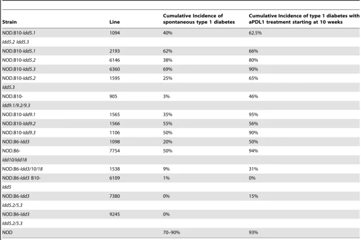
Table 2.Spontaneous incidence of type 1 diabetes at 7 months of age compared to anti PDL1 treatment of 10 week old mice.
Strain Line
Cumulative Incidence of spontaneous type 1 diabetes
Cumulative Incidence of type 1 diabetes with aPDL1 treatment starting at 10 weeks
NOD.B10-Idd5.1 1094 40% 62.5%
Idd5.2 Idd5.3
NOD.B10-Idd5.1 2193 62% 66%
NOD.B10-Idd5.2 6146 38% 80%
NOD.B10-Idd5.3 6360 69% 90%
NOD.B10-Idd5.2 1595 25% 65%
Idd5.3
NOD.B10- 905 3% 46%
Idd9.1/9.2/9.3
NOD.B10-Idd9.1 1565 35% 95%
NOD.B10-Idd9.2 1566 55% 56%
NOD.B10-Idd9.3 1106 50% 90%
NOD.B6-Idd3 1098 20% 50%
NOD.B6- 7754 50% 94%
Idd10/Idd18
NOD.B6-Idd3/10/18 1538 9% 31%
NOD.B6-Idd3B10- 6109 1% 0%
Idd5
NOD.B6-Idd3 7380 0% 15%
Idd5.2/5.3
NOD.B6-Idd3 9245 0%
Idd5.2/5.3
NOD 70–90% 93%
In the current study we examined the effects of 9Iddregions, alone and in combination, on accelerated type 1 diabetes following anti PDL1 treatment. These congenic and subcongenic mice have multiple protective alleles that mediate varying degrees of resistance to type 1 diabetes. We chose the NODIddcongenics (Idd3/5, Idd9, Idd3/10/18 and Idd3/5.2/5.3) that are almost completely protected from spontaneous diabetes occurrence due to allelic interactions between the candidate genes present in the loci and subloci. We also looked at mouse strains that are variations of the above-mentioned strains, containing the individ-ual locus or a combination of loci, where the disease progression is either moderate (Idd3, Idd9.1, Idd5) or relatively high but always lower than that of the NOD parental strain (Idd5.1, Idd5.2, Idd5.3, Idd5.2/5.3, Idd9.1, Idd9.2, Idd9.3, Idd10/18). Frequency of diabetes in these mice strains range from,5% to 65%.
PDL1 blockade is known to accelerate diabetes precipitation in NOD mice. Our aim was to determine if the interaction between the protective Idd loci in the different NOD congenics affects diabetes induction by PDL1 blockade. All except one among the NOD congenic strains tested here,Idd3/5, developed accelerated type 1 diabetes following anti PDL1 treatment. Our data show that PDL1 blockade is not enough to induce accelerated diabetes in the NODIdd3/5congenic mice strain that contains alleles for
Il2, Ctla4, Slc11a1andAcadl. The interaction between these alleles is able to protect the mice from diabetes induction by PDL1 blockade. We do not observe this in the case of any other congenic
strains. This is probably because Ctla4and Il2both modulate the survival and function of Treg cell population and the blockade of PD1-PDL1 pathway is not enough to limit the ability of these Tregs, and tolerance is maintained in theIdd3/5congenic mice. It is also worth mentioning that PDL1 andCtla4mediated tolerance induction functions through two distinct pathways. Allelic interaction ofCtla4with the other candidate genes in theIdd3/5
strain is able to overcome the effect of anti PDL1 treatment and maintain tolerance in these congenic mice. Slc11a1 plays an important part in antigen presenting function of DCs and may play a role in inducing tolerance to self antigens.Acadlis proposed to have a significant role in T cell function and survival by altering fatty acid metabolism. We therefore suggest that the combination of these four candidate genes and their interaction renders the
Idd3/5 congenics resistant to diabetes induction by PDL1 blockade. TheIdd3/5mice have also been shown to be resistant to other experimental autoimmunity [41].
The Idd10/18strain has an insulitis rate of 78%, and ,50%
rate of spontaneous diabetes development [37], in contrast to the
Idd3/10/18 strain which shows greater protection, with 19% of mice developing insulitis and 7% developing type 1 diabetes. The
Idd3/10/18strain demonstrates a median protection against anti PDL1 accelerated diabetes development with a 31% cumulative incidence as opposed to a cumulative incidence of 94% inIdd10/ 18,and 0% in theIdd3/5strain following treatment. TheIdd3/ 10/18strain is almost completely protected from diabetes, similar to theIdd3/5strain. However, the effect of PDL1 blockade results in significantly different outcomes in respect to diabetes induction. These data also imply that in the absence of negative costimulation by the PD1-PDL1 pathway, CTLA4 possibly maintains the self tolerance with the help of IL-2 (one of the candidate genes inIdd3) in case of theIdd3/5 strain. And IL-2 alone is not sufficient to prevent anti PDL1 mediated accelerated diabetes in case of the
Idd3/10/18 strain. Similarly in Idd3/5.2 and Idd3/5.3 strains where the congenic Idd5.1 locus containing the CTLA4 gene is absent, IL-2 alone cannot prevent diabetes induced by PDL1 blockade.
The Idd9strain including sub-congenic9.1, 9.2 and 9.3shows high levels of pancreatic infiltrates (90%), but does not develop diabetes at a high rate (3%) [21]. Nonetheless, this strain develops insulin autoantibodies [27]. The Idd9.1 region was identified to control type 1 diabetes development through TNF-a[42].Idd9.2
andIdd9.3regions were found to be responsible for preventing the expansion of islet specific CD8+T cells, providing an explanation for the dichotomy of high insulitis incidence and a low rate of actual diabetes development in theIdd9strain [24].
Remarkably, a profound increase of diabetes incidence (3% to 50%) was observed after PDL1 blockade inIdd9congenic mice. We studied the effect of anti PDL1 onIdd9subcongenics (Idd9.1,
Idd9.2andIdd9.3). Interestingly, almost 100% ofIdd9.1andIdd9.3
mice developed accelerated diabetes following anti PDL1 treat-ment, in contrast to theIdd9.2strain, which was partially protected (56% became diabetic). Findings in theIdd9strain suggest that in mice that already have infiltrating lymphocytes in target organs at 10 weeks of age, like Idd9.1 and Idd9.3 strains, diabetes development is exacerbated following anti PDL1 treatment.
Idd9.1andIdd9.3strains which develop accelerated diabetes, also show slightly higher scores of insulitis.Idd9.2mice had significantly less pancreatic infiltrates and only 56% developed diabetes after anti PDL1 treatment.
TheIdd9mice strain similar toIdd3/5andIdd3/10/18, is also resistant to spontaneous diabetes occurrence. However, the level of insulitis is much higher in theIdd9strain in comparison to that of
Idd3/5andIdd3/10/18. The difference in the pathogenicity of the
Figure 2. Insulitis scores in anti PDL1 treated mice.Iddcongenic mice were grouped into control treated (untx, white columns,Idd9.1
n = 7, Idd9.2n = 10, Idd9.3n = 9, Idd3n = 4, Idd10/18n = 5, Idd3/10/18
n = 18, Idd3/5 n = 3, NOD n = 7), anti PDL1 treated diabetic (Tx-D, checked columns, Idd9.2 n = 4, Idd3 n = 5, Idd10/18 n = 8, Idd3/10/18
n = 6, NOD n = 11) and anti PDL1 treated non-diabetic (Tx-ND, black columns,Idd9.2n = 5,Idd3n = 5,Idd10/18n = 1#,Idd3/10/18n = 6,Idd3/ 5n = 17) mice. The antibody treatment was given to 10-week old mice at day 0 (500mg), and days 2, 4, 6, 8 and 10 (250mg). The injections
were stopped once the mouse had become diabetic and had a glucose reading of .250 mg/dl on two consecutive days. H&E sections of pancreases were scored for degree of infiltrating lymphocytes in islets. Results are expressed as Mean6SD. P-values are expressed as * (P, 0.05), ** (P,0.01), *** (P,0.0001). # In case of the Idd10/18 mice, treatment with anti PDL1 resulted in 94% of diabetes incidence. Therefore, it was extremely difficult to increase the n of anti PDL1 treated non-diabetic group.
disease in the Idd9 strain is attributed to the Th2 type cellular response induced by the TNFR superfamily gene cluster present in theIdd9locus. The combination of three sub loci Idd9.1, Idd9.2
andIdd 9.3renders this strain resistant to the disease even though individually the candidate genes are susceptible. PDL1 blockade negates this interaction and in the absence of any other functional negative costimulatory pathway, disease progression is accelerated and pathogenicity is altered. Among the three subloci of Idd9, Idd9.2is considered the most potent region in providing protection against the disease by restraining autoreactive CD8+ T cells. Blockade of PDL1 is known to cause CD8+T cell exhaustion. This explains the similarity in the anti PDL1 induced diabetes incidence (approximately 50%) in theIdd9and Idd9.2 strains; whereas the
Idd9.1 and Idd9.3 strains are completely susceptible (90-95%) to anti PDL1 induced diabetes. We also observe a very low frequency of pancreatic infiltrates in theIdd9.2strain. This can be explained by the fact that the strains ofIdd9and its sub regions have a low frequency of autoreactive CD8+T cells in comparison to the NOD mice. Further, the genes in theIdd9sub regions prevent a massive expansion of these autoreactive CD8+
T cells during disease onset and progression [25]. However, extensive insulitis was observed in the group of Idd9.2 mice that become diabetic after PDL1 blockade suggesting that in some of the mice the low frequency of autoreactive cells can expand when this regulatory pathway is inhibited. The decreased cytokine and chemokine production in
Idd9andIdd9.2mice may also be related to the low frequency of
autoreactive T cells [25] that affects the quality of the infiltrating cells following PDL1 blockade as compared to mice not having protective alleles atIdd9.2.
Our study using treatment with anti PDL1 mAb indicates that sufficient numbers of effector cells are present in these congenic strains to mediate type 1 diabetes. The rapid onset of diabetes in some of theIddcongenic strains is probably due to auto-aggressive memory/effector T cells that are suddenly set free when PDL1 is blocked, as has been shown in a study in NOD mice [43].
Further, CD4+
Type II NKT cells were shown as regulators of diabetes and it was shown that these cells were sufficient in down-regulating diabetes, promoting activity of CD4+
BDC2.5 tg T cells
in vivo. Interestingly, blockade of ICOS and PDL1 was found to negate the regulatory effect of the CD4+
Type II NKT cells in the pancreatic lymph node leading to a sudden development of diabetes [44].
We used the Idd3/10/18 strain to further dissect anti PDL1 mediated diabetes. BDC2.5 Tg CD4+
T cells were transferred into NOD andIdd3/10/18mice treated with anti PDL1. Similar rates of T cell proliferation were observed in pancreatic LNs of both strains. Corresponding to CD4+
TCR Tg T cells tested above, CD8+
8.3 TCR Tg+
T cells divided more frequently, as seen by an increase in the number of CFSE-diluted CD8+
T cells inIdd3/10/ 18 mice (and NOD mice) that received anti PDL1 antibody as compared to mice that received control IgG. These data are similar to our findings in regular NOD mice [4] and suggest that
Figure 3. Quantitative PCR detection of cytokine, chemokine and transcription factor levels in pancreas tissue after anti PDL1 treatment inIdd9(n = 3) and subcongenicsIdd9.1(n = 3),Idd9.2(n = 5),Idd9.3(n = 5).Mice (10 weeks old) were treated with 500mg anti PDL1
on day 0 and 250mg anti PDL1 on days 2, 4, 6, 8 and 10 by i.p. injection. Pancreas tissue was harvested on day 30 or when mice had turned diabetic.
Horizontal lines show median value. P-values are expressed as * (P,0.05), ** (P,0.01), *** (P,0.0001) in figure. a): IFN-c:Idd9.1vs.Idd9.2p = 0.0039;
Idd9.1vs.Idd9.3p = 0.063. b): CCR2:Idd9vs.Idd9.1p = 0.0003;Idd9.1vs.Idd9.2p,0.0001;Idd9.2vs.Idd9.3p = 0.0431;Idd9vs.Idd9.3p = 0.0183. c): RANTES:Idd9vs.Idd9.1p = 0.022;Idd9.1vs.Idd9.2p = 0.0016;Idd9.2vs.Idd9.3p = 0.0284;Idd9vs.Idd9.3p = 0.0382. d): No significant differences in FoxP3 expression betweenIdd9and subcongenics. e): MIP-1a:Idd9vs.Idd9.1p = 0.0257;Idd9.2vs.Idd9.1p = 0.0017. f): TNF-a:Idd9vs.Idd9.1p = 0.019;
Idd9.2vs.Idd9.1p = 0.0004.
the lower susceptibility of Idd3/10/18 mice to develop type 1 diabetes following PDL1 blockade is probably not dependent on expansion of CD4 and CD8 T cells, rather that anti PDL1 treatment likely affects behavior of these cells which contributes to lowering susceptibility for developing disease. The role of PDL1 expression in the pancreas and its effect on resistance in this strain remains to be further investigated.
Correlation between the level of basal insulitis and the development of anti PDL1 induced diabetes also proved to be true in the case of Idd3/5 mice to which anti PDL1 was administered. These mice stayed diabetes free. TheIdd3/5strain exhibits profound resistance, has the lowest spontaneous diabetes incidence, and also shows the lowest levels of insulitis among allIdd
congenics.
Prominent genes associated withIdd3/5congenic strain areIl2
andIl21fromIdd3, andCtla4fromIdd5regions [37]. The role of IL-2 in diabetes has been previously demonstrated. NOD mice express less IL-2 than diabetes resistant mouse strains [45], and low dose IL-2 administered at the onset of type 1 diabetes can reverse established disease in NOD mice [46]. This mechanism has been attributed to an increase in regulatory T cell numbers in the pancreas, and to increased expression of FoxP3, CD25, CTLA-4, ICOS and GITR [46]. Lower levels of IL-2 were found to have an impact on antigen presenting cells like DCs, since low
IL-2 levels correlated with higher numbers of DCs and increased T cell stimulation and activation. The cellular mechanism of protection from T1D inIdd3/5 congenic mice strain is already defined by Hamilton-Williams et al. [9].
Further analysis of the Idd3/5region showed that removal of protective alleles at a subcongenic region from theIdd3/5region as in theIdd3/5.2/5.3strain results in a 15% incidence of disease upon anti PDL1 treatment in contrast to no incidence of disease in theIdd3/5group, which supports the role of Ctla4at the Idd5.1
locus in preventing diabetes in a concerted interplay withIdd3. AlthoughIdd3/5.2/5.3mice do not develop diabetes, an increase of insulitis inIdd3/5.2/5.3mice as compared toIdd3/5has been reported [33]. These observations support the hypothesis that the ability of PDL1 to accelerate diabetes relies on some minimal amount of effector cell accumulation that is normally manifested as at least a mild insulitis.
Conclusion
Taken together, our data show that PDL1 blockade destroys the genetic protection mediated by different protective alleles. We show a link between occurrence of insulitis and disease suscepti-bility through a break of tolerance induced by anti PDL1. We suggest that the presence of a functional CTLA4 allele is probably
Figure 4. Proliferation of adoptively transferred BDC2.5 Tg CD4 T and NY8.3 tg CD8+T cells in pancreatic LN and spleen of anti PDL1 treated NOD andIdd3/10/18mice.A) Representative CFSE dilution plot for each group is shown. Cells were gated on CD4+
Vbeta 4+
. B) A representative CFSE dilution plot of transferred NY8.3tg T cells for each group is shown. Cells were gated on CD8+Vbeta 8+.
responsible to prevent disease susceptibility induced by anti PDL1. Increased understanding of the mechanisms of gene-gene interac-tion, and discovering additional traits that play a role in type 1 diabetes will help identify novel treatments of this disease. The PD1-PDL1 pathway is currently studied for developing therapy for cancer and infectious diseases including HIV, since blockade of this pathway results in increased immune responses against tumor cells [1,47,48] and infectious agents [3,49,50]. However, we show that blockade of this pathway interrupts critical tolerance mechanisms that operate to prevent autoimmune diabetes. Acceleration of diabetes following PD1-PDL1 pathway blockade to treat disease underscores the need for caution before proceeding to a widespread use of this form of treatment, especially when used in combination with antiCTLA-4 (Ipilimumab) that is currently approved for use in melanoma. A combined blockade of CTLA4 and PD1-PDL1 will in all probability shift the balance from an effective immune response towards autoimmunity. It is important to note that our group had earlier shown that type 1 diabetes resistant NOR mice, which are congenic for the MHC locus to the
NOD mice, did not develop diabetes following anti PDL1 treatment [51]. The fact that these congenic mice were protected against type 1 diabetes post-anti PDL1 treatment suggests that PDL1 blockade may still prove suitable in human patients without HLA alleles associated with autoimmune disease such as type 1 diabetes.
Future research should focus on strategies to exploit enhanced immune responses by blocking the PD-PDL1 pathway and at the same time prevent the development of autoimmune disease as a consequence.
Author Contributions
Conceived and designed the experiments: IG. Performed the experiments: NMK AJK BZ LA. Analyzed the data: NMK ST AJK BZ. Wrote the paper: NMK ST. Reviewed and Edited the manuscript: MHS DLG LSW IG DR AR. Guarantor of this work and, as such, had full access to all the data in the study and takes responsibility for the integrity of the data and the accuracy of the data analysis: IG.
Figure 5. Proliferation of adoptively transferred BDC2.5 Tg CD4 T and NY8.3 tg CD8+T cells in pancreatic LN and spleen of anti PDL1 treated NOD andIdd3/10/18mice.a) Collective data from 3 out of 8 experiments of the percentage of CFSE-dividing cells (gated on CD4+
Vb4+) are shown. Horizontal lines express mean value. For CD4+T cells pLN, NOD, control vs. treated p = 0.0276; pLN,Idd3/10/18, control vs. treated
p = 0.0002. b) A representative experiment from 3 performed is shown. For CD8 T cells pLN,Idd3/10/18control vs. treated p = 0.0321, spleenIdd3/10/ 18control vs. treated p = 0.0185.
References
1. Blank C, Mackensen A (2007) Contribution of the PD-L1/PD-1 pathway to T-cell exhaustion: an update on implications for chronic infections and tumor evasion. Cancer Immunol Immunother 56: 739–745.
2. Zhou Q, Munger ME, Highfill SL, Tolar J, Weigel BJ, et al. Program death-1 signaling and regulatory T cells collaborate to resist the function of adoptively transferred cytotoxic T lymphocytes in advanced acute myeloid leukemia. Blood 116: 2484–2493.
3. Trautmann L, Janbazian L, Chomont N, Said EA, Gimmig S, et al. (2006) Upregulation of PD-1 expression on HIV-specific CD8+ T cells leads to reversible immune dysfunction. Nat Med 12: 1198–1202.
4. Guleria I, Gubbels Bupp M, Dada S, Fife B, Tang Q, et al. (2007) Mechanisms of PDL1-mediated regulation of autoimmune diabetes. Clin Immunol 125: 16– 25.
5. Fife BT, Pauken KE, Eagar TN, Obu T, Wu J, et al. (2009) Interactions between PD-1 and PD-L1 promote tolerance by blocking the TCR-induced stop signal. Nat Immunol 10: 1185–1192.
6. Wang J, Yoshida T, Nakaki F, Hiai H, Okazaki T, et al. (2005) Establishment of NOD-Pdcd1-/- mice as an efficient animal model of type I diabetes. Proc Natl Acad Sci U S A 102: 11823–11828.
7. Driver JP, Serreze DV, Chen YG (2011) Mouse models for the study of autoimmune type 1 diabetes: a NOD to similarities and differences to human disease. Semin Immunopathol 33: 67–87.
8. Wicker LS, Todd JA, Peterson LB (1995) Genetic control of autoimmune diabetes in the NOD mouse. Annu Rev Immunol 13: 179–200.
9. Hamilton-Williams EE, Cheung J, Rainbow DB, Hunter KM, Wicker LS, et al. (2012) Cellular mechanisms of restored beta-cell tolerance mediated by protective alleles of Idd3 and Idd5. Diabetes 61: 166–174.
10. Liu SM, Lee DH, Sullivan JM, Chung D, Jager A, et al. (2011) Differential IL-21 signaling in APCs leads to disparate Th17 differentiation in diabetes-susceptible NOD and diabetes-resistant NOD.Idd3 mice. J Clin Invest 121: 4303–4310. 11. Anderson AC, Chandwaskar R, Lee DH, Kuchroo VK (2008) Cutting edge: the
Idd3 genetic interval determines regulatory T cell function through CD11b+ CD11c- APC. J Immunol 181: 7449–7452.
12. Yamanouchi J, Rainbow D, Serra P, Howlett S, Hunter K, et al. (2007) Interleukin-2 gene variation impairs regulatory T cell function and causes autoimmunity. Nat Genet 39: 329–337.
13. D’Alise AM, Auyeung V, Feuerer M, Nishio J, Fontenot J, et al. (2008) The defect in T-cell regulation in NOD mice is an effect on the T-cell effectors. Proc Natl Acad Sci U S A 105: 19857–19862.
14. Rainbow DB, Moule C, Fraser HI, Clark J, Howlett SK, et al. (2011) Evidence that Cd101 is an autoimmune diabetes gene in nonobese diabetic mice. J Immunol 187: 325–336.
15. Fraser HI, Dendrou CA, Healy B, Rainbow DB, Howlett S, et al. (2010) Nonobese diabetic congenic strain analysis of autoimmune diabetes reveals genetic complexity of the Idd18 locus and identifies Vav3 as a candidate gene. J Immunol 184: 5075–5084.
16. Hamilton-Williams EE, Martinez X, Clark J, Howlett S, Hunter KM, et al. (2009) Expression of diabetes-associated genes by dendritic cells and CD4 T cells drives the loss of tolerance in nonobese diabetic mice. J Immunol 183: 1533– 1541.
17. Dai YD, Marrero IG, Gros P, Zaghouani H, Wicker LS, et al. (2009) Slc11a1 enhances the autoimmune diabetogenic T-cell response by altering processing and presentation of pancreatic islet antigens. Diabetes 58: 156–164. 18. Araki M, Chung D, Liu S, Rainbow DB, Chamberlain G, et al. (2009) Genetic
evidence that the differential expression of the ligand-independent isoform of CTLA-4 is the molecular basis of the Idd5.1 type 1 diabetes region in nonobese diabetic mice. J Immunol 183: 5146–5157.
19. Kissler S, Stern P, Takahashi K, Hunter K, Peterson LB, et al. (2006) In vivo RNA interference demonstrates a role for Nramp1 in modifying susceptibility to type 1 diabetes. Nat Genet 38: 479–483.
20. Irie J, Reck B, Wu Y, Wicker LS, Howlett S, et al. (2008) Genome-wide microarray expression analysis of CD4+ T Cells from nonobese diabetic congenic mice identifies Cd55 (Daf1) and Acadl as candidate genes for type 1 diabetes. J Immunol 180: 1071–1079.
21. Hamilton-Williams EE, Rainbow DB, Cheung J, Christensen M, Lyons PA, et al. (2013) Fine mapping of type 1 diabetes regions Idd9.1 and Idd9.2 reveals genetic complexity. Mamm Genome 24: 358–375.
22. Lyons PA, Hancock WW, Denny P, Lord CJ, Hill NJ, et al. (2000) The NOD Idd9 genetic interval influences the pathogenicity of insulitis and contains molecular variants of Cd30, Tnfr2, and Cd137. Immunity 13: 107–115. 23. Yamanouchi J, Puertas MC, Verdaguer J, Lyons PA, Rainbow DB, et al. (2010)
Idd9.1 locus controls the suppressive activity of FoxP3+CD4+CD25+regulatory T-cells. Diabetes 59: 272–281.
24. Matsuki N, Stanic AK, Embers ME, Van Kaer L, Morel L, et al. (2003) Genetic dissection of V alpha 14J alpha 18 natural T cell number and function in autoimmune-prone mice. J Immunol 170: 5429–5437.
25. Hamilton-Williams EE, Wong SB, Martinez X, Rainbow DB, Hunter KM, et al. (2010) Idd9.2 and Idd9.3 protective alleles function in CD4+ T-cells and nonlymphoid cells to prevent expansion of pathogenic islet-specific CD8+ T-cells. Diabetes 59: 1478–1486.
26. Cannons JL, Chamberlain G, Howson J, Smink LJ, Todd JA, et al. (2005) Genetic and functional association of the immune signaling molecule 4-1BB (CD137/TNFRSF9) with type 1 diabetes. J Autoimmun 25: 13–20. 27. Waldner H, Sobel RA, Price N, Kuchroo VK (2006) The autoimmune diabetes
locus Idd9 regulates development of type 1 diabetes by affecting the homing of islet-specific T cells. J Immunol 176: 5455–5462.
28. Robles DT, Eisenbarth GS, Dailey NJ, Peterson LB, Wicker LS (2003) Insulin autoantibodies are associated with islet inflammation but not always related to diabetes progression in NOD congenic mice. Diabetes 52: 882–886. 29. Podolin PL, Denny P, Armitage N, Lord CJ, Hill NJ, et al. (1998) Localization of
two insulin-dependent diabetes (Idd) genes to the Idd10 region on mouse chromosome 3. Mamm Genome 9: 283–286.
30. Penha-Goncalves C, Moule C, Smink LJ, Howson J, Gregory S, et al. (2003) Identification of a structurally distinct CD101 molecule encoded in the 950-kb Idd10 region of NOD mice. Diabetes 52: 1551–1556.
31. Wicker LS, Chamberlain G, Hunter K, Rainbow D, Howlett S, et al. (2004) Fine mapping, gene content, comparative sequencing, and expression analyses support Ctla4 and Nramp1 as candidates for Idd5.1 and Idd5.2 in the nonobese diabetic mouse. J Immunol 173: 164–173.
32. Hunter K, Rainbow D, Plagnol V, Todd JA, Peterson LB, et al. (2007) Interactions between Idd5.1/Ctla4 and other type 1 diabetes genes. J Immunol 179: 8341–8349.
33. Lin X, Hamilton-Williams EE, Rainbow DB, Hunter KM, Dai YD, et al. (2013) Genetic interactions among Idd3, Idd5.1, Idd5.2, and Idd5.3 protective loci in the nonobese diabetic mouse model of type 1 diabetes. J Immunol 190: 3109– 3120.
34. Luhder F, Katz J, Benoist C, Mathis D (1998) Major histocompatibility complex class II molecules can protect from diabetes by positively selecting T cells with additional specificities. J Exp Med 187: 379–387.
35. Yamazaki T, Akiba H, Iwai H, Matsuda H, Aoki M, et al. (2002) Expression of programmed death 1 ligands by murine T cells and APC. J Immunol 169: 5538– 5545.
36. Salomon B, Lenschow DJ, Rhee L, Ashourian N, Singh B, et al. (2000) B7/ CD28 costimulation is essential for the homeostasis of the CD4+CD25+ immunoregulatory T cells that control autoimmune diabetes. Immunity 12: 431–440.
37. Pearson T, Markees TG, Wicker LS, Serreze DV, Peterson LB, et al. (2003) NOD congenic mice genetically protected from autoimmune diabetes remain resistant to transplantation tolerance induction. Diabetes 52: 321–326. 38. Hill NJ, Lyons PA, Armitage N, Todd JA, Wicker LS, et al. (2000) NOD Idd5
locus controls insulitis and diabetes and overlaps the orthologous CTLA4/ IDDM12 and NRAMP1 loci in humans. Diabetes 49: 1744–1747.
39. Cameron MJ, Arreaza GA, Grattan M, Meagher C, Sharif S, et al. (2000) Differential expression of CC chemokines and the CCR5 receptor in the pancreas is associated with progression to type I diabetes. J Immunol 165: 1102– 1110.
40. Ridgway WM, Peterson LB, Todd JA, Rainbow DB, Healy B, et al. (2008) Gene-gene interactions in the NOD mouse model of type 1 diabetes. Adv Immunol 100: 151–175.
41. Bour-Jordan H, Thompson HL, Giampaolo JR, Davini D, Rosenthal W, et al. Distinct genetic control of autoimmune neuropathy and diabetes in the non-obese diabetic background. J Autoimmun 45: 58–67.
42. Chamberlain G, Wallberg M, Rainbow D, Hunter K, Wicker LS, et al. (2006) A 20-Mb region of chromosome 4 controls TNF-alpha-mediated CD8+T cell aggression toward beta cells in type 1 diabetes. J Immunol 177: 5105–5114. 43. Paterson AM, Brown KE, Keir ME, Vanguri VK, Riella LV, et al. (2011) The
programmed death-1 ligand 1:B7-1 pathway restrains diabetogenic effector T cells in vivo. J Immunol 187: 1097–1105.
44. Kadri N, Korpos E, Gupta S, Briet C, Lofbom L, et al. (2012) CD4(+) type II NKT cells mediate ICOS and programmed death-1-dependent regulation of type 1 diabetes. J Immunol 188: 3138–3149.
45. Tang Q, Adams JY, Penaranda C, Melli K, Piaggio E, et al. (2008) Central role of defective interleukin-2 production in the triggering of islet autoimmune destruction. Immunity 28: 687–697.
46. Grinberg-Bleyer Y, Baeyens A, You S, Elhage R, Fourcade G, et al. (2010) IL-2 reverses established type 1 diabetes in NOD mice by a local effect on pancreatic regulatory T cells. J Exp Med 207: 1871–1878.
47. Zhang L, Gajewski TF, Kline J (2009) PD-1/PD-L1 interactions inhibit antitumor immune responses in a murine acute myeloid leukemia model. Blood 114: 1545–1552.
48. Topalian SL, Hodi FS, Brahmer JR, Gettinger SN, Smith DC, et al. Safety, activity, and immune correlates of anti-PD-1 antibody in cancer. N Engl J Med 366: 2443–2454.
49. Freeman GJ, Wherry EJ, Ahmed R, Sharpe AH (2006) Reinvigorating exhausted HIV-specific T cells via PD-1-PD-1 ligand blockade. J Exp Med 203: 2223–2227.