CHRONIC PROGRESSIVE EXTERNAL OPHTHALMOPLEGIA
I. A QUANTITATIVE HISTOCHEMICAL STUDY OF SKELETAL MUSCLES
ELZA DIAS-TOSTA *
SUMMARY — This study quantitates the major morphological and cytochemical changes in limb muscle biopsies from 37 patients with the, syndrome of chronic progressive external ophthalmoplegia (CPEO). The aim was to assess the value of limb muscle biopsy in the diagnosis of this syndrome; to define the myopathological changes and to determine whether there were any specific clinico-pathological correlations. Patients were divided into three clinical groups — 11 patients with CPEO with facial and/or limb muscle weakness; 10 with CPEO with facial and/or limb muscle weakness and a positive family history; 16 with CPEO with one or more of the following: pigmentary retinopathy, cerebellar ataxia, pyra-midal signs and peripheral neuropathy. The following parameters were measured: the proportions of histochemical fibre types, the muscle fibre areas and the percentage of muscle fibres showing increased oxidative enzyme activity. Pooled results for each of the clinical categories were compared. Statistical analysis of fibre areas and the percentage of fibres with increased oxidative enzyme activity, showed that group 2 differed from the others (p < 0.05). Patients in group 2 showed the highest incidence of type 1 fibre hypertrophy, type 2A atrophy and the lowest incidence of fibres with increased oxidative activity. Fibre type disproportions occurred in all three groups but the differences were not significant.
Oftalmoplegia externa crônica progressiva: I. Estudo histoquímico quantitativo de músculos esqueléticos.
RESUMO — Estudaram-se quantitativamente as alterações morfológicas e histoquímicas de biópsias de músculos dos membros superiores ou inferiores em 37 pacientes com a síndrome de oftalmoplegia externa crônica e progressiva (OECP). O objetivo era determinar o valor da biópsia do músculo esquelético dos membros no diagnóstico desta síndrome, definir as alterações miopatológicas e determinar se havia alguma correlação clínico-patológica especí-fica. Os pacientes foram classificados em três grupos clínicos: 11 pacientes com OECP mais fraqueza de musculatura facial e/ou de membros; 10 pacientes com OECP, fraqueza muscuiar e história familiar positiva; 16 pacientes com OECP, fraqueza muscular e um ou mais dos seguintes sinais — retinopatia pigmentar, ataxia cerebelar, sinais piramidais e neuropatia periférica. Os seguintes parâmetros foram avaliados: proporção de tipos histoquímicos de fibras musculares, áreas destas fibras, percentagem de fibras mostrando aumento de ativi-dade enzimática oxidativa. Os resultados foram comparados entre os três grupos definidos clinicamente. Análises estatísticas das áreas das fibras e da percentagem de fibras com aumento da atividade enzimática oxidativa, mostram que o grupo 2 difere dos demais (p < 0,05). Os pacientes do grupo 2 mostraram a mais alta incidência de hipertrofia de fibras do tipo 1, atrofia do tipo 2A e a mais baixa incidência de fibras com aumento da atividade oxidativa. A desproporção de tipos de fibras ocorreu nos três grupos, mas sem diferença significativa.
Muscle Histochemistry Laboratory, National Hospital for Nervous Diseases, University of London, Queen Square, London, England: * M.D., Ph.D.
Chronic progressive external ophthalmoplegia (CPEO) is a descriptive term which has been applied to a group of patients who present with slowly progressive ptosis and limitation of ocular movements but with normal pupillary responses to light and convergence. They show no evidence of exophtalmos and the symptoms and signs do not show any significant fluctuation. Classical histopathological techniques did not permit the delineation of distinct pathological entities within the group of CPEO patients. With the additional aid of histochemistry and electronmicroscopy it became possible to identify some distinct morphological features such as mitochon-drial abnormalities9»!9 and rimmed v a c u o l e s8
. The mitochondrial abnormalities have been shown in the group of CPEO patients with multisystem involvement 7-15 while the presence of rimmed vacuoles was used to characterize the group of oculopha-ryngeal dystrophy 8.
The aim of this study was to obtain quantitative histochemical data on the myopathological changes in a group of patients presenting clinically with CPEO. It was intended to use such data to: define the value of limb muscle biopsies in the diagnosis of this syndrome; describe and define the myopathological changes; deter-mine whether this syndrome constitutes one or more clinico-pathological entities. Quantitative electronmicroscopy data will be reported in a subsequent paper.
MATERIAL AND METHODS
Patients — Thirty-seven patients with CPEO were studied. Eleven of them were selected because they were sporadic cases with slowly progressive ptosis and weakness of external ocular muscles, without pupillary changes cr significant diplopia. Ten other cases presented similar clinical features but had a positive family history. The other 16 cases exhibited one or more of the following additional features: atypical pigmentary retinopathy, cerebellar ataxia, pyramidal signs, peripheral neuropathy and cardiac conduction defects.
Muscle biopsies — Open biopsies were taken from one of the following limb muscles: 19 triceps, 9 biceps, 5 extensors in the forearm, 2 deltoid, 2 quadriceps. All samples were rapidly frozen in isopentane, cooled in liquid nitrogen, and cryostat sections 5-10 ^m, stained with the modified Gomori trichrome method ( i o ) , succinic dehydrogenase O7
), PAS, Sudan
black, DPNH-tetrazolium reductase and inenadione-linked ^glycerophosphate dehydroge-nase ( 2 0 ) . The myofibrillar adenosine triphosphatase (ATPase) reactions were used to identify the three major histochemical fibre types ( i , s ) . Quantitative studies — 33 specimens were
suitable for quantitative studies. Fibres with increased oxidative enzyme activity, as shown by the succinic dehydrogenase reactions, were unevenly distributed throughout the sections. For quantitative studies, the area with the highest density of these fibres was selected in each case and photographed. From these areas a sample of 100 muscle fibres was counted, and the percentage of those with increased oxidative enzyme activity was determined. These fibres were typed by comparison with similar areas in sections stained by ATPase reactions. The proportions of the three histochemical fibre types were determined in printed photo-graphies of 3 to 6 different areas from sections stained with the myofibrillar ATPase reactions (at pH 4.4, 4.5 or 4.6). The total area examined in each biopsy contained from 294 up to 348 fibres. The individual area of each muscle fibre was measured on the same sample used for determining the proportions of the histochemical fibre types. These measurements were made with the Reichert Manual Optical Analysis System AM03 (MOP system), and mean diameters were calculated from these areas. The reproducibility of the method of measurement was tested when: different magnifications of the same photographed area were used; the same observer measured the area twice; different observers measured the same area. The results were shown to be reproducible. Comparison between mean diameters obtained by our method using the MOP system and those obtained with the «lesser» diameter (8) were made. In view of the consistently larger fibre diameters ranges obtained using the MOP system of measurement, normal ranges for fibre diameters were considered as 40-80 ^ m for women, and from 50-90 ^m for men.
Data analysis — The following data were determined from each muscle: the percentage of each fibre type, the mean fibre diameter for each fibre type, together with their ranges, standard deviations ( S D ) , standard errors (SE) and relative standard errors (RSE). Histo-grams of the fibre sizes were constructed, and atrophy factors ( A F ) , hypertrophy factors (HF) and variability coefficients (v.c.) were calculated for each sample (2,3). The proportions
variables the following analyses were used: Student's «t» test to compare two means; one way analysis of variance for the comparison of more than two means; correlation coefficient (Pearson) to measure the level of linear association between two variables. Variables with a non-normal distribution were compared by using the Spearman rank correlation coefficient and the Kruskal-Wallis one way analysis of variances by ranks.
RESULTS
On the basis of the clinical features and the family history, patients were divided into three clinical groups — A, sporadic cases with muscle weakness only (11 patients); B, familial cases with muscle weakness only: 10; C, patients with clinical and laboratory evidence of multisystem disease: 16. The overall clinical findings are presented in Fig 1.
OVERALL CLINICAL FINOINGS
GROUP A In = I I I GROUP B (r, = 10) GROUP C (o * 16) OPHTHALMOPLEGIA
PTOSIS . . . .. ._.
\
1
1
• i Id C.P.E.O. \1
1 »i Id BULBAR SYMPTOMS WEAKNESS FACE 3 ' i \1
1_ J
1
NECK TRUNK UPPER LIM8S modvrat* • J Id LOWER LIMBS «od»rat• «i Id FATIGABILITY FAMILY HISTORY RETINOPATHY CEREBELLAR ATAXIA PYRAMIDAL SIGNS PER1PH. NEUROPATHY CARDIAC DEFECTS SHORT STATURE EARLY ONSET1
1
1
1
NECK TRUNK UPPER LIM8S modvrat* • J Id LOWER LIMBS «od»rat• «i Id FATIGABILITY FAMILY HISTORY RETINOPATHY CEREBELLAR ATAXIA PYRAMIDAL SIGNS PER1PH. NEUROPATHY CARDIAC DEFECTS SHORT STATURE EARLY ONSET
•
Z I
i~ l
Z )
— 11
] 1 NECK TRUNK UPPER LIM8S modvrat* • J Id LOWER LIMBS «od»rat• «i Id FATIGABILITY FAMILY HISTORY RETINOPATHY CEREBELLAR ATAXIA PYRAMIDAL SIGNS PER1PH. NEUROPATHY CARDIAC DEFECTS SHORT STATURE EARLY ONSET•
Z I
11
] 1 NECK TRUNK UPPER LIM8S modvrat* • J Id LOWER LIMBS «od»rat• «i Id FATIGABILITY FAMILY HISTORY RETINOPATHY CEREBELLAR ATAXIA PYRAMIDAL SIGNS PER1PH. NEUROPATHY CARDIAC DEFECTS SHORT STATURE EARLY ONSET•
Z I
1flnn
n
NECK TRUNK UPPER LIM8S modvrat* • J Id LOWER LIMBS «od»rat• «i Id FATIGABILITY FAMILY HISTORY RETINOPATHY CEREBELLAR ATAXIA PYRAMIDAL SIGNS PER1PH. NEUROPATHY CARDIAC DEFECTS SHORT STATURE EARLY ONSET•
Z I
1
i
Î0 10 60 80 100 ?0 40 60 80 100 X «.0 60 »0 100 7. OF CASES Y. OF CASES * OF CASES
Fig. 1 — The overall clinical findings.
Fibres with increased oxidative Fibre types enzyme activity Cases Muscle*
1 2A 2B 1 2A 2B Total
Al B 92 6 2 19
—
—
19A2 B 37 28 35 5 14 3 22
A3 T 27 42 31 4
—
—
4A4 T 42 46 12
—
—
-
-A6 D 71 27 2
—
—
—
—
A7 T 34 39 27 6 3
—
9A8 T 24 23 53
—
1 1 2A10 B 50 24 26
—
—
—
—
A l l T 64 26 10 20 5
—
25Bl T 62 23 15
—
—
—
—
B2 T 29 53 18
—
—
—
—
B3 Q 79 20 1
—
—
—
B4 E 98 1 1
—
—
—
—
B5 T 21 46 33
—
—
—
—
B6 B 74 25 1
—
—
—
-B7 B 70 26 4
—
—
—
—
B8 E 50 36 14
—
—
—
B9 D 83 17 < 1 3
—
—
3CI T 78 16 6 10 1
—
11C2 Q 28 35 37 4 1
—
5C3 T 41 48 11 1 6
—
7C4 T 33 27 40 6 3 1 10
C5 T 32 37 31
—
—
—
—
C6 E 95 5
—
17 5—
22C7 B 54 31 15 31 6
—
37C8 T 37 35 28 1
—
—
1CIO T 22 31 47
—
2 2 4C l l T 36 27 37 9
**
**
11C12 B 28 21 51 1
—
—
1C13 E 94 5 1 36 3
—
39C14 B 61 27 12 4 1
—
5C15 T 57 32 11 5 3
—
8C16 T 74 23 3 7 1
—
8Table 1 — Proportions of fibre types mid fibres with increased oxidative enzyme activity.
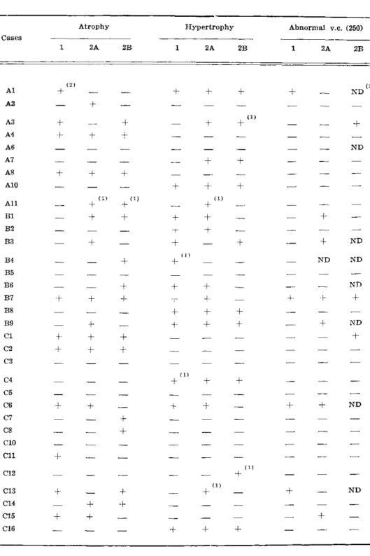
Table 2 — Fibre s\ze abnormalities: atrophy, hypertrophy and variability coefficient (v.c).
(1), 10% or more of the fibres had abnormal diameters but the corresponding atrophic factors (AF) or hypertrophic factors (HFj were not abnormal. (2), abnormal value for
from group B and one from group A, the atrophic angulated fibres were darker in DPNH-tetrazolium reductase and menadione linked a-glycerophosphatase. They were shown with ATPase stains to be of all fibre types. In cases A , B and B these fibres were arranged
1 0 6 8
in small groups. Occasional fibres undergoing necrosis and phagocytosis were seen in 4 cases from group A, three from group B and 5 from group C. A slight increase in the amount of endomysial connective tissue was seen in three out of 11 cases from group A, three out of 10 from group B and 4 out of 16 from group C.
Quantitative studies — The presence of fibres with increased oxidative enzyme activity was the predominant change in 21 of the 33 cases: 6 from group A, one from group B and 14 from group C. The percentage of the «ragged-red» fibres ranged from 1% to 39% in different cases. In the majority (13 cases) their incidence was between 5% and 25% and in 6 cases they contributed less than 5%, and in two cases more than 30% of the total fibre count. Fibre type proportions were normal in 18 cases. Type 1 predominance (in these series ranged from 61% to 98%) was always present in those 15 biopsies with fibre type dysproportion: three cases from group A, 6 from group B and 6 from group C. (Table 1). In no biopsy a complete fibre type uniformity was found.
Fibre measurements — Thirty-three biopsies were studied quantitatively and the abnormal results are listed in Table 2. In biopsies of patients in group A and C some fibres from all three types were found to have atrophy unselective for fibre type. In group B, however, atrophy of fibres 2A and 2B predominated. Only one case showed type 1 atrophy (Table 2). In addition, the three clinical groups differed in regard to the lower range diameter values for type 2A fibres, the lowest being found in group B. The type 1 fibre atrophy which occurred in some of our patients from group A and C, was neither a constant nor a selective finding.
Data analysis — Preliminary statistical analyses were performed to determine whether a number of differences between biopsies could have influenced the results obtained. The variables tested included: the site of the biopsy (biceps, deltoit, triceps, extensors in the forearm and quadriceps), the side (left or right) of the body from which the muscle specimens were taken, the patient's aee at biopsy and the relative proportions of males and females hi the samples. From the entire sample (n=33) the following results were obtained One way analysis of variance showed that the site of the biopsied muscle influenced the fibre type proportions. The patient's age at biopsy was positively correlated with the mean fibre sizes. The quantitative data were pooled according to the three clinical groups (A, B and C) and the grouped means were analysed. Because of the results stated above, when comparisons of fibre sizes were made between grouped sets of biopsies, we first examined whether there were ase differences between biopsy groups that might influence the results. W e also investigated whether the location of the biopsied muscle affected the results when the fibre type proportions were compared. Having certified that the age and the location of the biopsies in those grouped means were not influencing the results, further analysis were carried out between the groups in relation to the quantitative data obtained in light microscopy. The results showed a statistically significant difference (p < 0.05) between the groups in relation to the mean size of type 1 fibre, type 1 fibre hypertrophy factor, the proportion of large type 1 fibres, the lower size ranges of type 2A fibre and the proportion of fibres having increased oxidative enzyme activity. In the samples with «ragged-red» fibres the proportion of these fibres correlated positively with the proportion of type 1 fibre, and negatively with the proportions of type 2A and 2B fibres.
COMMENTS
140 Arq. Neuro-Psiquiat. (Sao Paulo J 46(2) 1988
with hereditary chronic progressive external ophthalmoplegias. Signs of myiopathic changes were present in 4 out of the 6 biopsies, with vacuolated fibres, with the exception of one case ( B5) with very mild clinical involvement. It is suggested that
the vacuoles represent the first sign of the degenerative myopathic process in those individual muscle fibres. As in hereditary muscular dystrophies, progress of the disease leads to loss of muscle fibres and their replacement by fat and connective tissue, with consequent clinical weakness. In our series, the advanced stage of this process was present in two cases ( B6 and B7) . The impression of a myopathic process
was reinforced by the abnormal variability coeficient. It was more frequently found in patients from group B and mostly due to variation in the size of fibre type 2A.
Hypertrophy of one or more fibre types was a major feature of biopsies from patients from group B, but was less common in group A and infrequent in group C. Although all fibre types were affected, hyperthophy most commonly involved type 1 and 2A. It is possible that the lower incidence of type 2B hypertrophy reflected a deficiency of this fibre type. In addition, the grouped mean size of type 1 fibres differed in the three clinical groups A, B and C, the largest mean value being found in group B. The difference between the grouped mean fibre sizes appeared not to be influenced by patient's age at the time of biopsy nor to be related to the duration of symptoms. In human pathological material, fibre hypertrophy has been described in denervation with or without reinnervation l, in some forms of muscular dystrophy, such as fascioscapulohumeral dystrophy 8, and also in carriers of Duchenne muscular dystrophy1 4
. In our series of cases, fibre hypertrophy was found in biopsies showing pathological features of myopathy exclusively, in those with pathological features of myopathy and denervation (small groups of atrophic angulated fibres), in cases in which "ragged-red" fibres were the only abnormality, and in cases lacking any other obvious abnormality. The presence of hypertrophy could not be correlated with clinical signs either; being found in clinically normal muscles ( A7) , in mildly weak
muscles ( C4, C6) , and in severely weak muscle ( B2, B7) . Furthermore, hypertrophy
could not be correlated with duration of disease, being found in muscle biopsies from patients in which the duration of the disease was as short as four years ( B5) or as
long as 53 years ( B2) . Moreover, no evidence of hypertrophy was found in case C3,
biopsied 30 years after the onset of symptoms. In Dubowitz and Brooke's series of 9 familial cases of CPEO, 6 had fibre 2A hypertrophy, and one type 1 hypertrophy. It is interesting that in their series the type 1 preferentially showed atrophy.
The three clinical groups differed also in regard to the lower range diameter values for type 2A fibres, the lowest being found in group B. The type 1 fibre atrophy which occurred in some of our patients from group A and C, was neither a constant finding, nor the selective fibre atrophy as seen in congenital fibre type disproportion 8. The special behaviour of type 1 fibres in our group B may be related to both its physiological properties and the nature of the disease process in this syndrome. In groups A and C, in which there was evidence of mitochondrial abnor-malities (fibres with increased succinic dehydrogenase activity) type 1 fibres which depend upon aerobic glycolysis, were generally atrophic. In group B type 2 atrophy appeared to predominate and to be associated with compensatory hypertrophy of type 1 fibres. In group B type 1 predominance was the most frequent disproportion found, and was combined with fibre type 1 hypertrophy in 6 out of 9 cases. This finding may have diagnostic value in this group of genetically determined myopathies. The three cases in group B without fibre type 1 predominance were B2, B3 and B8.
The former was clinically atypical for this group presentation, complaining of excessive fatigability and also had a very early onset of the disease. In case B5 the disease
was clinically very mild and the muscle biopsy was suggestive of an early stage in the pathological process.
Although it is accepted that fibres type characteristics are influenced by neural activity 6,13, in the majority of human pathological conditions fibre type 1 predomi-nance has been associated with myopathic processes, mainly those genetically deter-mined 12. Changes in the proportion of fibre type have generally been attributed to either fibre type transformations or to selective fibre type atrophy *2,16. Studies in human muscle n have suggested the possibility of interconversion of fibre types 1
remaining fibre types. However, in cases Alf A6, C1 6 in whose biopsies type 1 predominance was evident, there was no evidence of fibre type 2A or 2B atrophy or necrosis which would be the expected intermediate stage before fibre disappearance.
In conclusion, clinical evidence has been presented to justify the classification of patients with CPEO in three groups. In these muscle biopsies analyses our data suggest that this series of patients contains two clinicopathological entities: CPEO witn fibres with increased oxidative enzyme activity and CPEO with occasional vacuo-lated muscle fibres. Features of non-specific myopathy was found in some patients from both groups. The quantitative studies reinforced the clinical and qualitative impression that group B patients are different from the other two groups (A and C ) . The group B patients showed the highest incidence of type 1 fibre hypertrophy, the smallest range of size of type2A fibres, and the lowest incidence of fibre with increased oxidative enzyme activity. The clinical differences found between groups A and C, and within patients from group C was suggested by the quantitative studies to be due to either a heterogeneity in the disease process or to different degrees of severity of the same disease.
Acknowledgements — I am grateful to Dr. J.A. Morgan-Hughes and Dr. D.N. Landon for their constant interest and helpful criticism during the preparation of this work, and also made available to me the laboratory facilities in their departments. I also like to thank Dr. A. Pullen and Miss E. Paul for their helps with the statistics, Miss M. Ellison for excelent technical assistance. Dr. Tosta was supported during preparation of this work by the Brazilian Government.
REFERENCES
1. Bernat JL, Ochoa J L — Muscle hypertrophy after partial denervation: a human case. J Neurol Neurosurg Psychiat 41 : 719, 1978.
2. Brooke MH, Engel W K — The histographic analysis of human muscle biopsies with regard to fibre types: 1. Adult male and female. Neurology 19 : 221, 1969.
3. Brooke MH, Engel W K — The histographic analysis of human muscle biopsies with regard to fibre types: 2. Diseases of the upper and lower motor neuron. Neurology 19 : 378, 1969.
4. Brooke MH, Kaiser K K — Muscie fibre types : how many and what kind? Arch Neurol 23 : 369, 1970.
5. Brooke MH, Kaiser K K — Three «Myosin Adenosine Triphosphatase» systems: the nature of their p H lability and sulphydryl dependence. J Histochem Cytochem 18 : 670, 1970. 6. Buller AJ, Pope R — Plasticity in mammalian skeletal muscle. Philos Soc London
(Biol) 278 : 295, 1977.
7. Castaigne P, Laplane D, Fardeau M, Dordain G, Autret A, Hirt L — Myopathie avec anomalies mitochondriales localisées aux fibres de type 1: documents cliniques, histo-¬ chimiques et ultrastructuraux concernant une forme atrophique diffuse à debut oculaire. Rev Neurol 126 : 81, 1972.
13. Lömo T — The role of activity in the control of membrane and contractile properties of skeletal muscle. In: Thesleff S ( e d ) : Motor Innervation of Muscle. Academic Press, New York, 1976, pg 289.
14. Maunder-Sewry CA, Dubowitz V. — Needle muscle biopsy for carrier detection in Duchenne muscular dystrophy: 1. Light microscopy, histology, histochemistry and quantitation. J Neurol Sci 305, 1981.
15. Morgan-Hughes JA, Mair WGP — Atypical muscle mitochondria in oculoskeletal myopathy. Brain 96 : 215, 1973.
16. Munsat TL, McNeal D, Waters R — Effects of nerve stimulation of human muscle. Arch Neurol 33 : 608, 1976.
17. Nachlas MM, Tsou KC, de Souza E, Cheng CS, Seligman AM — Cytochemical demons-tration of succinic dehydrogenase by the use of a new p-nitrophenyl substituted dite-trazole. J Histochem Cytochem 5 : 420, 1957.
18. Novikoff AB, Shin MY, Drucker J — Mitochondrial localization of oxidative enzymes: staining results with two tetrazolium salts. J Biophys Biochem Cytol 9 : 47, 1961. 19. Olson W, Engel W K , Walsh GO, Einaugler R — Oculocraniosomatic neuromuscular
disease with «ragged-red» fibres: histochemical and ultrastructural changes in limb muscles of a group of patients with idiopathic progressive external ophthalmoplegia. Arch Neurol 26 : 193, 1972.