Cop
yright
© ABE&M t
odos os dir
eit
os r
eser
vados
.
Congenital leptin deiciency:
diagnosis and effects of leptin
replacement therapy
Deiciência congênita de leptina: diagnóstico e efeitos da terapia de reposição
Gilberto Paz-Filho1, Claudio Mastronardi1, Tuncay Delibasi2, Ma-Li Wong1, Julio Licinio1
SUMMARY
To describe our 10-year experience in treating leptin-deficient humans. Three adults and one boy presented with childhood-onset morbid obesity, hypogonadism and family history of obesity and early death. Serum leptin was inappropriately low. A recessive C105T leptin gene mutation was identified. Metabolic and endocrine assessments were conducted, before and while on and off leptin. The adults’ body mass index decreased from 51.2 ± 2.5 to 29.5 ± 2.8 kg/m2. Serum lipids normalized, insulin resistance decreased, and one of the initially diabetic females became normoglycemic. Hypogonadotropic hypogonadism was reversed, and other changes were observed in the adrenal, sympathetic, somatotropic and thyroid functions. Leptin replacement therapy reverses endocrine and metabolic alterations associated with leptin deficiency. Some of these results may be extrapolated to other diseases. Arq Bras Endocrinol Metab. 2010;54(8):690-7
SUMÁRIO
Descrever nossa experiência de 10 anos tratando pacientes deficientes em leptina. Três adultos e um menino apresentaram obesidade mórbida com início na infância, hipogonadismo e histó-ria familiar de obesidade e morte precoce. A leptina sérica era inaprophistó-riadamente baixa. A mu-tação recessiva C105T no gene da leptina foi identificada. Avaliações metabólicas e endócrinas foram realizadas antes e durante o tratamento. O índice de massa corporal dos adultos baixou de 51,2 ± 2,5 para 29,5 ± 2,8 kg/m2. Houve normalização dos lipídios séricos, a resistência in-sulínica diminuiu e a paciente que era diabética se tornou normoglicêmica. O hipogonadismo hipogonadotrópico foi revertido e outras alterações foram observadas nas funções adrenal, simpática, somatotrópica e tireoidiana. A reposição de leptina reverte as alterações endócrinas e metabólicas associadas com a deficiência de leptina. Alguns desses resultados podem ser extrapolados para outras doenças. Arq Bras Endocrinol Metab. 2010;54(8):690-7
1 John Curtin School of Medical
Research, The Australian National University, Canberra, Australia
2 Department of Endocrinology
and Metabolism, Ankara Diskapi Training and Research Hospital Ankara, Turkey
Correspondence to: Julio Licinio
Garran Rd, building 131 Acton, ACT 0200 Australia
Received on Jul/27/2010 Accepted on Sept/27/2010
INTRODUCTION
L
eptin is a hormone produced mainly by the whiteadipose tissue, with multiple actions in the endo-crine and immune systems, including glucose homeo-stasis, reproduction, bone formation, tissue remodeling and inlammation (1). Leptin is a key regulator of en-ergy homeostasis, by regulating enen-ergy intake and ex-penditure through its actions on the arcuate nucleus of the hypothalamus (2,3). Leptin levels are positively cor-related with fat mass, being increased in obesity (4,5).
Among mice that are leptin-deicient (ob/ob) or
leptin-resistant (db/db), blunted metabolic rate and
hyperphagia are characteristic features and obesity is a
hallmark feature (6-10). In particular, ob/ob mice also
have cellular immune deiciency, hypogonadotropic hypogonadism, hypercortisolism, decreased levels of growth hormone, type 2 diabetes and central hypothyroidism. This phenotype is prevented or even reversed with the administration of exogenous leptin.
Cop
yright
© ABE&M t
odos os dir
eit
os r
eser
vados
.
Congenital leptin deiciency
absent leptin levels due to abnormal deposition and distribution of adipose tissue. In addition, congenital human leptin deiciency may also be caused by mutations in the leptin gene.
These forms of leptin deiciency due to mutations in the leptin gene are extremely rare, with 20 patients having been identiied in the world to date. These are patients of Pakistani (n = 12) (8,11,12), Turkish (n = 5) (7,13,14), Egyptian (n = 2) (15) and Austrian (n = 1) (16) background. The evaluation of those pa-tients before and during leptin replacement therapy has unveiled the importance of leptin in the homeostasis of several systems, such as the brain, immunity and glu-cose metabolism.
In this manuscript, we have summarized the diagnostic approach and the phenotypic indings of ive patients from a unique Turkish leptin-deicient family with a mutation in the leptin gene. Moreover, we describe the endocrine and metabolic effects of leptin replacement therapy in these patients.
PATIENTS AND METHODS
In 1998, investigators from the Gulhane Medical School in Ankara, Turkey and from the Cochin In-stitute of Molecular Genetics, University of Paris VII identiied two adults (male A, 22-year-old; female B, 34-year-old) and one 6-year-old girl from Turkey, who presented with morbid obesity starting at early childhood, hypogonadotropic hypogonadism (in the adults), and extremely and unappropriately low serum leptin levels (0.9 ng/mL for the adult male, 1.6 ng/ mL for the adult female, and 1.1 ng/mL for the girl). Due to this unique phenotype, the diagnosis of leptin deiciency was suspected.
Subsequently, a Mendelian recessive leptin gene
mutation was identiied, consisting of a C→T
substi-tution in codon 105 of the leptin gene, resulting in
an Arg→Trp replacement in the mature protein. That
substitution abolishes an MspI restriction site,
allow-ing rapid PCR screenallow-ing for the mutation (7). This is
the same mutation that is observed in the ob/ob mouse
where it causes a premature stop codon.
For the identiication of the mutation, the cod-ing region of exon 3 is ampliied by PCR. Primers derived from the human sequence (GenBank acces-sion no. NM_000230.2) were used. The sense primer
(5′-CAGTCAGTCTCCTCCAAACA-3′)
correspond-ed to nucleotides 202-221, and the antisense primer
(5′-CTTAACGTAGTCCTTGCAGG-3′) was
comple-mentary to nucleotides 580-600.
Subsequently, a restriction enzyme assay is per-formed with DNA of the PCR-ampliied coding region
of exon 3, digested by MspI. Since the mutation causes
the disappearance of one of the two MspI sites, patients
that are homozygous for the mutation will show only two bands of 29 and 370 bp whereas patients without the mutation will have three bands of 29, 139, and 231 bp in the agarose gel. With regard to heterozygous pa-tients, they will display 4 bands of 29, 139, 231 and 370 bp (Figure 1). That particular mutation leads to the synthesis of a truncated protein that is not secreted into the medium, as evaluated in early functional stu-dies (7).
Figure 1. Agarose gel of homozygous, heterozygous and wild-type patients for the C105T mutation of the leptin gene. Adapted from (7) with permission.
Patients 1 and 2 are homozygous, patients 3 and 4 are heterozygous, and patients 5 and 6 do not carry the C105T mutation of the leptin gene.
Samples 2, 4 and 6 were submitted to MspI digestion.
In 1999, in collaboration with Ozata, we identiied in this extended family a new adult homozygous female (patient C, 30-year-old) (13), who was severely obese and amenorrheic (6). Subsequently, a 5-year-old boy (patient D) from the same pedigree was diagnosed with leptin deiciency. In this family, all but one wild-type and heterozygous individual had normal body weight, or were overweight. One heterozygous male individual
(patients D’s father) is obese (BMI of 32 kg/m2),
dys-lipidemic (total cholesterol = 226 mg/dL, triglycerides = 340 mg/dL, HDL = 30 mg/dL and LDL = 128 mg/dL) and has been recently diagnosed with type 2 diabetes in two oral glucose tolerance tests (95 mg/dL at baseline and 206 mg/dL 2h after 75 g of glucose).
Eight members of this family, whom we presume to have been leptin-deicient, had severe obesity and died during childhood due to infections. This number in-cludes the 6 year-old girl, who died before the initiation of treatment. The pedigree chart is illustrated in igure 2.
M 1 2 3 4 5 6
Cop
yright
© ABE&M t
odos os dir
eit
os r
eser
vados
.
Figure 2. Pedigree chart of the Turkish cohort. Patient A: 34-year-old male; patient B: 44-year-old female; patient C: 49-year-old female; patient D: 10-year-old boy (actual ages); patient E: 9-year-10-year-old deceased girl.
LEPTIN REPLACEMENT THERAPY
At the diagnosis, clinical assessments of the endocrine, sympathetic, and immune functions were performed. Subsequently, over 10 years, we evaluated the effect of leptin replacement therapy with daily injection of re-combinant methionyl human leptin (r-metHuLeptin, Amylin Pharmaceuticals, San Diego, USA). In addi-tion, we assessed the effects of brief periods of leptin withdrawal and reinstitution.
Treatment with r-metHuLeptin was initiated at ages 5 (boy – patient D), 27 (adult male − patient A), 30 and 40 (females B and C). Leptin replacement therapy is undertaken at low physiological doses, start-ing at 0.02-0.04 mg/kg/day given subcutaneously at 6 pm. A daily subcutaneous injection in the evening was chosen to mimic leptin’s normal circadian rhythm, which peak occurs at night (17). For the adults, this dose increases serum leptin to levels that are normally observed in adult males with 20% body fat, or in adult females with 30% body fat. The child’s initial dose was calculated to increase the peak serum leptin to 70 ng/ mL (18,19). Subsequently, doses were recalculated and scaled-down, avoiding excessively rapid weight loss.
The adults’ initial mean dose was 4.1 ± 1.2 mg/ day: 2.8 mg for male patient A, 4.2 mg for female pa-tient B, and 5.3 mg for female papa-tient C. The current dose is 1.4 ± 1.9 mg/day: 0.3 mg for A, 0.45 mg for B,
and 3.6 mg for C. The child’s initial dose was 1.36 mg/ day, and the current dose is 0.95 mg. More signiicant decreases in leptin dose for patient C resulted in weight gain possibly because that patient presents higher leptin resistance, associated with common obesity.
PHENOTYPE BEFORE AND DURING TREATMENT
Body composition, food intake and energy expenditure
The most evident phenotypic inding in our patients was morbid obesity. The adults’ initial mean body mass index
(BMI) was 51.2 ± 2.5 kg/m2 (51.4, 46.7, and 55.4 kg/m2,
for patients A, B, and C respectively). After 18 months of treatment, the patients reached a stable mean BMI of
26.9 ± 2.1 kg/m2 (24.8, 26.1, and 31.3 kg/m2 for A, B,
and C respectively) (Figures 3 and 4). Since early 2003, the patients’ weight has remained fairly stable, and the
latest BMI (as of March 2010) was 29.5 ± 2.8 kg/m2.
The boy also lost a signiicant amount of weight, going
from a BMI of 39.6 kg/m2 before treatment at age 5,
to 22.6 kg/m2 at age 9 (Figure 5). Most of the decrease
in BMI was attributed to fat mass loss, as measured by DXA (13). In 2001, the initial total body fat percentages were 43.7, 45.7, and 49.9% for patients A, B, and C, re-spectively. In 2007, those percentages were equal to 7.0, 36.4, and 45.0%, respectively for patients A, B, and C.
A C
B
Cop
yright
© ABE&M t
odos os dir
eit
os r
eser
vados
.
Congenital leptin deiciency
Figure 3. Adult patients before (A) and 18 months after treatment (B). Adapted from (13) with permission.
From left to the right: patients C, B and A (blurred faces), before (a) and 18 months after r-metHuLeptin treatment (b).
Figure 4. Weights (A), energy intake (B), and r-metHuLeptin dose (C) over the first 18 months of treatment. Adapted from (13) with permission.
Before treatment, patient A had low bone mineral density (BMD) at the lumbar spine (BMD of L2–L4,
0.924 g/cm2; T-score -1.96; Z-score -2.36), and the
females had normal BMDs at all sites. After 6 years, the male’s BMD at the lumbar spine increased by 11%
(BMD of L2–L4, 1.042 g/cm2; T-score -1.5; Z-score
-1.1). The females’ BMDs remained within normal range, without signiicant changes.
Weight loss was achieved without any instruction on dietary changes or on increase in physical activity. In the adults, leptin replacement decreased energy intake from 2,384 ± 946 kcal/day to 1,179 ± 790 kcal/day (Figure 4). In addition, leptin replacement changed the macronutrient content of the patients’ diet, with an increase in carbohydrate and a decrease in fat con-sumption (20). In the child, after two years, caloric in-take decreased from 2,709 ± 370 to 2,194 ± 292 kcal/ day, which is 106% of the recommended caloric intake for a boy this age, height and weight (14). Activity lev-els were measured in the adults by actigraphy, which showed progressive and linear increases (13).
Before treatment, 24-h energy expenditure and 24-h fat oxidation in the adults were comparable to those of age-, sex- and weight-matched controls. During treat-ment, the weight loss-associated decrease in energy ex-penditure was less pronounced in our patients than in controls under a weight loss program. In addition, fat oxidation was also higher in our patients. Therefore, leptin replacement prevented the reduction in meta-bolic rate that is associated with weight loss (21).
Lipid and glucose metabolism
Before treatment, all patients had low HDL-cholesterol and normal or high triglycerides and insulin (Table 1). In addition, the older female was diagnosed with type 2 diabetes (6,13). Leptin replacement normalized serum lipid, glucose and insulin levels, leading to the resolu-tion of type 2 diabetes in patient C (13). These changes are sustained until the present time.
A A
B
C B
Patient A Patient B Patient C
C A B
Weight (kg)
140
120 100
80 60
40 -2 0 2 4 6 8 10 12 14
Months
16 18
Energy intake (kcal/d)
3.000
2.000
1.000
0
-2 0 2 4 6 8 10 12 14 Months
16 18
r-metHuLeptin (mg/d)
6
4
2
0
-2 0 2 4 6 8 10 12 14 Months
Cop
yright
© ABE&M t
odos os dir
eit
os r
eser
vados
.
Figure 5. Patient’s D growth and body mass index charts. The patient experienced substantial weight gain in the last evaluation due to a brief period of shortage of supplies.
Table 1. Serum lipids, glucose and insulin before and after treatment
Total cholesterol (mg/dL)
HDL-c (mg/dL)
LDL-c (mg/dL)
Triglycerides (mg/dL)
Insulin (uU/mL)
Glucose ng/dL Before After# Before After# Before After* Before After# Before After# Before After#
A 137 96 29.9 53.3 82 29 125 69 4.8 1.8 91 78
B 115 110 36.8 51.2 62 49 79 47 3.8 1.8 88 84
C 181 125 28.6 38.4 103 64 247 115 7.5 3.1 131 86
D* 166 155 36 65 87 66 216 120 21 7 79 87
* After 2 years; # After 18 months.
By measuring glucose, insulin and C-peptide and mathematically modeling glucose homeostasis, dur-ing a 24-h period of intensive blood sampldur-ing (every 7 minutes; 207 samples in total/24h), with standardized meals used as tolerance tests, we observed that leptin replacement increased insulin sensitivity by at least 5.7-fold, decreased pancreatic insulin secretion calculated by deconvolution of plasma C-peptide levels and also decreased hepatic extraction of insulin (22) estimated
as the instantaneous differences between insulin secre-tion and post hepatic delivery rates.
During the 4th, 5th, and 6th year of treatment, the
Cop
yright
© ABE&M t
odos os dir
eit
os r
eser
vados
.
clamps) while off leptin, as the newly acquired adipose tissue absorbed glucose in excess (23).
Gonadal and reproductive function
Before treatment, the adults were hypogonadic. The adult male was prepuberal: no beard, bilateral gyneco-mastia, scanty pubic and axillary hair, small penis, small testis, and azoospermia. Patient C had spontaneous menarche at age 35, and had scanty pubic and axillary hair, small uterus and ovaries, and no mammary tissue. The younger female had spontaneous menarche at age 29, with normal pubic and axillary hair, small ovaries and borderline uterus, and diminished mammary tis-sue. Gonadotropin responses to GnRH stimulation were normal, which favors the diagnosis of hypogo-nadotropic hypogonadism (Table 2).
After treatment, menstrual periods became regular in both patients, with serial midluteal phase progester-one measurements > 10 ng/mL, which are indicative of ovulation. The male adult’s testosterone and free testosterone levels reached normal values for adults. All adults fully developed secondary sexual characteristics and developed normal sexual function (13).
Adrenals and sympathetic tone
Contrary to ob/ob mice, leptin-deicient humans do not
present with hypercortisolemia. Our patients had nor-mal levels of free urinary cortisol. Serum cortisol levels were 11.5, 22.3, and 26.4 μg/dL for patients A, B, and C respectively. Those levels were suppressed to less
than 5 μg/dL with 1 mg of dexamethasone (6). Patient A was submitted to frequent blood sampling over 24 hours, which showed that leptin replacement increases the mean 24-hour levels of serum cortisol, from 4.04 ± 0.22 μg/dL to 5.97 ± 0.30 μg/dL. Leptin also al-tered the circadian rhythms of cortisol, by decreasing the number of pulses from 25 to 19, increasing their amplitude, increasing the morning peak, and increasing regularity (13).
Low sympathetic tone was shown in the adults (pa-tients A, B, and C) and in the girl (patient E), evalu-ated by cold pressor response tests, by orthostatic hy-potension tests, and by skin response tests. Results in the leptin-deicient patients (Table 3) were signiicantly different from 15 age- and sex-matched healthy con-trols, in which systolic cold pressor response was 10.6 ± 0.37 mmHg (z = -4.74; P < 0.001) and diastolic cold pressor response was 12.0 ± 0.38 mmHg (z = -4.754; P < 0.001). Sympathetic function was normal in hetero-zygous and wild-type subjects (6).
In patient A, there were no aldosterone and renin responses to the postural test: supine position: aldoste-rone < 10 pg/mL; renin, 0.3 ng/mL; upright posi-tion: aldosterone < 10 pg/mL; renin, 0.3 ng/mL. In patient B, responses to the postural test were adequate: supine position: aldosterone 50.3 pg/mL; renin 1.4 ng/mL; upright position: aldosterone 92.3 pg/mL; renin 4.5g/mL. In patient C, the response was also ad-equate: supine position: aldosterone 25 pg/mL; renin, 0.3 ng/mL; upright position: aldosterone 127.5 pg/ mL; renin, 4.8 ng/mL.
Congenital leptin deiciency
Table 2. Sex hormones before treatment
FSH LH
Estrogen Total testosterone Before GnRH Peak after GnRH Before GnRH Peak after GnRH
A 9 18.4 4.4 23.2 80.8 80.0
B 6 9.6 5.3 18.4 53.0 53.0
C 2.6 5.1 1.6 14.7 31.0 31.0
Normal reference ranges for FSH (IU/L): 1.4-18.1; LH (IU/L): 1.5-9.3; estrogen (pg/mL): 21-76; total testosterone (ng/mL): 241-827 (male reference range).
Table 3. Cold pressor blood pressure and orthostatic hypotension tests is leptin-deficient humans before treatment.
Cold pressor blood pressure response Orthostatic hypotension test
Systolic Diastolic Systolic and diastolic pressure before Systolic and diastolic pressure after
A 7.2 ± 0.16 6.95 ± 0.12 132.5 ± 2.88 / 95.0 ± 4.08 91.2 ± 2.5 / 61.2 ± 2.5
B 7.22 ± 0.17 7.42 ± 0.17 171.2 ± 3.5 / 108.7 ± 2.9 143.2 ± 5.3 / 80.0 ± 4.1
C 7.62 ± 0.12 7.2 ± 0.13 123.7 ± 2.9 / 72.5 ± 2.8 88.7 ± 4.7 / 61.7 ± 2.3
E 7.57 ± 0.17 7.05 ± 0.13 113.7 ±3.5/ 70.5 ± 4.2 86.7 ± 2.2 / 62.7 ± 2.2
Cop
yright
© ABE&M t
odos os dir
eit
os r
eser
vados
.
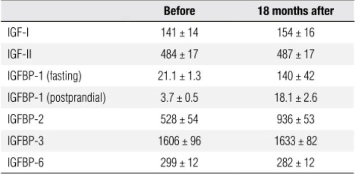
Table 4. Mean insulin-like growth factors and insulin-like growth factor binding globulins, before and after treatment
Before 18 months after
IGF-I 141 ± 14 154 ± 16
IGF-II 484 ± 17 487 ± 17
IGFBP-1 (fasting) 21.1 ± 1.3 140 ± 42
IGFBP-1 (postprandial) 3.7 ± 0.5 18.1 ± 2.6
IGFBP-2 528 ± 54 936 ± 53
IGFBP-3 1606 ± 96 1633 ± 82
IGFBP-6 299 ± 12 282 ± 12
Reference range values: IGF-I: 120-400 ng/mL; IGF-II: 290-730 ng/mL; IGFBP-1 (fasting): 13-120 ng/mL; IGFBP-1 (postprandial): 10-30 ng/mL; IGFBP-2: 360-1020 ng/mL; IGFBP-3: 1500-3600 ng/mL; IGFBP-6: 100-340 ng/mL.
Somatotropic axis
The adult patients had no history of delayed or im-paired growth, and their heights were within the family mean height. Growth hormone levels were ≤ 0.01 ng/ mL, and responses to insulin-induced hypoglycemia and exercise tests were absent in the male (nadir glu-cose level, 37 mg/dL; GH of 0.1 ng/mL in both tests) and in the younger adult female (nadir glucose level, 35 mg/dL; GH, 0.1 ng/mL in the hypoglycemia test and 0.3 ng/mL in the exercise tests). Given the absence of clinical features of growth hormone deiciency, these results are probably attributed to obesity (6). Before
treatment, the boy’s height was at the 50th percentile
in the growth chart. Weight gain and growth
decelera-tion was observed over 2 years (from the 50th to the
10th percentile), possibly due to inadequate dose
ad-justments. After leptin dose increases, the child is now
between the 10th and the 25th percentile, within the
tar-geted height (Figure 5).
In the adults, all of the IGF-related parameters were within normal range, except for postprandial IGFBP-1 (13). After 18 months of leptin replacement therapy (Table 4), we observed signiicant increases in pre and postprandial IGFBP-1 levels. We also observed signii-cant increases in mean IGFBP-2, possibly attributed to the decrease in insulin levels (24).
of TSH pulsatile and circadian rhythms (25). Those data conirm that leptin has a role in regulating TSH secre-tion in humans (26), and its absence may lead to thyroid dysfunction. Leptin replacement did not increase free T4 or T3 (17), as previously observed in other studies (12).
DISCUSSION
Leptin replacement is currently the only successful hormonal treatment for a monogenic form of human obesity. In a cohort of Turkish leptin-deicient patients, our 10-year experience of leptin replacement therapy showed that treatment leads to substantial effects on body composition, food intake and energy expendi-ture, lipid and glucose metabolism, sympathetic tone, and gonadal, adrenal, somatotropic and thyroid func-tions. Additional results also suggest that leptin has important roles on non-endocrine parameters, such as inlammation and coagulation (27), immunity (11,28), and brain structure and function (14,29-33).
Although leptin therapy has proven ineffective in treating patients with common and complex obesity, patients with severe forms of obesity and metabolic syn-drome may present relatively low levels of leptin, cor-rected by adipose tissue mass (34). It is also known that speciic polymorphisms in the leptin gene are associated with higher or lower leptin levels (35,36). Therefore, obese patients with relatively low leptin levels, or with polymorphisms associated with lower leptin, might beneit from leptin therapy.
Although leptin deiciency is an extremely rare dis-ease, our results provide new insight into the long-term effects of leptin in humans through a unique model of obesity presenting with leptin deiciency, without leptin resistance. By understanding the physiology of leptin, we will be able to design future studies that will evaluate the effects of leptin in other diseases, such as common obesity, lipodystrophy syndromes, diabetes, hypotha-lamic amenorrhea, anorexia nervosa, mood and cogni-tive disorders, immune deiciencies, and lipotoxicity.
Disclosure: no potential conlict of interest relevant to this article was reported.
Acknowledgements: We would like to thank Amgen Inc., and Amylin Pharmaceuticals for graciously providing recombinant methionyl human leptin at no cost. We also thank Dr. Amhet Yesilyurt for designing the pedigree chart. This work was fun-ded by the National Institutes of Health Grants RR017365 and DK063240 (to M.-L.W.), RR016996 and DK058851 (to J.L.), and by The Australian National University institutional funds.
Thyroid function
Cop
yright
© ABE&M t
odos os dir
eit os r eser vados .
REFERENCES
1. Kelesidis T, Kelesidis I, Chou S, Mantzoros CS. Narrative review: the role of leptin in human physiology: emerging clinical applica-tions. Ann Intern Med. 2010;152:93-100.
2. Friedman JM. Leptin, leptin receptors, and the control of body weight. Nutr Rev. 1998;56:s38-46.
3. Velloso LA. The hypothalamic control of feeding and thermoge-nesis: implications on the development of obesity. Arq Bras En-docrinol Metabol. 2006;50:165-76.
4. Boucher J, Castan-Laurell I, Daviaud D, Guigne C, Buleon M, Car-pene C, et al. Adipokine expression profile in adipocytes of diffe-rent mouse models of obesity. Horm Metab Res. 2005;37:761-7. 5. Feitosa AC, Mancini MC, Cercato C, Villares SM, Halpern A.
Meta-bolic profile according to leptin levels in obese patients. Arq Bras Endocrinol Metabol. 2007;51:59-64.
6. Ozata M, Ozdemir IC, Licinio J. Human leptin deficiency caused by a missense mutation: multiple endocrine defects, decreased sympathetic tone, and immune system dysfunction indicate new targets for leptin action, greater central than peripheral resistan-ce to the effects of leptin, and spontaneous correction of leptin-mediated defects. J Clin Endocrinol Metab. 1999;84:3686-95. 7. Strobel A, Issad T, Camoin L, Ozata M, Strosberg AD. A leptin
missense mutation associated with hypogonadism and morbid obesity. Nature Genet. 1998;18:213-5.
8. Montague CT, Farooqi IS, Whitehead JP, Soos MA, Rau H, Wa-reham NJ, et al. Congenital leptin deficiency is associated with severe early-onset obesity in humans. Nature. 1997;387:903-8. 9. Farooqi IS, Jebb SA, Langmack G, Lawrence E, Cheetham CH,
Prentice AM, et al. Effects of recombinant leptin therapy in a child with congenital leptin deficiency. N Engl J Med. 1999;341:879-84. 10. Zhang Y, Proenca R, Maffei M, Barone M, Leopold L, Friedman
JM. Positional cloning of the mouse obese gene and its human homologue. Nature. 1994;372:425-32.
11. Farooqi IS, Matarese G, Lord GM, Keogh JM, Lawrence E, Agwu C, et al. Beneficial effects of leptin on obesity, T cell hyporespon-siveness, and neuroendocrine/metabolic dysfunction of human congenital leptin deficiency. J Clin Invest. 2002;110:1093-103. 12. Gibson WT, Farooqi IS, Moreau M, DePaoli AM, Lawrence E,
O’Rahilly S, et al. Congenital leptin deficiency due to homozygo-sity for the Delta133G mutation: report of another case and eva-luation of response to four years of leptin therapy. J Clin Endocri-nol Metab. 2004;89:4821-6.
13. Licinio J, Caglayan S, Ozata M, Yildiz BO, de Miranda PB, O’Kirwan F, et al. Phenotypic effects of leptin replacement on morbid obe-sity, diabetes mellitus, hypogonadism, and behavior in leptin-deficient adults. Proc Natl Acad Sci U S A. 2004;101:4531-6. 14. Paz-Filho GJ, Babikian T, Asarnow R, Delibasi T, Esposito K, Erol
HK, et al. Leptin replacement improves cognitive development. PLoS One. 2008;3:e3098.
15. Mazen I, El-Gammal M, Abdel-Hamid M, Amr K. A novel homo-zygous missense mutation of the leptin gene (N103K) in an obese Egyptian patient. Mol Genet Metab. 2009;97:305-8.
16. Fischer-Posovszky P, von Schnurbein J, Moepps B, Lahr G, Strauss G, Barth TF, et al. A new missense mutation in the leptin gene causes mild obesity and hypogonadism without affecting T cell responsiveness. J Clin Endocrinol Metab. 2010;95:2836-40. 17. Paz-Filho G, Delibasi T, Erol HK, Wong ML, Licinio J. Congenital
leptin deficiency and thyroid function. Thyroid Res. 2009;2:11. 18. Licinio J, Mantzoros C, Negrao AB, Cizza G, Wong ML, Bongiorno
PB, et al. Human leptin levels are pulsatile and inversely related to pituitary- adrenal function. Nature Medicine. 1997;3:575-9. 19. Mantzoros CS, Ozata M, Negrao AB, Ziotopoulou M, Caglayan S,
Suchard M, et al. Synchronicity of frequently sampled TSH and
leptin concentrations in healthy adults and leptin deficient sub-jects: evidence for possible partial TSH regulation by leptin in hu-mans. J Clin Endocrinol Metab. 2001;86:3284-91.
20. Licinio J, Ribeiro L, Busnello JV, Delibasi T, Thakur S, Elashoff RM, et al. Effects of leptin replacement on macro- and micronutrient preferences. Int J Obes (Lond). 2007;31:1859-63.
21. Galgani JE, Greenway FL, Caglayan S, Wong ML, Licinio J, Ra-vussin E. Leptin replacement prevents weight loss-induced me-tabolic adaptation in congenital leptin-deficient patients. J Clin Endocrinol Metab. 2010;95:851-5.
22. Andreev VP, Paz-Filho G, Wong ML, Licinio J. Deconvolution of insulin secretion, insulin hepatic extraction post-hepatic delivery rates and sensitivity during 24-hour standardized meals: time course of glucose homeostasis in leptin replacement treatment. Horm Metab Res. 2009;41:142-51.
23. Paz-Filho G, Esposito K, Hurwitz B, Sharma A, Dong C, Andreev V, et al. Changes in insulin sensitivity during leptin replacement therapy in leptin-deficient patients. Am J Physiol Endocrinol Me-tab. 2008;295:E1401-8.
24. Wheatcroft SB, Kearney MT. IGF-dependent and IGF-independent actions of IGF-binding protein-1 and -2: implications for metabo-lic homeostasis. Trends Endocrinol Metab. 2009;20:153-62. 25. Mantzoros CS, Ozata M, Negrao AB, Suchard MA, Ziotopoulou M,
Caglayan S, et al. Synchronicity of frequently sampled thyrotro-pin (TSH) and leptin concentrations in healthy adults and leptin--deficient subjects: evidence for possible partial TSH regulation by leptin in humans. J Clin Endocrinol Metab. 2001;86:3284-91. 26. Moura EG, Moura CC. Regulation of thyrotropin synthesis and
secretion. Arq Bras Endocrinol Metabol. 2004;48:40-52.
27. Paz-Filho GJ, Andrews D, Esposito K, Erol HK, Delibasi T, Wong ML, et al. Effects of leptin replacement on risk factors for car-diovascular disease in genetically leptin-deficient subjects. Horm Metab Res. 2009;41:164-7.
28. Paz-Filho GJ, Delibasi T, Erol HK, Wong ML, Licinio J. Cellular im-munity before and after leptin replacement therapy. J Pediatr En-docrinol Metab. 2009;22:1069-74.
29. Baicy K, London ED, Monterosso J, Wong ML, Delibasi T, Sharma A, et al. Leptin replacement alters brain response to food cues in genetically leptin-deficient adults. Proc Natl Acad Sci U S A. 2007;104:18276-9.
30. Matochik JA, London ED, Yildiz BO, Ozata M, Caglayan S, De-Paoli AM, et al. Effect of leptin replacement on brain structure in genetically leptin-deficient adults. J Clin Endocrinol Metab. 2005;90:2851-4.
31. Farooqi IS, Bullmore E, Keogh J, Gillard J, O’Rahilly S, Fletcher PC. Leptin regulates striatal regions and human eating behavior. Science. 2007;317:1355.
32. Lieb W, Beiser AS, Vasan RS, Tan ZS, Au R, Harris TB, et al. Asso-ciation of plasma leptin levels with incident Alzheimer disease and MRI measures of brain aging. JAMA. 2009;302:2565-72. 33. Paz-Filho G, Wong ML, Licinio J. Leptin levels and Alzheimer
dise-ase. JAMA. 303:1478; author reply -9.
34. da Paz-Filho GJ, Volaco A, Suplicy HL, Radominski RB, Bo-guszewski CL. Decrease in leptin production by the adipose tis-sue in obesity associated with severe metabolic syndrome. Arq Bras Endocrinol Metabol. 2009;53:1088-95.
35. Hinuy HM, Hirata MH, Forti N, Diament J, Sampaio MF, Armaga-nijan D, et al. Leptin G-2548A promoter polymorphism is asso-ciated with increased plasma leptin and BMI in Brazilian women. Arq Bras Endocrinol Metabol. 2008;52:611-6.
36. Hinuy HM, Hirata MH, Sampaio MF, Armaganijan D, Arazi SS, Sa-lazar LA, et al. Relationship between variants of the leptin gene and obesity and metabolic biomarkers in Brazilian individuals. Arq Bras Endocrinol Metabol. 54:282-8.